PLUSH ‘BEAUTY BLEND’ : Youthful Skin Restoration / Ultimate Anti Wrinkle Agent 200:1
July 19, 2021
JING FORCE : Kidney Restoration Tonic 200:1
October 22, 2021LIVER REGENERATOR 200:1 – Master Hepatoprotective
$275.00
introducing
Liver REGENERATOR

MASTER Hepatoprotective FORMULA
200:1 Concentration
Featuring: (−)-4′-O-Methylepigallocatechin • 3,5-Di-O-Caffeoyl Quinic Acid • 5,7,4′-Trihydroxy-3′- Methoxyisoflavone • Aburs Cantoniensis Extract • Acacia Confusa Merr. (Leguminosae), • Actinoscirpus Grossus Tubers • Adenosine Triphosphate • Agathisflavone • Amaranthus Spinosus L • Amorphophallus Campanulatus Roxb. Tubers • Anastatin A • Anastatin B • Andrographolide • Angelica Sinensis (Oliv.)Diels Root Extract • Arjunolic Acid • Aronia Melanocarpa • Artemisia Capillaris • Artemisia Iwayomogi • Artemisia Scoparza Waldst.Et Kit Extract • Asparagus Racemosus Willd. Root Extract • Astragalus Membranaceus Extract • Astragalus Mongholicus Bunge Extract • Berberine • Beta Vulgaris Linn.(Bv, Chenopodiaceae) Leaves • Betalain • Bupleurum Chinense Root Extract • Cassia Seed Extract • Catechin • Chestnut (Castanea Crenata) Inner Shell Extract • Chrysanthemum Indicum L. Extract • Cichorium Inthybus L. • Cichorium Intybus Root Extract • Cimicifuga Foetida L.Root Extract • Cinnamomum Cassia Presl Extract • Coptis Chinensis Franch Root Extract • Cordyceps Sinensis( Berk.)Sacc. Extract • Cortex Dictamni Aqueous Extract • Crataegus Pinnatifi Da Bge. Var. Major Leaves Extract • Curcuma Longa L. Extract • Curcuma Phaeocaulis Radix • Curcumin • Curdrania Tricuspidata Leaves • Dendropanax Morbifera Leveille Leaves • Desmodium Triquetrum Leaf • Dl-Methionine • Dropwort (Oenanthe Javanica) • Eclipta Alba • Epicatechin • Forsythia Suspensa Extract • Fructus Schisandrae (Wuweizi In Chinese) • Galangin • Ganoderma Lucidum • Garlic (Aged Black) • Genistein • Gentiana Scabra Bunge Extract • Glycyrrhiza Glabra • Glycyrrhiza Uralensis Fisch. Extract • Glycyrrihizin • Gossypin • Halichrysin A And B • Hedyotis Diffusa Extract • Hibiscus Vitifolius Root Extract • Houttuynia Cordata Thunb Extract • Hovenia Dulcis Extract • Hypophyllanthin • Imperata Cylindrica Beauv.Var. Major( Nees)C.E.Hubb. Root Extract • Isatis Indigotica Fort.Root Extract • Isoquercitrin • Kaempferol • Kolaviron • Kutkoside • Ligularia Fischeri • Ligusticum Chuanxiong Hort Extract • Limonium Tetragonum • Lithospermum Erythrorhizon Sieb. Et Zucc Extract • Lonicera Japonica Thunb. Extract • Luteolin • Lycium Barbarum L. Extract • Lysimachia Christinae Hance Extract • Mangiferin • Murraya Koenigii • Naringenin • Nelumbo Nucifera Gaertn. • Neoandrographolide • Nepenthes Khasiana Hook. F • Onitin • Ornithine Aspartate • Oxymatrine • Paeonia Lactiflora Pall Root Extract • Panax Ginseng C. A. Meyer Extract • Panaxnotoginseng(Burk.)F.H.Chen Root Extract • Patrinia Scabiosaefolia Extract • Penthorum Chinense Pursh Extract • Phellodendri Cortex (Pc; Bark Of Phellodendron Amurense Rupr.) • Phloridzin • Phyllanthin • Phyllanthus Emblica L. (Amla) • Phyllanthus Niruri • Picrorhiza Kurroa • Picroside • Plumbagin • Polydatin • Polyene Phosphatidyl Choline • Polygonum Cuspidatum Sieb.Et Zucc Extract • Polyporus Umbellatus Extract • Poria Cocos • Psoralea Corylifolia L. (Pc) Seeds Extract (Pce) • Puerarin (Pr), An Active Component Extracted From The Kudzu Root • Quercetin • Radix Puerariae • Rehm,Annia Glutinosa Libosch. Extract • Rheum Palmatum L Extract • Rubiadin • Rutin • Salidroside • Salvia Miltiorrhiza Bge Root Extract • Sanguisorba Officinalis L Extract • Schisandra Chinensis(Turcz.)Baill. Extract • Scopoletin • Scutellaria Barbata D. Don Extract • Sedum Sarmentosum Bunge Extract • Semen Hoveniae • Sida Cordifolia Linn. (Malvaceae) • Silybin, Isosilybin, Silychristin, Silydianin • Silybum Marianum (L.) Gaertn. Extract • Spatholobus Suberectus Dunn Extract • Stephania Tetrandra S.Moore Root Extract • Superoxide Dismutase [Cu-Zn] • Tarax Acum Mongolicum Extract • Taraxacum Officinale (Dandelion) Extract • Troxerutin • Vitexin 7-O-Β-L-Glucopyranoside & Vitexin 2″-O-Β-Glucopyranoside • Vitis Amurensis Seeds (Procyanidins From Wild Grape) • Wedelolactone • Wighteone • Withaferin A • Wu-Zi-Yan-Zong-Wan • Β-Sitosterol •
Introduction to Liver
In the human body, the Liver plays a crucial role in regulating many biological processes, such as detoxification, metabolism of fat, carbohydrate, and protein, secretion of bile, removal of components from the blood circulation, and storage of vitamins. Through these physiological actions, the Liver supports the maintenance, performance, and regulation of the human body’s homeostasis. It also involves multiple biochemical pathways related to energy provision, fighting against disorders/diseases, nutrient supply, growth, and reproduction. The Liver is also an abundant source of essential (histidine and threonine) and nonessential (aspartate, alanine, glycine, glutamate, and serine) amino acids.
From the 1970s, several researchers analyzed the ability of the Liver to synthesize useful agents to detoxify drugs, waste substances, xenobiotics, and toxic metabolites from portal circulation by using various models in humans and animals. This detoxification of the Liver makes it liable to preserve attacks on offending endogenous and/or exogenous substances, causing Liver dysfunction. Generally, Liver disease is defined as an injury to hepatic cells and tissues. The hepatic diseases are mainly categorized into two subtypes, namely acute and chronic hepatitis, associated with inflammatory complications like hepatosis. It is estimated that about two million hepatic disease–related deaths are recording annually across the world. Of these, 50% of the cases are caused by cirrhosis (Liver fibrosis), and the rest are the result of hepatitis and hepatocellular carcinoma. Overall, hepatic-related deaths accounted for ∼3% of all the deaths annually worldwide, highest in Latin America and Africa, followed by Asia.
Mostly, hepatic diseases are caused by viral infections (Hepatitis A–E), autoimmune diseases, high doses of drug consumptions (i.e., paracetamol, acetaminophen, and antibiotics), hazardous compound exposure (aflatoxin, CCl4, GalN, dimethylnitrosamine, chlorinated hydrocarbons, peroxidized oil, and thioacetamide), and excessive intake of Alcohol. Etiological studies identified that Liver dysfunction is chiefly induced by lipid peroxidation in the Liver, followed by cirrhosis and hepatitis. Despite broad advances in current medicine, there are no effective treatments of choice for stimulating hepatic function or aid in rejuvenating hepatic cells. Moreover, hepatic disease treatments are controversial because most of the current synthetic drugs for managing these diseases cause severe side effects. Therefore, it is in great demand to search for alternative medicine to treat hepatic diseases with less toxicity and adverse effects. This current review aimed to provide sufficient information about hepatic diseases and their current diagnostic methods. Additionally, the use of bioflavonoids in the treatment of hepatic diseases was discussed.
As the Liver is close to the gastrointestinal system, it is susceptible to viral infections and toxic substances due to its unique metabolism. Usually, the Liver gets drained with the concentrated forms of drugs and xenobiotic substances from the gastrointestinal organs’ blood. The accumulated xenobiotics and toxic substances get activated in the Liver and produce reactive metabolic species as byproducts.
These unstable species induce hepatic damage or worsen the injury by several mechanisms. Also, hepatotoxic chemicals such as amiodarone, ethanol, GalN, halothane, isoniazid, CCl4, phenytoin, methyldopa, paracetamol, and inorganic compounds (arsenic, copper, phosphorus, and iron) harm the hepatic cells mostly by inducing lipid peroxidation in the Liver. The various types of hepatotoxicity conditions are mentioned below.
Alcoholic Liver Disease : It is evidenced that excessive Alcohol consumption causes the chronic hepatic disease that damages the Liver and buildups of inflammation, fats, and scars. Usually, Alcoholic Liver disease has four phases—Alcoholic fatty Liver disease, Alcoholic hepatitis, fibrosis, and cirrhosis. In early stage, Alcoholic Liver disease can be diagnosed by abdominal pain, diarrhea, and vomiting. Later stages include edema of the lower limbs, jaundice, ascites, fever, itchy skin, clubbing, blood in stools and vomit, and significant bodyweight reduction. Avoiding excessive Alcohol consumption has been noted as the best and only way to avoid Alcoholic Liver disease.

Various types of hepatic diseases.
Cirrhosis: Long-term inflammation of the Liver creates a life-threatening condition called cirrhosis. During cirrhosis, fibrosis is formed in the Liver leading to scarring and loss of organ function. It is well known that all chronic forms of hepatic disease finally end with cirrhosis, characterized by the reduced capability to rejuvenate hepatic cells, decreased blood flow, and loss of hepatic cells. In the beginning, cirrhosis shows no abnormalities on imaging or in blood tests. There is a loss of hepatic function due to internal bleeding and fluid accumulation in the abdominal cavity in the long run. To date, Liver transplantation is the only treatment for last-stage cirrhosis.
Hepatocellular carcinoma: Hepatocellular carcinoma is the most common kind of hepatic cancer in humans with chronic hepatic diseases. It is categorized into two major subtypes, namely hepatocellular carcinoma and intrahepatic cholangiocarcinoma. Hepatic cancer is diagnosed by blood tests, imaging, and Liver biopsy. Current treatments include chemotherapy, surgery, immunotherapy, radiation therapy, and Liver transplantation.
Hepatitis: Hepatitis is an inflammatory disorder of the hepatic system mostly caused by viruses (Hepatitis A, B, C, D, or E) and rarely by an autoimmune reaction (autoimmune hepatitis). Hepatitis A and E are acute (short term), while others are chronic forms (on-going). Hepatitis A is transmitted via food and/or water or contaminated feces from the infected person. Hepatitis B and C are transmitted via infectious body fluids or sex with an infected person. Hepatitis D (Δ-hepatitis) is the rarest form of hepatitis and is transmitted mainly by direct contact with infected blood. Hepatitis E, on the other hand, is waterborne and very dangerous to pregnant women. The main symptoms of hepatitis are flu-like symptoms, dark urine, yellow skin and eyes, signs of jaundice, pale stool, and abdominal pain. The treatment of hepatitis varies depending on the means of viral transmission. However, hepatitis is prevented by lifestyle precautions and immunizations.
NonAlcoholic fatty Liver disease: NonAlcoholic fatty Liver is the most common disorder that refers to a higher accumulation of fat in the hepatic cells due to obesity and diabetes in people who do not abuse Alcohol. Although the presence of fat in the Liver is abnormal, it does not harm the Liver. The majority of the patients with fatty Liver show no signs and symptoms, except abdominal pain in some cases. Epidemiological studies suggest that for only a few people, fatty Liver may lead to non-Alcoholic steatohepatitis, a severe condition involving inflammation in fatty Liver cells. The clinicopathological studies show that fatty Liver is a significant cause of cryptogenic hepatocellular carcinoma, cryptogenic cirrhosis, and hepatic transaminases. However, clinical studies on fatty Liver suggest that it could be treated by controlled weight loss and physical activities.
Plants and their phytochemicals used in hepatic diseases: From prehistoric times, plants have been the primary source for managing several ailments due to the presence of high bioactive compounds with low toxicity. Nearly 80% of the world’s population has employed plant materials (also known as traditional medicine) for healthcare. The etiology of hepatic diseases is diverse, and a variety of medicinal plants has been reported to have Hepatoprotective activity through various mechanisms, such as reducing peroxidation, improvement of antioxidant defense (CAT, GPx, and SOD activity), antioxidant properties, reversed hepatic fibrosis, activation of hepatic stellate cells, anti-inflammatory activity, and antifibrotic properties.
Moreover, basic clinical research has exposed the mechanism of actions by which a few plants produce their beneficial properties in regards to cirrhosis, Liver regenerating effects, fatty Liver, radiation toxicity, ischemic injury, viral hepatitis, and toxic hepatitis. Several medicinal plants were subjected to chemical examination to identify potential bioactive metabolites having Hepatoprotective properties, which led to the identification of phytochemicals of phenolics, terpenoids, flavonoids, lignans, carotenoid, steroids, amino acids, proteins, carotenoid, tannins, glycosides and alkaloids classes.
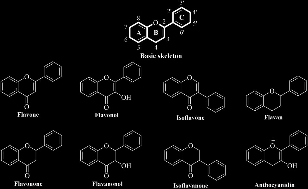
Bioflavonoids with Hepatoprotective activity: Bioflavonoids or flavonoids are low-molecular-weight phenolics that possess a basic C6–C3–C6structure of an aromatic ring (A) attached to a benzene ring (B) fused to a heterocyclic ring (C) with a single C–C bond. Flavonoids can be divided into a number of groups that differ from each other according to the degree of oxidation on ring C, the degree of unsaturation, and the number and position of –OH groups on flavones, flavanones, flavonols, flavanonols, isoflavones, isoflavanones, flavans, and anthocyanidins.. Flavonoids are often bound to glycosides, such as arabinose, d-glucose, galactose, l-rhamnose, glucorhamnose, etc., and are termed as flavonoid-glycosides. Similarly, glycoside forms of anthocyanidins are called as anthocyanins. On the other hand, flavonoids bound to lignans are termed as flavonolignans.
Biologically, flavonoids are found in various plant parts and are mostly used for plant growth and defensive mechanisms. These molecules also act as a UV filter to plants and protect them from different abiotic and biotic stresses. They have mostly been identified in fruits, flowers, vegetables, leaves, cocoa, tea, onions, and dietary products, and they have been reported to have multiple bioactivities such as antioxidant, anticancer, and antidiabetic anti-inflammation, antimicrobial, immunomodulatory, and Hepatoprotective properties. They are also well acknowledged to have potent inhibitory properties for different enzymes such as cyclooxygenase, α-amylase, phosphoinositide, lipoxygenase, xanthine oxidase, α-glucosidase, and aldose reductase. Flavonoids have been long credited for their beneficial effects on human health and recently tested for their chemoprevention and disease therapy. To date, nearly 6000 flavonoids have been identified from fruits, seeds, flowers, stems, roots, buds, and vegetables of various medicinal plants. Of these, approximately 100 bioflavonoids have been reported for their Hepatoprotective activites.
INGREDIENTS & SCIENCE:
(−)-4′-O-Methylepigallocatechin
These results suggest that the antioxidative activity of the principal phenolic compounds is involved in the Hepatoprotective activity of S. reticulata.
3,5-Di-O-caffeoyl quinic acid
In Vitro Hepatoprotective compounds from Suaeda glauca
Bioassay-guided fractionation of the MeOH extract of Suaeda glauca yielded four phenolic compounds, methyl 3,5-di-O-caffeoyl quinate (1) and 3,5-di-O-caffeoyl quinic acid (2), isorhamnetin 3-O-β-D-galactoside (3), and quercetin 3-O-β-D-galactoside (4). Compounds 1 and 2 were Hepatoprotective against tacrine-induced cytotoxicity in human Liver-derived Hep G2 cells with the EC50 values of 72.7±6.2 and 117.2±10.5 μM, respectively. Silybin as a positive control showed an EC50 value of 82.4±4.1 μM.
The water extract of propolis (PWE) showed a strong Hepatoprotective activity against CCl4-toxicity in rats and D-galactosamine (GalN)/lipopolysaccharide (LPS)-induced Liver injury in mice.
5,7,4′-Trihydroxy-3′- methoxyisoflavone
New Isoflavones and Pterocarpane with Hepatoprotective Activity from the Stems of Erycibe expansa
The methanolic extract from the stems of Erycibe expansa was found to show a Hepatoprotective effect on D-galactosamine-induced cytotoxicity in primary cultured mouse hepatocytes. By bioassay-guided separation, two new prenylisoflavones and a pterocarpane, erycibenins A (1), B (2), and C (3), were isolated from the active fraction (the EtOAc-soluble fraction) together with ten isoflavones (4 – 13) and seven pterocarpanes (14 – 20).
The stereostructures of the new compounds were determined on the basis of chemical and physicochemical evidence including modified Mosher’s method. In addition, the isolated constituents, erycibenin A (1, IC50 = 79 μM), genistein (6, 29 μM), orobol (7, 36 μM), and 5,7,4′-trihydroxy-3′-methoxyisoflavone (8, 55 μM) exhibited inhibitory activity on D-galactosamine-induced cytotoxicity in primary cultured mouse hepatocytes.
Hepatoprotective potential of bioflavonoids
The Liver controls the body’s internal environment via various physiological and metabolic processes, either independently or together with otfher organs. Liver disease is defined as an injury to the hepatic cells and tissues, mainly caused by viral infections, toxic compounds, high doses of drugs, and excessive Alcohol intake. There are an estimated two million hepatic disease–associated deaths recorded annually worldwide with a diverse etiology.
Numerous medicinal plants and their phytochemicals have been reported to display Hepatoprotective activity. Among those, bioflavonoids, low-molecular-weight phenolics possessing a basic C6–C3–C6 structure, have been reported to have the ability to treat hepatic diseases. In the current study, we aimed to summarize hepatic diseases and strategies to treat them and added a brief note on the bioflavonoids that have been tested against hepatic diseases.
Aburs cantoniensis extract
Protective Effect of Ethanol Extraction of Abrus cantoniensis on Non-Alcoholic Fatty Liver in Rats
CONCLUSION: Ethanol extraction of A.cantoniensis can reduce the content of ALT and AST in serum and Liver index,and relieve the pathologic damage and the degree of fatty degeneration of hepatic tissue.Ethanol extraction of A.cantoniensis can improve fatty Liver induced by CCl4 and high-fat feeding in rats.
Abrusamide A and B, two Hepatoprotective isomeric compounds from Abrus mollis Hance
Two isomeric compounds, abrusamide A (1) and abrusamide B (2), were isolated from the leaves of Abrus mollis Hance. Their structures were well defined by means of UV, HR–ESI–MS, 1H NMR, 13C NMR and 2D NMR techniques. From a biogenetic point of view, these two compounds including a cyclobutane basic core should be considered as a [2+2] dimerization product of (E)-N-(4-hydroxycinnamoyl) tyrosine (3), which was also isolated from the plant for the first time. They were also tested for their Hepatoprotective effects on human L-02 cells and displayed significant promote effects on the proliferation of L-02 cells and significant Hepatoprotective effects on CCl4-induced injury of L-02 cells.
The antihepatotoxic activities of soyasaponin I and kaikasaponin III, triterpenoidal saponins isolated from Abri Herba, the whole plant of Abrus cantoniensis, were studied on Liver injury induced by CCl4 in primary cultured rat hepatocytes. The antihepatotoxic activities of these saponins and glycyrrhizin (positive control) were demonstrated by measuring the levels of glutamic pyruvic transaminase (GPT) and glutamic oxaloacetic transaminase (GOT). Soyasaponin I inhibited the elevation of GOT and GPT activities. The activities were comparable to those of glycyrrhizin.
On the other hand, kaikasaponin III was more effective than soyasaponin I and glycyrrhizin. Kaikasaponin III showed the antihepatotoxic activity at less than 100 µg/ml. Furthermore, the highest activity was observed even in the lower doses (50, 100 µg/ml). However, soyasaponin I and kaikasaponin III showed some toxicity at the highest dose (500 µg/ml), though glycyrrhizin did not show toxicity at any dose.
It has been claimed that consumptions of Abrus cantoniensis (AC) and Abrus mollis (AM) as folk beverages and soups are good to cleanse Liver toxicants and prevent Liver diseases. There is scant information on the phytochemical profiles and antioxidant activities of these two varieties. Five major phytochemicals in these two cultivars were qualitatively and quantitatively compared using UPLC-PDA. A high level of total phenolic content (TPC) and total flavonoid content (TFC) was found in AC and AM. AC, in general, showed some antioxidant activities comparable to that of BHT, and stronger radical scavenging activities and higher reducing power than that of AM (p < 0.05). When principal component analysis (PCA) was applied, high correlation between TPC, TFC and their antioxidant activities was found. Hence, this study proved that, both AC and AM could serve as antioxidant-rich component in foods or beverages to promote health function.
Acacia confusa Merr. (Leguminosae)
These results suggested that the ACBE and gallic acid exhibit potent hepatoprotection against CCl4-induced Liver damages in rats, and the Hepatoprotective effects of ACBE and gallic acid may be due to the modulation of antioxidant enzymes activities and inhibition of lipid peroxidation and CYP2E1 activation.
A review of antioxidant and pharmacological properties of phenolic compounds in Acacia confusa
In the present review article, the phytochemical, antioxidant and pharmacological studies are congregated and summarized concerning the current knowledge of the phenolic compounds of a traditional medical plant Acacia confusa in Taiwan.
This plant is native to Taiwan and South-East Asia. It possesses major pharmacological activities, including antioxidant and radical scavenging activity, Hepatoprotective effect, xanthine oxidase inhibition, semicarbazide-sensitive amine oxidase inhibition, angiotensin I converting enzyme inhibition, antihyperuricemic effect and anti-inflammatory activity.
Anti-hepatitis C virus activity of Acacia confusa extract via suppressing cyclooxygenase-2
Chronic hepatitis C virus (HCV) infection continues to be an important cause of morbidity and mortality by chronic hepatitis, cirrhosis and hepatocellular carcinoma (HCC) throughout the world. It is of tremendous importance to discover more effective and safer agents to improve the clinical treatment on HCV carriers. Here we report that the n-butanol–methanol extract obtained from Acacia confusa plant, referred as ACSB-M4, exhibited the inhibition of HCV RNA replication in the HCV replicon assay system, with an EC50 value and CC50/EC50 selective index (SI) of 5 ± 0.3 μg/ml and >100, respectively. Besides, ACSB-M4 showed antiviral synergy in combination with IFN-α and as HCV protease inhibitor (Telaprevir; VX-950) and polymerase inhibitor (2′-C-methylcytidine; NM-107) by a multiple linear logistic model and isobologram analysis.
A complementary approach involving the overexpression of COX-2 protein in ACSB-M4-treated HCV replicon cells was used to evaluate the antiviral action at the molecular level. ACSB-M4 significantly suppressed COX-2 expression in HCV replicon cells. Viral replication was gradually restored if COX-2 was added simultaneously with ACSB-M4, suggesting that the anti-HCV activity of ACSB-M4 was associated with down-regulation of COX-2, which was correlated with the suppression of nuclear factor-kappaB (NF-κB) activation. ACSB-M4 may serve as a potential protective agent for use in the management of patients with chronic HCV infection.
Actinoscirpus grossus tubers
Thus, tubers of A. grossus plant may serve as reservoir of natural products having diverse functions and could be utilised as a potent Hepatoprotective agent.
Agathisflavone
Agathisflavone: Botanical sources, therapeutic promises, and molecular docking study
CONCLUSION: Agathisflavone may be one of the promising plant-derived lead compounds in the treatment of oxidative stress, inflammatory diseases, microbial infection, hepatic and neurological diseases and disorders, and cancer.
Structure and Hepatoprotective Activity of a Biflavonoid from Canarium manii
Canarium manii (Burseraceae) was chemically investigated and the presence of the biflavonoid agathisflavone is reported for the first time from this plant. Pharmacologically, this biflavonoid in doses 50.0 mg and 100.0 mg given orally exhibited dose-dependent Hepatoprotective activity against experimentally-induced carbon tetrachloride-hepatotoxicity in rats and mice.
Hepatoprotective potential of bioflavonoids
The Liver controls the body’s internal environment via various physiological and metabolic processes, either independently or together with other organs. Liver disease is defined as an injury to the hepatic cells and tissues, mainly caused by viral infections, toxic compounds, high doses of drugs, and excessive Alcohol intake. There are an estimated two million hepatic disease–associated deaths recorded annually worldwide with a diverse etiology.
Numerous medicinal plants and their phytochemicals have been reported to display Hepatoprotective activity. Among those, bioflavonoids, low-molecular-weight phenolics possessing a basic C6–C3–C6 structure, have been reported to have the ability to treat hepatic diseases. In the current study, we aimed to summarize hepatic diseases and strategies to treat them and added a brief note on the bioflavonoids that have been tested against hepatic diseases.
Research on the Scientific Evolution of the Flavonoid Agathisflavone
CONCLUSION: Although agathisflavone has been known in the literature since at least 1969, only 23 of the eligible articles found evaluated its possible therapeutic effects. The demonstrated biological activities of agathisflavone range from antiprotozoal to neurogenesis and neuroprotection, however, the molecule needs to be better studied at the in vivo and human level.
A review of natural products with Hepatoprotective activity
Liver diseases are a major worldwide health problem, with high endemicity in developing countries. They are mainly caused by chemicals and some drugs when taken in very high doses. Despite advances in modern medicine, there is no effective drug available that stimulates Liver function, offer protection to the Liver from damage or help to regenerate hepatic cells. There is urgent need, therefore, for effective drugs to replace/supplement those in current use. The plant kingdom is undoubtedly valuable as a source of new medicinal agents. The present work constitutes a review of the literature on plant extracts and chemically defined molecules of natural origin with Hepatoprotective activity. The review shows 107 plants, their families, geographical distribution, plant parts utilized, type of assay and inducer of Liver damage. It also includes 58 compounds isolated from higher plants, classified into appropriate chemical groups. This work intends to aid researchers in the study of natural products useful in the treatment of Liver diseases.
A quest for staunch effects of flavonoids: Utopian protection against hepatic ailments
The role of flavonoids as the major red, blue and purple pigments in plants has gained these secondary products a great deal of attention over the years. Flavonoids are polyphenols and occur as aglycones, glycosides and methylated derivatives. Flavonoids are the main components of a healthy diet containing fruits and vegetables and are concentrated especially in tea, apples and onions. Till date, more than 6000 flavonoids have been discovered, out of which 500 are found in free state.
They are abundant in polygonaceae, rutaceae, leguminosae, umbelliferae and compositae. Flavonoids are powerful antioxidants. In addition to their role in nutrition, flavonoids possess many types of pharmacological activities, including anti-inflammatory, antioxidative, Hepatoprotective, vasorelaxant, antiviral and anticarcinogenic effects. The present review is focused on flavonoids derived from natural products that have shown a wise way to get a true and potentially rich source of drug candidates against Liver ailments.
The present review initially highlights the current status of flavonoids and their pharmaceutical significance, role of flavonoids in hepatoprotection, therapeutic options available in herbal medicines and in later section, summarizes flavonoids as lead molecules, which have shown significant Hepatoprotective activities.
Amaranthus spinosus L
A review for discovering Hepatoprotective herbal
drugs with least side effects on kidney
The Liver is a vital organ which plays a major role in the metabolism and excretion of xenobiotics from the body, and Liver disease is a worldwide health problem. The currently available synthetic drugs to treat Liver disorders cause further damage to the Liver and kidney so it is imperative to find new drugs with least side effects. There are a number of treatment combinations which are derived from medicinal plants and commonly administered as tonic for the Liver.
In this review, we have introduced most important medicinal plants that are used in Liver disorders and have least side effects on kidney. In this regards, we have focused on their active constituents, effects and trial studies, mechanisms of action, pharmacokinetic characteristics, dosages, and toxicity.Amaranthus spinosusL.,Glycyrrhiza glabra,Cichorium inthybusL. Phyllanthus species (amarus,niruri,emblica),Picrorhiza kurroa, andSilybum marianumhave been extensively administered for the treatment of Liver disorders. The introduced medicinal plants can be used for production of new drugs via antioxidant-related properties, Hepatoprotective activities and least side effects on kidney for the prevention and treatment of Liver disorders
Our results provide the first experimental evidence that a novel fatty acid isolated from A. spinosus exhibits significant antiproliferative activity mediated through the induction of apoptosis in HepG2 cells. These encouraging results may facilitate the development of A. spinosus fatty acid for the prevention and intervention of hepatocellular carcinoma.
CONCLUSION: The Hepatoprotective potential of ASS could be explained by its high phenolic content, antioxidant properties and phytochemical contents.
CONCLUSION: This presence of amino acids, flavonoids and phenolic compounds in the MEAS might be responsible for its marked chemoprotective and antioxidant activities in paracetamol induced-Liver damage in Wistar rats.
Hepatoprotective activity of Amaranthus spinosus in experimental animals
The results of this study strongly indicate that whole plants of A. spinosus have potent Hepatoprotective activity against carbon tetrachloride induced hepatic damage in experimental animals. This study suggests that possible mechanism of this activity may be due to the presence of flavonoids and phenolics compound in the ASE which may be responsible to Hepatoprotective activity.
Results of this study revealed that A. spinosus extract could afford a significant protection against d-GalN/LPS-induced hepatocellular injury.
Amorphophallus campanulatus Roxb. tubers
CONCLUSION: The results of this study strongly indicate that ethanolic extract of Amorphophallus campanulatus has potent Hepatoprotective action against ethanol induced hepatic damage particularly by scavenging free radicals and combating oxidative stress. Further investigation can lead to the development of phytomedicines of therapeutic significance against oxidative stress. Keywords: Amorphophallus campanulatus, Ethanol, Hepatoprotection, Oxidative stress, Phytomedicine
CONCLUSION: From the results it can be concluded that Amorphophallus paeoniifolius possesses Hepatoprotective effect against paracetamol-induced Liver damage in rats.
The results of this study strongly indicate that Amorphophallus campanulatus (Roxb.) tubers have potent Hepatoprotective action against carbon tetrachloride induced hepatic damage in rats. The ethanolic extract was found Hepatoprotective more potent than the aqueous extract. The antioxidant activity was also screened and found positive for both ethanolic and aqueous extracts.
This study suggests that possible mechanism of this activity may be due to free radical scavenging potential caused by the presence
of flavonoids in the extracts.
Anastatin A
New skeletal flavonoids, anastatins A and B, were isolated from the methanolic extract of an Egyptian medicinal herb, the whole plants of Anastatica hierochuntica. Their flavanone structures having a benzofuran moiety were determined on the basis of chemical and physicochemical evidence. Anastatins A and B were found to show Hepatoprotective effects on d-galactosamine-induced cytotoxicity in primary cultured mouse hepatocytes and their activities were stronger than those of related flavonoids and commercial silybin.
New skeletal flavonoids, anastatins A and B, were isolated from the methanolic extract of an Egyptian medicinal herb, the whole plants of Anastatica hierochuntica. Their flavanone structures having a benzofuran moiety were determined on the basis of chemical and physicochemical evidence. Anastatins A and B were found to show Hepatoprotective effects on d-galactosamine-induced cytotoxicity in primary cultured mouse hepatocytes and their activities were stronger than those of related flavonoids and commercial silybin.
The results show that most of these flavonoid compounds have good antioxidant activity and low cytotoxicity in vitro. Among them, the most potent compound was 38c, which exhibited a protective effect on CCl4-induced hepatic injury by suppressing the amount of CYP2E1. These findings indicate that anastatin flavonoid derivatives have potential therapeutic utility against oxidative hepatic injury.
CONCLUSION: Biochemical observations, supplemented by histopathological examination revealed that AH affords extract-depending protection against CCl_4-hepatotoxicity.
Anti-Hepatotoxic Effect of the Methanolic Anstatica
Hierochuntica Extract In Ccl 4- Treated Rats
From the above results, it is concluded for the first time that methanolic Anastatica hierochuntica extract offers protective effect against CCl4-induced hepatotoxicity in experimental rats.
New skeletal flavonoids, anastatins A and B, were isolated from the methanolic extract of an Egyptian medicinal herb, the whole plants of Anastatica hierochuntica. Their flavanone structures having a benzofuran moiety were determined on the basis of chemical and physicochemical evidence. Anastatins A and B were found to show Hepatoprotective effects on d-galactosamine-induced cytotoxicity in primary cultured mouse hepatocytes and their activities were stronger than those of related flavonoids and commercial silybin.
Anastatin B
New skeletal flavonoids, anastatins A and B, were isolated from the methanolic extract of an Egyptian medicinal herb, the whole plants of Anastatica hierochuntica. Their flavanone structures having a benzofuran moiety were determined on the basis of chemical and physicochemical evidence. Anastatins A and B were found to show Hepatoprotective effects on d-galactosamine-induced cytotoxicity in primary cultured mouse hepatocytes and their activities were stronger than those of related flavonoids and commercial silybin.
New skeletal flavonoids, anastatins A and B, were isolated from the methanolic extract of an Egyptian medicinal herb, the whole plants of Anastatica hierochuntica. Their flavanone structures having a benzofuran moiety were determined on the basis of chemical and physicochemical evidence. Anastatins A and B were found to show Hepatoprotective effects on d-galactosamine-induced cytotoxicity in primary cultured mouse hepatocytes and their activities were stronger than those of related flavonoids and commercial silybin.
Andrographolide
The results suggest that andrographolide is the major active antihepatotoxic principle present in A. paniculata.
Taken together, our findings unveil a novel mechanism of drug action by andrographolide in Liver cancer cells and suggest that andrographolide may represent a promising novel agent in the treatment of Liver cancer.
Enhanced protective activity of nano formulated andrographolide against arsenic induced Liver damage
CONCLUSION: The results of this study suggest that NA could be beneficial against arsenic-induced Liver toxicity.
In Summary, Nrf2 is critically involved in preventing Liver fibrosis induced by long-term administration of APAP in mice, and Andro alleviates APAP-induced Liver fibrosis by attenuating Liver oxidative stress injury via inducing Nrf2 activation. This study points out the potential application of Andro in the treatment of Liver fibrosis in clinic.
Andrographolide impairs alpha-naphthylisothiocyanate-induced cholestatic Liver injury in vivo
These data suggest that andrographolide may impair cholestatic Liver injury via anti-inflammatory and anti-oxidative stress.
Protective effect of andrographolide against concanavalin A-induced Liver injury
Thus, our results indicate that Ag prevents Con-A-induced Liver injury and reduced the hepatic oxidative stress response. The hepatic protective effect of Ag indicates that Ag supplementation may be beneficial in the treatment of immune-mediated Liver injury.
Our results showed that IAN could be a promising lead to treat NAFLD with comparatively low toxicity and improved efficacy.
AP can effectively prevent Liver injury induced by CCl4, which may be due to inhibition of oxidative stress and inflammatory responses.
CONCLUSION: Thus, andrographolide probably by binding to adenosine A2a receptor activates Nrf-2 transcription and also inhibits its exclusion from the nucleus by inactivating GSK-3β, together resulting in activation of HO-1.
We speculate that andrographolide can be used as a therapeutic drug to combat oxidative stress implicated in pathogenesis of various diseases such as diabetes, osteoporosis, neurodegenerative diseases etc.
These results demonstrate that Andro prevents Liver inflammation and fibrosis, which is in correlation with the inhibition of the TGF-β1/Smad2 and TLR4/NF-κB p50 pathways, highlighting Andro as a potential therapeutic strategy for Liver fibrosis.
Andrographolide enhances redox status of Liver cells by regulating microRNA expression
Andrographolide thus, can play a beneficial role in modulating antioxidant defense in oxidative stress induced diseases such as diabetes, ageing etc.
CONCLUSION: ADH protected against LPS/D-GalN-induced Liver injury by inhibiting NF-κB and activating Nrf2 signaling pathway.
CONCLUSION: ADH protected against LPS/D-GalN-induced Liver injury by inhibiting NF-κB and activating Nrf2 signaling pathway.
Oral andrographolide nanocrystals protect Liver from paracetamol induced injury in mice
Pharmacodynamic studies on mice showed that AGNC exerted Hepatoprotective activity at a lower dose against paracetamol induced Liver injury, in comparison to crude AG. The work highlighted that nanocrystal technology can be considered as one platform for circumventing the biopharmaceutical limitations of AG. This also ensures significant successes in biological applications.
Angelica sinensis (Oliv.)Diels root extract
Protective effect of polysaccharides-enriched fraction from Angelica sinensis on hepatic injury
These findings suggest that the pathogenic mechanisms of both acetaminophen and CCl4 are different. AP is more effective in the protection against Liver damage induced by acetaminophen, which is associated with the glutathione depletion and nitric oxide synthase activation in the Liver.
Histological examination clearly showed that ASP reduced lipid accumulation in the Liver and attenuated hepatic steatosis in HFD-fed mice. In addition, ASP markedly alleviated serum and Liver lipid disorders and fatty Liver via the upregulation of PPARγ expression and the activation of adiponectin-SIRT1-AMPK signaling. Furthermore, ASP also significantly relieved severe oxidative stress, demonstrating that ASP might attenuate nonAlcoholic fatty Liver disease via a “two-hit” mechanism.
In addition, ASP reduced blood glucose levels and ameliorated insulin resistance via the regulation of related metabolic enzymes and by activating the PI3K/Akt pathway in HFD-fed mice. Our findings revealed that ASP might be used as an alternative dietary supplement or health care product to ameliorate metabolic syndrome in populations that consistently consume HFDs.
Taken together, our findings reveal that ASP has potential to be used as a Hepatoprotective agent for the management of APAP-induced Liver injury.
Angelica sinensis polysaccharide attenuates CCl4-induced Liver fibrosis via the IL-22/STAT3 pathway
CONCLUSION: ASP effectively alleviates chronic Liver fibrosis by inhibiting HSC activation through the IL-22/STAT3 pathway.
According to the results, ACNPs may serve as a promising Liver-targeting drug deLivery carrier for Liver disease prevention.
Our results showed that ASP had beneficial effects in preventing hyperglycemia, stimulating insulin secretion, promoting hepatic glycogen synthesis, regulating adipokine release, reducing Liver fat accumulation, and attenuating Liver injury.
Moreover, mechanistic studies illustrated that ASP could upregulate the expression of PPARγ and Liver insulin signaling proteins, including IRS-2, PI3K, Akt, p-Akt and GLUT2, increase anti-apoptotic protein Bcl-2, decrease pro-apoptotic protein Bax expressions, and protect the mice against hepatic damage. These findings revealed the potential mechanisms of ASP-mediated therapeutic effects in diabetic mice. It suggested that ASP might be used in prescriptions or functional foods for the prevention or treatment of diabetes and Liver diseases.
Results revealed that low MW ASP dose-dependently decreased TG, TC in vitro and TG, TC, ALT, HDL-C, and LDL-C in vivo. Oil Red O-positive area and Nile red fluorescence intensity decreased in ASP treatment groups both in vitro and in vivo which suggested ASP could reduce lipid accumulation and fatty regeneration.
Hematoxylin–eosin staining results shown a decrease in hepatocytes ballooning indicating that ASP could ameliorate Liver lipid degeneration. Briefly, a novel polysaccharide with low MW was successfully obtained which can prospectively act as NAFLD therapy.
Protective effect of polysaccharides-enriched fraction from Angelica sinensis on hepatic injury
AP is more effective in the protection against Liver damage induced by acetaminophen, which is associated with the glutathione depletion and nitric oxide synthase activation in the Liver.
Results revealed that AS significantly improved Liver and kidney function as assessed by organ index and functional parameters. In addition, AS pretreatment effectively ameliorated the histological deterioration. AS attenuated the MDA level and markedly enhanced the activities and gene expressions of antioxidative enzymes, namely Cu, Zn-SOD, CAT, and GPx.
Furthermore, AS markedly inhibited the D-gal-mediated increment of expressions of inflammatory cytokines iNOS, COX-2, IκBα, p-IκBα, and p65 and promoted the IκBα expression level in both hepatic and renal tissues. In sum, AS pretreatment could effectively guard the Liver and kidney of mice from D-gal-induced injury, and the underlying mechanism was deemed to be intimately related to attenuating oxidative response and inflammatory stress.
Our results showed that the lipid-lowering effect of ASP might result from the dual inhibition of lipid synthesis and CD36-mediated lipid uptake. The antioxidation of ASP might be attributed to the reversal of Alcohol metabolic pathways from CYP2E1 catalysis to ADH catalysis. Taken together, the study demonstrated the direct role of ASP in lipid metabolism for the first time and revealed the underlying mechanism of reducing ROS, providing an available strategy for ASP as a potential agent to treat AFLD.
The results of the pathological changes observed in the Liver, the biochemical parameters in plasma, and the metabolomics of the plasma and Liver homogenate all showed that Liver injury was successfully reproduced, ASP exhibited Hepatoprotective effect, and the medium dose of ASP exhibited the best. Nine endogenous metabolites in the Liver homogenate and ten endogenous metabolites in the plasma were all considered as potential biomarkers.
They were considered to be in response to Hepatoprotective effects of ASP involved in the amino acids metabolism, energy metabolism, and lipids metabolism. Therefore metabolomics is a valuable tool in measuring the efficacy and mechanisms of action of traditional Chinese medicines.
CONCLUSION: NO production may play a role in the LPS-induced hepatotoxicity. Angelica Sinensis Polysaccharides can alleviate the immune Liver injury by modulating the expression of cNOS, iNOS, Bax, Bcl-2.
CONCLUSION: cDNA microarray technique is effective in screening the differentially expressed genes between two different kinds of tissue. Further analysis of those obtained genes will be helpful to understand the molecular mechanism of hepatic immunological injury and to study the intervention of drug. Both ASP and GdCl3 can decrease the number of the differentially expressed genes in Liver tissue of mice with hepatic immunological injury.
CONCLUSION: Changes of NO production and TNF-α mRNA may play an important role in ILI. The mechanism of ASP in intervening ILI may be through modulation on cNOS, iNOS, bax, bcl-2 expression to block the damage of BCG vaccine and LPS on hepatocytes.
CONCLUSION: ASP can antagonize the Liver injury induced by D-galactose in aging mice, and its mechanism may be related to the inhibition of oxidative stress.
Arjunolic acid
Results showed that Cd could trigger both intrinsic and extrinsic apoptotic pathways. In addition, Cd markedly increased NF-κB nuclear translocation in association with IKKα/β phosphorylation and IκBα degradation. Simultaneous treatment with AA (200 μM), however, reduced Cd-induced oxidative stress, attenuated the nuclear translocation of NF-κB and protects the hepatocytes from Cd-induced apoptotic death.
Combining, data suggest that Cd-induced hepatic dysfunction and apoptosis might be supported by the ROS formation and mediated via the activation of NF-κB. AA treatment, on the other hand, reduced Cd-induced oxidative stress, attenuated the activation of NF-κB and mitochondrion-dependent and independent apoptotic signaling pathways.
Protection of Arsenic-Induced Hepatic Disorder by Arjunolic Acid
CONCLUSION: The results suggest that arjunolic acid possesses the ability to attenuate arsenic-induced oxidative stress in murine Liver probably via its antioxidant activity.
Arjunolic acid: A new multifunctional therapeutic promise of alternative medicine
In recent years, a number of studies describing the effective therapeutic strategies of medicinal plants and their active constituents in traditional medicine have been reported. Indeed, tremendous demand for the development and implementation of these plant derived biomolecules in complementary and alternative medicine is increasing and appear to be promising candidates for pharmaceutical industrial research.
These new molecules, especially those from natural resources, are considered as potential therapeutic targets, because they are derived from commonly consumed foodstuff and are considered to be safe for humans.
CONCLUSION: The data suggest that arjunolic acid affords protection against acetaminophen-induced hepatotoxicity through inhibition of P450-mediated APAP bioactivation and inhibition of JNK-mediated activation of mitochondrial permeabilization
Curative effect of arjunolic acid from Terminalia arjuna in non-Alcoholic fatty Liver disease models
The prevalence of Non Alcoholic Fatty Liver Disease (NAFLD) is increasing globally. Terminalia arjuna W. & Arn. (Combretaceae) is an endemic tree found in India and Sri Lanka and used traditionally for its cardioprotective and Hepatoprotective effects. Arjunolic acid (AA) is an oleanane triterpenoid found mainly in the heartwood of T. arjuna. This study was aimed to evaluate the Hepatoprotective effect of AA using cellular and rodent models of NAFLD. AA was isolated from the ethyl acetate extract of the heartwood of T. arjuna. The structure of AA was confirmed by physical and spectroscopic data. Steatosis was induced in HepG2 cells using palmitate-oleate mixture and the effects of AA on triglyceride accumulation and lipotoxicity were assessed. In vivo effect of AA on NAFLD was assessed using HFD fed rats. The treatment with AA did not affect the cell viability upto 100 μM and showed GI25 value of 379.9 μM in HepG2 cells. The treatment with AA significantly lowered the ORO concentration by 35.98% and triglyceride accumulation by 66.36% at 50 μM concentration (P < 0.005) compared to the vehicle treated group.
The treatment with AA also reduced the leakage of ALT and AST by 61.11 and 48.29% in a significant manner (P < 0.005). The in vivo findings clearly demonstrated that the animals treated with AA at 25 and 50 mg/kg concentrations showed a significant decrease in the levels of transaminases, phosphatase and GGT (P < 0.005). In the Liver, the expression of PPARα and FXRα expressions were upregulated, while PPARγ expression was downregulated by the treatment with AA. The Liver histology of the animals showed reduction in steatosis and MNC infiltration.
These preliminary evidences suggested that AA might be a promising lead to treat NAFLD. Future robust scientific studies on AA will lead to tailoring it for the treatment of NAFLD.
Results of immunofluorescence (using anti-caspase-3 and anti-Apaf-1 antibodies), DAPI/PI staining, and DNA ladder formation and information obtained from FACS analysis confirmed the apoptotic cell death in diabetic Liver tissue. Histological studies also support the experimental findings. AA treatment prevented or ameliorated the diabetic Liver complications and apoptotic cell death. The effectiveness of AA in preventing the formation of ROS, RNS, HbA1C, AGEs, and oxidative stress signaling cascades and protecting against PARP-mediated DNA fragmentation can speak about its potential uses for diabetic patients.
Collectively, our results convey that AA can substantially mitigate NAFLD via indirectly activating Sirt1/AMPK signaling, inducing autophagy and restoring gut barrier, and will be considered as a promising candidate for NAFLD therapy.
Results showed that Cd could trigger both intrinsic and extrinsic apoptotic pathways. In addition, Cd markedly increased NF-κB nuclear translocation in association with IKKα/β phosphorylation and IκBα degradation. Simultaneous treatment with AA (200 μM), however, reduced Cd-induced oxidative stress, attenuated the nuclear translocation of NF-κB and protects the hepatocytes from Cd-induced apoptotic death.
Combining, data suggest that Cd-induced hepatic dysfunction and apoptosis might be supported by the ROS formation and mediated via the activation of NF-κB. AA treatment, on the other hand, reduced Cd-induced oxidative stress, attenuated the activation of NF-κB and mitochondrion-dependent and independent apoptotic signaling pathways.
This study explored for the first time the Arj Hepatoprotective effect against CP-induced hepatotoxicity through its antioxidant, anti-inflammatory, and antiapoptotic activities.
Results suggest that AA could effectively and extensively counteract these adverse effects and might protect Liver and kidney from ATO-induced severe tissue toxicity.
Aronia melanocarpa
Altogether, AM prevented Alcohol-induced Liver injury, potentially by suppressing oxidative stress via the Nrf2 signaling pathway.
These results suggest that oral ingestion of LF and AM mixed composite is able to protect Liver against Alcohol-induced injury by increasing Alcohol metabolism activity and antioxidant system along with decreasing inflammatory responses.
These results showed that AMPs, as a bioactive substance, could enhance the intestinal barrier function and modulate the gut microbiota of LPS-induced Liver diseases.
The results allow us to conclude that the consumption of aronia products under exposure to Cd may offer protection against oxidative injury of the main cellular macromolecules in the Liver, including especially lipid peroxidation, and in this way prevent damage to this organ.
Artemisia capillaris
CONCLUSION: These results support the relevance in clinical use of Artemisia capillaris for Alcohol-associated hepatic disorders. The underlying mechanisms may involve both enhancement of antioxidant activities and modulation of proinflammatory cytokines.
A Survey of Therapeutic Effects of Artemisia capillaris in Liver Diseases
Artemisia capillaris has been recognized as an herb with therapeutic efficacy in Liver diseases and widely used as an alternative therapy in Asia. Numerous studies have reported the antisteatotic, antioxidant, anti-inflammatory, choleretic, antiviral, antifibrotic, and antitumor activities of A. capillaris. These reports support its therapeutic potential in various Liver diseases such as chronic hepatitis B virus (HBV) infection, cirrhosis, and hepatocellular carcinoma. In addition, several properties of its various constituents, which provide clues to the underlying mechanisms of its therapeutic effects, have been studied.
This review describes the scientific evidence supporting the therapeutic potential of A. capillaris and its constituents in various Liver diseases.
Artemisia capillaris extract protects against bile duct ligation-induced Liver fibrosis in rats
Artemisia capillaris has been widely used as a traditional herbal medicine in the treatment of Liver diseases. However, no previous study has investigated whether A. capillaries alone is effective in treating pathological conditions associated with cholestatic Liver injury. In the present study, we evaluated the anti-hepatofibrotic effects of A. capillaris (aqueous extract, WAC) in a bile duct ligation (BDL)-induced cholestatic fibrosis model. After BDL, rats were given WAC (25 or 50 mg/kg) or urosodeoxycholic acid (UDCA, 25 mg/kg) orally for 2 weeks (once per day). The serum cholestatic markers, malondialdehyde, and Liver hydroxyproline levels were drastically increased in the BDL group, while administering WAC significantly reduced these alterations.
Administering WAC also restored the BDL-induced depletion of glutathione content and glutathione peroxidase activity. Cholestatic Liver injury and collagen deposition were markedly attenuated by WAC treatment, and these changes were paralleled by the significantly suppressed expression of fibrogenic factors, including hepatic alpha-smooth muscle actin (α-SMA), platelet-derived growth factor (PDGF), and transforming growth factor beta (TGF-β). The beneficial effects of WAC administration are associated with antifibrotic properties via both upregulation of antioxidant activities and downregulation of ECM protein production in the rat BDL model.
These results suggest that the inhibitory effect of AEAC on the expression of inflammatory proteins involves suppression of NF-κB activation.
These observations clearly indicate that ACWE contains antioxidant catechins capable of ameliorating the AAPH-induced hepatic injury by virtue of its antioxidant activity.
The essential oil of Artemisia capillaris protects against CCl4-induced Liver injury in vivo
To study the Hepatoprotective effect of the essential oil of Artemisia capillaris Thunb., Asteraceae, on CCl4-induced Liver injury in mice, the levels of serum aspartate aminotransferase and alanine aminotransferase, hepatic levels of reduced glutathione, activity of glutathione peroxidase, and the activities of superoxide dismutase and malondialdehyde were assayed.
Administration of the essential oil of A. capillaris at 100 and 50 mg/kg to mice prior to CCl4 injection was shown to confer stronger in vivo protective effects and could observably antagonize the CCl4-induced increase in the serum alanine aminotransferase and aspartate aminotransferase activities and malondialdehyde levels as well as prevent CCl4-induced decrease in the antioxidant superoxide dismutase activity, glutathione level and glutathione peroxidase activity (p < 0.01). The oil mainly contained β-citronellol, 1,8-cineole, camphor, linalool, α-pinene, β-pinene, thymol and myrcene.
This finding demonstrates that the essential oil of A. capillaris can protect hepatic function against CCl4-induced Liver injury in mice.
CONCLUSION: This study shows the effect of β-sitosterol on the TGF-β -or DMN-induced hepatofibrosis. Hence, we demonstrate the β-sitosterol as a potential therapeutic agent for the hepatofibrosis.
CONCLUSION: Our results show that AI exerts greater Hepatoprotective and anti-fibrotic effects as compared with AC via enhancing antioxidant capacity and downregulating fibrogentic cytokines.
The results implicated that scoparone possesses a wide spectrum of pharmacological activities, including anti-inflammatory, antioxidant, anti-apoptotic, anti-fibrotic and hypolipidemic properties. Pharmacokinetic studies have addressed that isoscopoletin and scopoletin are major primary metabolites of scoparone. Moreover, hepatic dysfunction might promote bioavailability of scoparone due to limited intrinsic clearance. On the other hand, the bioavailability of multi-component including scoparone in certain TCM formula can also be enhanced by applying this formula at a high dose on account of their interacted effects.
In view of good pharmacological actions, scoparone is anticipated to be a potential drug candidate for various Liver diseases, such as acute Liver injury, fulminant hepatitis, Alcohol-induced hepatotoxicity, non-Alcoholic fatty Liver disease and fibrosis. However, further studies are warranted to clarify its molecular mechanisms and targets, elucidate its toxicity, and identify its interplay with other active ingredients of classical TCM formula in clinical settings.
Taken together, HEAC and scoparone exerted protective effect against CCl4-induced Liver injury by attenuating hepatic lipid depots and reducing oxidative stress.
CONCLUSION: Taken together, these data indicate that exposure to AC reduces the oxidative stress by inhibiting the expression of NADPH oxidase (NOX2 and p22phox) through the Nrf2 signaling pathway. We therefore propose the potential of AC for the prevention and treatment of TAA-induced Liver injury.
The Hepatoprotective properties of Artemisiae Capillaris Herba (AC) and Picrorrhiza Rhizoma (PR) are well known. The aim of this study was to determine the optimal composition of AC and PR mixtures for better complimentary or alternative regimens in reducing the level of hepatic fibrosis. Ten weeks of carbon tetrachloride injections caused subacute hepatic damage, manifested as significantly less body weight gain and hepatic protein content, and a higher Liver weight, serum aspartate aminotransferase and alanine aminotransferase levels, hepatic malondialdehyde (an index for lipid peroxidation), and hydroxyproline (an index for collagen synthesis) concentrations. The carbon tetrachloride–induced toxic effects were inhibited by 11 different AC/PR mixtures as well as the single AC or PR treatment.
More favorable effects were detected in all mixed-formulation groups compared with the AC or PR single formulations. Moreover, the AC/PR 2:1 formulation showed the most favorable Hepatoprotective activity. The AC and PR mixtures showed good synergic Hepatoprotective activity that was attributed to increasing free-radical scavenging ability. Among the 11 types of mixed formula tested in this study, the AC/PR 2:1 formulation had the most impressive synergic effects on inhibiting the subacute hepatic damage induced by carbon tetrachloride in rats and showed more favorable effects than with an equal dose of silymarin.
CONCLUSION: these results suggest that AE treatment alleviates hepatic fibrosis by reducing oxidative stress through the activation of the NF-E2-related factor 2 (Nrf2) pathway.
CONCLUSION: ACF ameliorates high-fat diet-induced hepatosteatosis and dyslipidemia in rats by altering lipid metabolism-related gene expression, specifically of FAS, ACC, and CPT.
Artemisia iwayomogi
These results indicate that AIE and ACE exhibit Hepatoprotective and hypolipidemic properties by enhancing hepatic Alcohol, antioxidant, and lipid metabolism. AIE seemed to have more potent Hepatoprotective effects than ACE.
CONCLUSION: Our results show that AI exerts greater Hepatoprotective and anti-fibrotic effects as compared with AC via enhancing antioxidant capacity and downregulating fibrogentic cytokines.
Our data suggest that WAI may have antifibrotic properties via both improvement of antioxidant activities and inhibition of ECM protein production in the rat model of BDL.
The ethanol-soluble part of the hot-water extract from A. iwayomogi inhibited fibrosis and lipid peroxidation in rats with Liver fibrosis induced by carbon tetrachloride. Both hot-water extract (either ethanol-soluble or -insoluble) and methanol extract of A. iwayomogi also lowered serum cholesterol levels in fibrotic rats.
These results indicate that AIE and ACE exhibit Hepatoprotective and hypolipidemic properties by enhancing hepatic Alcohol, antioxidant, and lipid metabolism. AIE seemed to have more potent Hepatoprotective effects than ACE.
In these results, AIWE seems to enhance hepato-protective and recoverable effect on induced hepatotoxicity in rats.
CONCLUSION: we found synergic effects of A. iwayomogi and C. longa on NASH, supporting the clinical potential for fatty Liver disorders. In addition, modulation of ER stress-relative molecules would be involved in its underlying mechanism.
Extract of Hericium erinacium Cultivated with Artemisia iwayomogi
The improvement efficacy of Erinacol∨®, water extract of Hericium erinacium cultivated with Artemisia iwayoomogi, in comparison with that of Artemisia iwayomogi extract (AIE), on Alcoholic fatty Liver was investigated using Alcohol and high-fat diet combination model in rats. Male Sprague-Dawley rats were orally administered with 8 ml/kg (20 ml/kg as 40% in water) of ethanol for 1 week, and then with 6 ml/kg (15 ml/kg as 40%) for additional 3 weeks, under simultaneous feeding of a high-fat diet containing 5% corn oil, 1% cholesterol and 0.1% cholic acid for all 4 weeks. Erinacol∨® (33, 100 or 300 mg/kg) or their vehicle (water, 1 ml/kg) were orally treated 30 min prior to ethanol administration everyday.
Effect of Artemisia Iwayomogi water extract on hepatic injury by carbon tetrachloride in rats I
CONCLUSION: AIWE did not affect normal Liver function and had property of antioxidant, due to reduced lipid peroxidation by $CCl_4$. AIWE seems to have Hepatoprotective effects rather than direct preventive effects to $CCl_4$-induced necrotic degeneration of Liver cell, cholestasis and damages in metabolism of lipid.
CONCLUSION: AIME enhanced the amelioration process from CCl4-induced lipid peroxidation, degeneration of Liver cell, and impairment of protein and bilirubin metabolisms.
CONCLUSION: AIWE did not affect normal Liver function and had Hepatoprotective effects rather than direct preventive effects to CCl4-induced cholestasis, damages in metabolisms of glucose, protein and bilirubin.
In vitro and in vivo anti-aging effects of compounds isolated from Artemisia iwayomogi
In Korean folk medicine, Artemisia iwayomogi has largely been employed for the improvement of diabetic complications and hepatic function as well as in the treatment of female diseases and skin whitening. Accordingly, the present study sought to assess cosmeceutical activity of Artemisia iwayomogi.
Artemisia scoparza Waldst.et Kit extract
Preliminary Screening of Active Part of Artemisia Scoparia Waldst.et Kit.in the Liver Protection
CONCLUSION: The chloroform extract is the effective part of Artemisia scoparia Waldst.et Kit.for Liver protection.
CONCLUSION: This study suggests that SCO may attenuate Liver lipid accumulation in DIO mice. Contributing mechanisms were postulated to include promotion of adiponectin expression, inhibition of hepatic lipogenesis, and/or enhanced insulin and AMPK signaling independent of FGF21 pathway.
Protective effect of Artemisia scoparia extract against acetaminophen-induced hepatotoxicity
These results indicate that Artemisia scoparia contains Hepatoprotective constituents and this study rationalizes the traditional use of this plant in hepatobiliary disorders.
Artemisia scoparia: Traditional uses, active constituents and pharmacological effects
CONCLUSION: As an important Chinese medicinal plant, modern pharmacological studies have demonstrated that A. scoparia has diverse bioactivities, especially on Liver protection and anti-inflammatory activities. These prominent bioactivities highlight prospects on new drug development. Nevertheless, the comprehensive evaluation, long-term in vivo toxicity, and clinical efficacy of A. scoparia require further in-depth research.
The Hepatoprotective activity of crude extract of artemisia scoparia (aerial parts) was investigated against experimentally produced hepatic damage using carbon tetrachloride (CCl4) as a model hepatotoxin. CCl4 at the dose of 1.5 ml/kg, produced Liver damage in rats as manifested by the rise in serum levels of AST and ALT to 395 +/- 110 and 258 +/- 61 IU/l (mean +/- SEM; n = 10) respectively, compared to control values of 106 +/- 15 and 26 +/- 04. Pretreatment of rats with plant extract (150 mg/kg) significantly lowered (P < 0.01), the respective serum GOT and GPT levels to 93 +/- 05 and 27 +/- 03 IU/l, indicating Hepatoprotective action.
Pentobarbital sodium (75 mg/kg)-induced sleeping time in mice was found to be 140.8 +/- 1.5 min (n = 10) which was similar (P > 0.05) to that obtained in the group of animals pretreated with the plant extract (139.9 +/- 1.8 min). CCl4 treatment extended the pentobarbital sleeping time to 212.2 +/- 19.1 min and pretreatment of animals with plant extract reversed the CCl4-induced prolongation in pentobarbital sleeping time to 143.9 +/- 5.5 min (P < 0.001) which further confirms the protective action of the plant extract against CCl4-induced Liver damage.
These data indicate that the plant artemisia scoparia is Hepatoprotective and validate the folkloric use of this plant in Liver damage.
This study is aimed to synthesize zinc oxide nanoparticles (ZnO NPs) by Artemisia scoparia extract and explore their cytotoxic behavior against Huh-7 Liver cancer cells. The green-synthesized ZnO NPs were investigated using UV–Vis, FT-IR, XRD, TEM, FESEM, EDX, DLS, and Zeta potential. The anti-cancer activity of ZnO NPs and plant extract against cancer cells was evaluated by MTT assay and flow cytometry. The expression of apoptotic genes was assessed by qPCR.
The average size of the ZnO NPs was 9.00 ± 4.00 nm. The A. scoparia extract and spherical ZnO NPs both inhibited cell growth and induced apoptosis in Huh-7 cancer cells in a time- and dose-dependent manner. The IC50 values of ZnO NPs and extract were 10.26 and 310.24 µg/mL, respectively. The ZnO NPs also upregulated pro-apoptotic genes while downregulating anti-apoptotic genes. Considering the anti-cancer features of the ZnO NPs, it seems that the green-synthesized ZnO NPs have strong anti-cancer potential.
Heptoprotective-Role-of-Artemisia-scoparia-Waldst-and-Kit-Against-CCl4-induced-Toxicity-in-Rats
The findings of this study demonstrate that Artemisia scoparia plant extract plays a significant role in preventing the hepatic damages instigated with CCl4 and can be used as a protective agent against oxidative stress-associated disorders.
The Hepatoprotective activity of crude extract of artemisia scoparia (aerial parts) was investigated against experimentally produced hepatic damage using carbon tetrachloride (CCl4) as a model hepatotoxin. CCl4 at the dose of 1.5 ml/kg, produced Liver damage in rats as manifested by the rise in serum levels of AST and ALT to 395 +/- 110 and 258 +/- 61 IU/l (mean +/- SEM; n = 10) respectively, compared to control values of 106 +/- 15 and 26 +/- 04.
Pretreatment of rats with plant extract (150 mg/kg) significantly lowered (P < 0.01), the respective serum GOT and GPT levels to 93 +/- 05 and 27 +/- 03 IU/l, indicating Hepatoprotective action. Pentobarbital sodium (75 mg/kg)-induced sleeping time in mice was found to be 140.8 +/- 1.5 min (n = 10) which was similar (P > 0.05) to that obtained in the group of animals pretreated with the plant extract (139.9 +/- 1.8 min). CCl4 treatment extended the pentobarbital sleeping time to 212.2 +/- 19.1 min and pretreatment of animals with plant extract reversed the CCl4-induced prolongation in pentobarbital sleeping time to 143.9 +/- 5.5 min (P < 0.001) which further confirms the protective action of the plant extract against CCl4-induced Liver damage.
These data indicate that the plant artemisia scoparia is Hepatoprotective and validate the folkloric use of this plant in Liver damage.
Asparagus racemosus Willd. root extract
Hence our results indicate that extracts from A. racemosus have potent antioxidant properties in vitro in mitochondrial membranes of rat Liver.
Hypolipidemic and antioxidant activities of Asparagus racemosus in hypercholesteremic rats
CONCLUSION:The present study demonstrates that addition of Asparagus racemosus root powder at 5 g% and 10 g% level as feed supplement reduces the plasma and hepatic lipid (cholesterol) levels and also decreases lipid peroxidation.
These results suggest that A. racemosus extract exerts its Hepatoprotective activity by inhibiting the production of free radicals and acts as a scavenger, reducing the free radical generation via inhibition of hepatic CYP2E1 activity, increasing the removal of free radicals through the induction of antioxidant enzymes and improving non-enzymatic thiol antioxidant GSH.
The results of the biochemical determinations also show that pretreatment of Wistar rats with the aqueous extract of Asparagus racemosus leads to the amelioration of oxidative stress and hepatotoxicity brought about by treatment with DEN.
These results prove that the aqueous extract of the roots of Asparagus racemosus has the potential to act as an effective formulation to prevent hepatocarcinogenesis induced by treatment with DEN.
These results indicate that the compounds present in the ethanol fraction of AR possesses Hepatoprotective activity against acetaminophen induced hepatotoxicity in rats.
Plant profile, phytochemistry and pharmacology of Asparagus racemosus (Shatavari): A review
Recently few reports are available demonstrating beneficial effects of Alcoholic and water extract of the roots of A. racemosus in some clinical conditions and experimentally indused disease e.g. galactogougue affects, antihepatotoxic, immunomodulatory effects, immunoadjuvant effect, antilithiatic effect and teratogenicity of A. racemosus. The present artical includes the detailed exploration of pharmacological properties of the root extract of A. racemosus reported so far.
Hepatoprotective effect of asparagus racemosus in paracetamol induced hepatotoxicity in rats
The results indicate that the Hepatoprotective properties of Asparagus racemosus against paracetamol-induced hepatotoxicity in rats
A Review on Pharmacological and Phytochemical Profile of Asparagus raecemosus
Ayurveda, an orthodox as well as main stream system of medicine has been with a source of new concepts and products for healthcare. Asparagus racemosus Willd. is an advantageously authenticated medicinal plant as observable from the literature. A. racemosus has also been used successfully by some Ayurvedic practitioners for inflammation, hepatic disorders, neurological disorder and certain infectious diseases. This study is a collective information concerning the ethnobotany, pharmacology, phytochemistry and biological activities of the Asparagus racemosus Willd.
A Comprehensive Review of the Pharmacological Actions of Asparagus Racemosus
Astragalus membranaceus extract
Inhibition of Astragalus membranaceus polysaccharides against Liver cancer cell HepG2
Consequently, the results of the in vitro assays suggest that the A. membranaceus polysaccharides possesses strong antitumour activities, which is benefical to treatment of Liver cancer.
The interaction between immune cells and hepatic stellate cells (HSCs) can modulate the development of hepatic fibrosis. It can also regulate hepatic fibrosis and Liver cirrhosis caused by excessive deposition of extracellular matrix (ECM). This article reviews the action mechanism of immune cells on Liver fibrosis and the effect of Astragalus membranaeus and its active components on immune cells.
In-depth study of interaction between immune cells and HSCs on the pathogenesis of Liver fibrosis, and the regulatory effect of Astragalus membranaeus and its active components on immune mechanism will provide new insights in the treatment of Liver fibrosis.
These results suggested that PAE significantly inhibited the progression of hepatic fibrosis induced by CCl4, and the inhibitory effect of PAE on hepatic fibrosis might be associated with its ability to scavenge free radicals, decrease the level of TGF-β1 and inhibit collagen synthesis and proliferation in HSCs.
Ageing is an unavoidable, universal, biological phenomenon affecting all organisms, which involves variable declines of individuals motor and memory capabilities. This study aimed to investigate the potential ameliorating effects of curcumin C3 complex, Astragalus membranaceus and blueberry on certain age-related biochemical alterations in rat Liver. Four groups of rats, aged 12 months-old, were used. The first group; aged control group in which rats were left without any treatment until the age of 17 months. The other three groups received daily by oral gavage for 5 months the following supplements; curcumin C3 complex (110 mg/kg), Astragalus membranaceus (100 mg/kg) and blueberry (100 mg/kg) respectively.
Additionally, a fifth group of rats, aged 5 months-old, was used as an adult control group. Our supplements alleviated ageing-induced redox state imbalance and inflammation as evidenced by reduction of hepatic thiobarbituric acid reactive substances and 8-hydroxydeoxyguanosine levels, restoration of total antioxidant capacity and nitric oxide contents, and lessening of lipofuscin deposition.
All supplements decreased hepatic interlukin-6 gene expression and serum levels. Notably, Astragalus membranaceus and blueberry upregulated hepatic telomerase reverse transcriptase gene expression and increased telomere length. Our findings recommend the use of these natural Hepatoprotective supplements for the elderly to promote healthy ageing and minimize the risk of age-related Liver diseases.
These results suggest that P. lactiflora and A. membranaceus have a protective effect on BCG/LPS-induced Liver injury mice, which might be associated with the antioxidant properties, ability to reduce nitric oxide production and suppression of Kupffer cell activity and pro-inflammatory mediator and cytokines production.
Specifically, constituents of the dried roots of Astragalus spp. (Radix Astragali) provide significant protection against heart, brain, kidney, intestine, Liver and lung injury in various models of oxidative stress-related disease. Different isolated constituents of Astragalus spp., such as astragalosides, flavonoids and polysaccharides also displayed significant prevention of tissue injury via antioxidant mechanisms. In this article, the antioxidant benefits of Astragalus spp. and its isolated components in protecting tissues from injury are reviewed, along with identification of the various constituents that possess antioxidant activity.
Inhibition of Astragalus membranaceus polysaccharides against Liver cancer cell HepG2
Consequently, the results of the in vitro assays suggest that the A. membranaceus polysaccharides possesses strong antitumour activities, which is benefical to treatment of Liver cancer
hese findings suggest that Astragalus membranaceus-polysaccharides may be used to ameliorate metabolic stress-induced diabesity and the subsequent neuroinflammation, which improved the behavior performance in metabolically stressed transgenic mice.
The results showed that PAE displays antifibrotic effects in rats induced by PS, the mechanism by which might be associated with its ability to scavenge free radicals, decreasing the expression of PDGFR-β, inhibition of HSC proliferation and MAPK activation. These findings indicate that PAE is a potential agent for the prevention of Liver fibrosis.
In CONCLUSION, our results indicate that in ovo injection of Astragalus membranaceus polysaccharides 1.5-4.5 mg in broiler eggs significantly improved serum ALT, AST, AP, creatinine enzymes, antioxidant activity, and immune function.
Moreover, the results of AL mix are well-matched with the effects of standard drug levothyroxine in the present study. Therefore, appropriate dosage of AL mix will be promising as new medicinal food for preventing thyroid dysfunctions and its related Liver damages.
CONCLUSION:SM and AM could improve portal hypertension effectively in Liver cirrhosis patients, one of the mechanism may be related with the improvement of Liver fibrosis.
Results showed that H2O2 decreased antioxidant activity, and increased ROS level and expression of CYP2E1. The above oxidative stress status had been changed with protections of the three components of Astragalus membranaceus (compared with oxidative stress group, P < 0.05, P < 0.01), which taken as a whole had equivalent effects as the drug of positive control group( bifendate). Taken together, three Astragalus membranaceus ingredients all had significant or extremely significant inhibiting effects on oxidative damaged Chang Liver cells which were induced by H2O2, and the oxidative damage of Chang Liver cells had been relieved.
Suppressive effect of Astragalus membranaceus Bunge on chemical hepatocarcinogenesis in rats
Astragalus membranaceus (AM) has been widely used for treating Liver diseases in traditional Chinese medicine. Experimental evidence indicates that it has antitumor potential. In this study, the effect of AM on hepatocarcinogenesis induced by diethylnitrosamine (DEN), two-thirds partial hepatectomy, and 2-acetylaminofluorene (2-AAF) (DEN-PH-AAF) was evaluated using glutathione S-transferase placenta form (GST-P) as marker. First, rats were injected intraperitoneally (i.p.) with DEN (200 mg/kg in saline), a two-thirds partial hepatectomy was carried out 2 weeks later, and the rats were then placed on a basal diet containing 0.02% AAF from week 3 to week 8 to induce hepatocarcinogenesis. The rats were given AM (90 mg/kg or 180 mg/kg body weight) by gavage from week 3 to week 8 (treatment groups). The formation of GST-P-positive foci and the expression of GST-P protein and mRNA caused by DEN-PH-AAF were reduced in the treatment groups, which clearly suggests that AM is effective in delaying DEN-PH-AAF-induced hepatocarcinogenesis.
MAP showed statistically significant reduction in ballooning degeneration, vascular steatosis, cytoplasmic or nuclear condensation, and shrinkage, as well as inflammations when compared to vehicle-treated Alcohol-induced Liver toxicity model. Mice treated with MAP showed statistically significant reduction in ASH scoring when compared to vehicle control. Therefore, the composition MAP could be potentially utilized as an effective hepatic-detoxifying agent for the protection of Liver damage caused by Alcohol consumptions.
Historically, botanicals have been reported to possess good antioxidative activities as demonstrated by their free radical scavenging property rendering their usage in Liver protection. In this study, we describe the potential use of MAP, a standardized blend comprising three extracts from Myristica fragrans, Astragalus membranaceus, and Poria cocos, in ameliorating chemically induced acute Liver toxicities. Acetaminophen (APAP) and carbon tetrachloride (CCl4)-induced acute Liver toxicity models in mice were utilized. Hepatic functional tests from serum collected at T24, histopathology analysis, and merit of blending three standardized extracts were evaluated. MAP administered at doses of 150-400 mg/kg showed statistically significant and dose-correlated inhibitions of serum alanine aminotransferase (ALT) ranging from 30.8% (P ≤ .05) to 88.1% (P = .0001) in the APAP and 66.9% (P = .002) to 83.7% (P = .0002) in the CCl4 models, respectively. Moreover, MAP resulted in up to 75.7%, 60.9%, and 33.3% reductions in serum aspartate aminotransferase (AST), bile acid, and total bilirubin, respectively. Mice treated with oral doses of composition of MAP at 300 mg/kg showed statistically significant reduction in hepatocyte necrosis when compared with vehicle control. Unexpected synergistic protection of Liver damage was also observed. Therefore, the composition, MAP, could be potentially utilized as an effective hepatic detoxifying agent for the protection of Liver damage.
These results clearly demonstrated that PAE induced hepatoma cell apoptosis through increasing the Bax-to-Bcl-2 ratio and upregulating the activation of caspase-3. In addition, the results of wound healing assay and Matrigel invasion assay showed that PAE displayed inhibitory activity on the migration and invasion of HCC cells. Taken together, the present data provides evidence that PAE is a potent antineoplastic drug candidate for the treatment of HCC.
Biological analysis of herbal medicines used for the treatment of Liver diseases
Herbal medicines have been used to treat Liver disorders for thousands of years in the East and have now become a promising therapy internationally for pathological Liver conditions. Biological analysis of Hepatoprotective herbs is an important issue from the pharmacokinetic perspective in developing new therapeutic managements for Liver disease. The biological analysis focuses on the pretreatment methods, separation and quantification of herbal medicines in biological samples. We have compiled and discuss the biological analytical method of six herbal medicines for Liver protection containing Silybum marianum(silymarin), Glycyrrhiza glabra, Scutellaria baicalensis, Schisandra chinensis, Salvia miltiorrhiza and Astragalus membranaceus. This review provides a convenient reference for researchers to reduce time-consuming method optimization.
Anti-Aging Implications of Astragalus Membranaceus (Huangqi): A Well-Known Chinese Tonic
Pharmacological research indicates that the extract component of Astragalus membranaceus can increase telomerase activity, and has antioxidant, anti-inflammatory, immunoregulatory, anticancer, hypolipidemic, antihyperglycemic, Hepatoprotective, expectorant, and diuretic effects. A proprietary extract of the dried root of Astragalus membranaceus, called TA-65, was associated with a significant age-reversal effect in the immune system.
Our review focuses on the function and the underlying mechanisms of Astragalus membranaceus in lifespan extension, anti-vascular aging, anti-brain aging, and anti-cancer effects, based on experimental and clinical studies.
Astragalus mongholicus Bunge extract
Suppressive effect of Astragalus membranaceus Bunge on chemical hepatocarcinogenesis in rats
Astragalus membranaceus (AM) has been widely used for treating Liver diseases in traditional Chinese medicine. The formation of GST-P-positive foci and the expression of GST-P protein and mRNA caused by DEN-PH-AAF were reduced in the treatment groups, which clearly suggests that AM is effective in delaying DEN-PH-AAF-induced hepatocarcinogenesis.
CONCLUSION: A. mongholicus polysaccharides may offer good protection against oxidative stress.
CONCLUSION:Our results indicated that quercetin, as the main active component in AMB, exerts an anti-NAFLD effect by regulating the AMPK/MAPK/TNF-α and AMPK/ACC/CPT1α signaling pathways to inhibit inflammation and alleviate lipid accumulation.
Therefore, mAPS supplementation ameliorates hepatic inflammation and lipid accumulation in NAFLD by modulating the gut microbiota and SCFA-GPR signaling pathways. The present study provides new evidence for mAPS as a natural active substance in the treatment of NAFLD.
CONCLUSION:In Summary, our research predicted the potential targets and pathways of LMCC, and experimentally demonstrated that AC might inhibit the growth and Liver metastasis in colon cancer by regulating EMT via the CXCL8/CXCR2 axis and PI3K/AKT/mTOR signaling pathway, which may facilitate the discovery of mechanisms and new therapeutic strategies for LMCC.
CONCLUSION: Our results demonstrate that APS plays a critical role in APAP-induced Liver injury and autophagy, providing a novel mechanism for the protective effects of APS on APAP-induced Liver injury.
Astragalus on the anti-fatigue effect in hypoxic mice
CONCLUSION: Astragalus can alleviate physical fatigue in mice under simulated plateau environment. It has an obvious anti-fatigue effect and it’s worthy of further study.
Berberine
Berberine attenuates nonAlcoholic hepatic steatosis through the AMPK-SREBP-1c-SCD1 pathway.
CONCLUSION: BBR reduces Liver TG synthesis and attenuates hepatic steatosis through the activation of AMPK-SREBP-1c-SCD1 pathway.
The pharmacological activity of berberine, a review for Liver protection
Liver plays an important role in bile synthesis, metabolic function, degradation of toxins, new substances synthesis in body. However, hepatopathy morbidity and mortality are increasing year by year around the world, which become a major public health problem. Traditional Chinese medicine (TCM) has a prominent role in the treatment of Liver diseases due to its definite curative effect and small side effects.
The Hepatoprotective effect of berberine has been extensively studied, so we comprehensively summarize the pharmacological activities of lipid metabolism regulation, bile acid adjustment, anti-inflammation, oxidation resistance, anti-fibrosis and anti-cancer and so on. Besides, the metabolism and toxicity of berberine and its new formulations to improve its effectiveness are expounded, providing a reference for the safe and effective clinical use of berberine.
Update on Berberine in NonAlcoholic Fatty Liver Disease
Berberine (BBR), an active ingredient from nature plants, has demonstrated multiple biological activities and pharmacological effects in a series of metabolic diseases including nonAlcoholic fatty Liver disease (NAFLD). The recent literature points out that BBR may be a potential drug for NAFLD in both experimental models and clinical trials.
This review highlights important discoveries of BBR in this increasing disease and addresses the relevant targets of BBR on NAFLD which links to insulin pathway, adenosine monophosphate-activated protein kinase (AMPK) signaling, gut environment, hepatic lipid transportation, among others. Developing nuanced understanding of the mechanisms will help to optimize more targeted and effective clinical application of BBR for NAFLD.
Molecular updates on berberine in Liver diseases: Bench to bedside
Liver diseases are life-threatening illnesses and are the major cause of mortality and morbidity worldwide. These may include Liver fibrosis, Liver cirrhosis, and drug-induced Liver toxicity. Liver diseases have a wide prevalence globally and the fifth most common cause of death among all gastrointestinal disorders. Several novel therapeutic approaches have emerged for the therapy of Liver diseases that may provide better clinical outcomes with improved safety. The use of phytochemicals for the amelioration of Liver diseases has gained considerable popularity. Berberine (BBR), an isoquinoline alkaloid of the protoberberine type, has emerged as a promising molecule for the treatment of gastrointestinal disorders. Accumulating studies have proved the Hepatoprotective effects of BBR. BBR has been shown to modulate multiple signaling pathways implicated in the pathogenesis of Liver diseases including Akt/FoxO2, PPAR-γ, Nrf2, insulin, AMPK, mTOR, and epigenetic pathways. In the present review, we have emphasized the important pharmacological activities and mechanisms of BBR in Liver diseases.
Further, we have reviewed various pharmacokinetic and toxicological barriers of this promising phytoconstituent. Finally, formulation-based novel approaches are also summarized to overcome the clinical hurdles for BBR.
Efficacy of Berberine in Patients with Non-Alcoholic Fatty Liver Disease
CONCLUSION: BBR ameliorates NAFLD and related metabolic disorders. The therapeutic effect of BBR on NAFLD may involve a direct regulation of hepatic lipid metabolism.
These results further demonstrate the potential of berberine as a therapeutic agent in the emerging list of cancer therapies with novel mechanisms.
Inhibitory effect of berberine on tert-butyl hydroperoxide-induced oxidative damage in rat Liver
These results lead us to speculate that berberine may play a chemopreventive role via reducing oxidative stress in living systems.
Berberine improves glucogenesis and lipid metabolism in nonAlcoholic fatty Liver disease
CONCLUSION: BBR improved NAFLD by inhibiting glucogenesis and comprehensively regulating lipid metabolism, and its effect on inhibiting hepatic lipogenesis was much stronger. The improvement may be partly mediated by weight loss. Berberine might be a good choice for patients with NAFLD and glucose metabolic disorder. Future clinical trials need to be conducted to confirm these effects.
These results suggest that berberine could be used to prevent experimental Liver fibrosis through regulation of the anti-oxidant system and lipid peroxidation.
These data indicate that DNA methylation of the MTTP promoter likely contributes to its downregulation during HFD-induced NAFLD and, further, that berberine can partially counteract the HFD-elicited dysregulation of MTTP by reversing the methylation state of its promoter, leading to reduced hepatic fat content.
CONCLUSION:Berberine altered circulating ceramides, which may underlie the improvement in fatty Liver disease.
Berberine induces apoptosis via the mitochondrial pathway in Liver cancer cells
Results showed that berberine induced the apoptosis of Liver cancer cells through procaspase-9, and its effector caspases, procaspase-3 and procaspase-7. Flow cytometry revealed that berberine caused cell cycle arrest at the M/G1 phase. The results of reverse transcription-polymerase chain reaction showed that berberine increased the expression of Bax, which resulted in the activation of the caspase cascade. The present findings demonstrated that berberine induces the apoptosis of Huh7 cells via the mitochondrial pathway.
CONCLUSION: According to analysis result, berberine has positive efficacy on blood lipids, blood glucose, Liver function, insulin resistance, and fatty Liver condition of NAFLD patients. However, due to the limitation of number and quality of trials included, more clinical randomized controlled trials with high quality are needed for further verification of the efficacy of berberine on NAFLD patients.
Hepatoprotective effect of berberine against methotrexate induced Liver toxicity in rats
Our results indicated that BBR might be useful for prevention of the hepatotoxicity induced by MTX via ameliorative effects on biochemical and oxidative stress indices.
Liver-target nanotechnology facilitates berberine to ameliorate cardio-metabolic diseases
Our results provide proof-of-concept for a Liver-targeting strategy to ameliorate CMD using natural medicines facilitated by Nano-technology.
These results suggest that the effects of berberine on up-regulation of P-TEFb expression, antioxidant and antilipid peroxidation may be related to its protective potential on diabetes.
The Potential Mechanisms of Berberine in the Treatment of NonAlcoholic Fatty Liver Disease
NonAlcoholic fatty Liver disease (NAFLD) is a globally observed metabolic disease with high prevalence both in adults and children. The immunologic mechanism of BBR in the treatment of NAFLD, development of berberine derivative, drug combinations, deLivery routes, and drug dose can be considered in the future research.
Our studies firstly demonstrated that BBR exerted Hepatoprotective effects possibly via activation of AMPK, blocking Nox4 and Akt expression. Our findings may benefit the development of new strategies in the prevention of chronic Liver disease.
Taken together, these results suggest that BBR actions on improving aspects of NAFLD are largely attributable to BBR suppression of inflammation, which is independent of AMPK.
Berberine Inhibits Growth of Liver Cancer Cells by Suppressing Glutamine Uptake
In Summary, we have uncovered an unexpected effect of BBR-SLNs on hepatosteatosis treatment through the inhibition of lipogenesis and the induction of lipolysis in the Liver of db/db mice.
Our results indicate that berberine may improve insulin resistance of NAFLD by up-regulating mRNA and protein levels of IRS-2, a key molecule in the insulin signaling pathway, suggesting that berberine may be used to treat NAFLD.
Hepatoprotective effects of berberine on carbon tetrachloride-induced acute hepatotoxicity in rats
CONCLUSION:The present study demonstrates that berberine possesses Hepatoprotective effects against CCl4-induced hepatotoxicity and that the effects are both preventive and curative. Berberine should have potential for developing a new drug to treat Liver toxicity.
Berberine Inhibits Hepatic Stellate Cell Proliferation and Prevents Experimental Liver Fibrosis
Thus, berberine was able to prevent Liver fibrosis by inhibition of hepatic stellate cell proliferation.
CONCLUSION: These results clearly suggested that BBR could be effective in reducing Liver PAP, lipid abnormality, Liver triglyceride and lateral side effects of hyperlipidemia.
Anti-inflammatory activity of berberine in non-Alcoholic fatty Liver disease via the Angptl2 pathway
CONCLUSION: Our findings demonstrate that BBR might attenuate the Liver inflammatory response in the Livers of rats with high-fat diet-induced NAFLD through the regulation of the Angptl2 pathway.
Hepatoprotective role of berberine against paraquat-induced Liver toxicity in rat
BR treatment caused significant decrease in PQ-induced cell death, ROS formation, and LDH release. On the other hand, it was found that BBR inhibits cellular glutathione depletion in PQ-treated hepatocytes. Also, BBR treatment significantly diminished PQ-induced the Liver function enzyme elevation. These data mention the potential Hepatoprotective effect of BBR with therapeutic capability against PQ-induced Liver damage.
CONCLUSION: BBR attenuated MTX-induced oxidative stress and apoptosis, possibly through up-regulating Nrf2/HO-1 pathway and PPARγ. Therefore, BBR can protect against MTX-induced Liver injury.
In CONCLUSION, BBR may alleviate hepatic oxidative stress in rats with NAFLD, which may be partly attributed to the activation of the Nrf2/ARE signalling pathway.
In CONCLUSION, our findings suggest that BBR serves as a potential agent for preventing or treating human ALF by inhibiting inflammation and mitochondria-dependent apoptosis.

Beta vulgaris Linn.(BV, Chenopodiaceae) leaves
PROTECTIVE ROLE OF BETA VULGARIS L. LEAVES EXTRACT
AND FRACTIONS ON ETHANOL-MEDIATED HEPATIC TOXICITY
The present study concluded that BVBF possess potent Hepatoprotective effect against ethanol-induced hepatic toxicity and it may have a great potential role in the
management of Alcoholic Liver disease
Liver-protecting effects of table beet (Beta vulgaris var. rubra) during ischemia-reperfusion
As a result of all the morphological and biochemical findings obtained, it was concluded that the extract of this plant has a protective effect on the Liver in diabetes mellitus.
In CONCLUSION, RBR prevented CPF-induced Liver injury via attenuation of oxidative stress, inflammation and apoptosis. RBR enhanced antioxidant defenses, suggesting that it could be used as a potential therapeutic intervention to minimize CPF hepatotoxicity.
The presence of flavonoids, such as Vitexin derivatives in beet stalks and leaves can help the Liver damage induced by HF diet.
Hepatoprotective activity of Beta vulgaris against CCl4-induced hepatic injury in rats
Ethanolic extract of Beta vulgaris roots given orally at doses of 1000, 2000 and 4000 mg/kg exhibited significant dose-dependent Hepatoprotective activity against carbontetrachloride (CCl4)-induced hepatotoxicity in rats. Hepatotoxicity and its prevention were assessed by serum markers viz. cholesterol, triglyceride, alanine amino transferase and alkaline phosphatase.
Chard is a plant used as an alternative hypoglycemic agent by diabetic people in Turkey. The aim of this study was to examine the molecular mechanism of hypoglycemic effects of chard extract. Male Sprague-Dawley rats (6–7 months old) were divided into five groups for this investigation: (1) control, (2) hyperglycemic, (3) hyperglycemic + chard, (4) hyperglycemic + insulin, (5) hyperglycemic + chard + insulin. Fourteen days after animals were rendered hyperglycemic by intraperitoneal injection of 60 mg/kg streptozotocin, the chard water extract (2 g/kg/day) or/and insulin (6 U/kg/day) was administered for 45 days. Hypoglycemic effect of chard extract was demonstrated by a significant reduction in the fasting blood glucose and increased glycogen levels in Liver of chard extract-treated hyperglycemic rats. Moreover, activity of adenosine deaminase, which is suggested as an important enzyme for modulating the bioactivity of insulin, was decreased by chard treatment. Immunostaining analysis showed increased nuclear translocation of Akt2 and synthesis of GLUT2 in the hepatocytes of chard or/and insulin-treated hyperglycemic rats. The oxidative stress was decreased and antioxidant defense was increased by chard extract or/and insulin treatment to hyperglycemic rats according to the decreased malondialdehyde formation, the activities of catalase, superoxide dismutase, myeloperoxidase and increased glutathione levels. These findings suggest that chard extract might improve glucose response by increasing GLUT2 through Akt2 and antioxidant defense in the Liver.
In CONCLUSION, chronic feeding of BE to STZ/HFD-induced T2DM in rats prevents hepatic steatosis and Liver damage by its hypoglycemic and insulin-sensitizing effects and its ability to upregulate antioxidants and PPARα.
Beet root (Beta vulgaris) protects lipopolysaccharide and Alcohol-induced Liver damage in rat
These results indicated that BVEE would have protective effects in hepatotoxicity by altering various indicators related to the Liver damage induced by LPS or Alcohol.
Although beet stalks and leaves are not consumed and are usually discarded, they are an important source of bioactive flavonoids possessing antioxidant and anti-inflammatory activity. The aim of this study was to assess the effect of supplementation with beet stalks and leaves on metabolic parameters and glucose homeostasis in mice exposed to a high-fat diet. Six-week-old male Swiss mice were randomly divided into five experimental groups submitted to either standard diet (CT) or high-fat diet (HF), and HF-fed mice were subdivided into three treatment groups supplemented with oven-dehydrated beet stalks and leaves (SL), lyophilized beet stalks and leaves (Ly) or beet stalk and leaf extract (EX). Supplementation with SL promoted a mild improvement in the glucose homeostasis and decreased the protein levels of TNFα with no alterations in hepatic triglyceride content. It remains to be clarified if the enhancement in the glucose homeostasis observed in HFSL could be a consequence of improvement in pancreatic insulin secretion and/or glucose uptake from skeletal muscle and white adipose tissues.
Red Beet (Beta vulgaris) Impact on Human Health
The last decade is characterized by an explosive growth of interest in the impact of red beet root on human health. In the review information on the chemical composition and nutritional value of red beet as well as pigments biological effects are presented. Analyzed reports abound on antioxidant, anti-inflammatory and chemo-preventive Beta vulgaris phytochemical activity, its impact on gastrointestinal and cardiovascular system as well as endurance exercise performance. I
n details the red beet nitrates bioconversion and its role in blood pressure regulation have been described. The first information on red beetroot juice impact on iron metabolism is summarized. Beet processing methods, which led to the appearance of a lot of conventional red beet products, functional food, and dietary supplements, are described. Fractionated red beetroot juice on the molecular mass basis is prospective for senile sarcopenia as well as senile cognitive decline and Alzheimer’s disease prevention.
The aim of this review is to discuss red beetroot biological effects and new trends in the studies, targeted on development of new functional food products as well as medicines.
Beta vulgaris subspecies cicla var. flavescens (Swiss chard)
The novel flavonoids, 2″,2″‘-di-O-α-rhamnopyranosyl-vicenin II, a di-C-glycosyl flavone, and herbacetin 3-O-β-xylopyranosyl- (1″‘ –> 2″)-O-β-glucopyranoside, were isolated from the leaves of Beta vulgaris subspecies cicla L. var. flavescens, an edible plant which is consumed in the Mediterranean areas, additional to the known flavonoids, 6-C-glucosyl isoscutellarein, vitexin-(1″‘ –> 2″)-O-β-xylopyranosyl, vitexin-(1′” –> 2″)-O-α-rhamnopyranosyI and vitexin.
All metabolites were established by conventional methods of analysis and their structures were confirmed by spectroscopic analysis, including 1 D and 2D-NMR and by HR-ESIMS, as well. The extract of the plant leaves shows Hepatoprotective effects in rats intoxicated by administration of acetaminophen and exhibits hypolipidemic activity in rats with high-fat-diet induced hypercholesterolemia. The evaluation was done through measuring the Liver function enzymes (aspartate and alanine aminotransferases and alkaline phosphatase, the lipid profile (total cholesterol, high density lipoprotein cholesterol, low density lipoprotein cholesterol and triglycerides) and histopathological analysis of Liver slides.
betalain
Medicinal plants with Hepatoprotective activity in Iranian folk medicine
There are a number of medicinal combinations in the Iranian traditional medicine which are commonly used as tonic for Liver. In this review, we have introduced some medicinal plants that are used mainly for the treatment of Liver disorders in Iranian folk medicine, with focus on their Hepatoprotective effects particularly against CC14 agent. In this study, online databases including Web of Science, PubMed, Scopus, and Science Direct were searched for papers published from January 1970 to December 2013. Search terms consisted of medicinal plants, traditional medicine, folk medicine, Hepatoprotective, Iran, Liver, therapeutic uses, compounds, antioxidant, CC14, anti-inflammatory, and antihepatotoxic, hepatitis, alone or in combination. Allium hirtifolium Boiss., Apium graveolens L., Cynara scolymus, Berberis vulgaris L., Calendula officinalis, Nigella sativa L., Taraxacum officinale, Tragopogon porrifolius, Prangos ferulacea L., Allium sativum, Marrubium vulgare, Ammi majus L., Citrullus lanatus Thunb, Agrimonia eupatoria L. and Prunus armeniaca L. are some of the medicinal plants that have been used for the treatment of Liver disorders in Iranian folk medicine.
Out of several leads obtained from plants containing potential Hepatoprotective agents, silymarin, β-sitosterol, betalain, neoandrographolide, phyllanthin, andrographolide, curcumin, picroside, hypophyllanthin, kutkoside, and glycyrrhizin have been demonstrated to have potent Hepatoprotective properties. Despite encouraging data on possibility of new discoveries in the near future, the evidence on treating viral hepatitis or other chronic Liver diseases by herbal medications is not adequate.
Therapeutic Application of Betalains: A Review
Anthocyanins, betalains, riboflavin, carotenoids, chlorophylls and caramel are the basic natural food colorants used in modern food manufacture. Betalains, which are composed of red–violet betacyanin and yellow betaxanthins, are water-soluble pigments that color flowers and fruits. Betalains are pigments primarily produced by plants of the order Caryophyllales. Because of their anti-inflammatory, cognitive impairment, anticancer and anti-hepatitis properties, betalains are useful as pharmaceutical agents and dietary supplements. Betalains also exhibit antimicrobial and antimalarial effects, and as an example, betalain-rich Amaranthus spinosus displays prominent antimalarial activity.
Studies also confirmed the antidiabetic effect of betalains, which reduced glycemia by 40% without causing weight loss or Liver impairment. These findings show that betalain colorants may be a promising alternative to the synthetic dyes currently used as food additives.
(+)-Dehydrovomifoliol (1), 3-hydroxy-5α,6α-epoxy-β-ionone (2), vitexin 7-O-β-D-glucopyrano-side (3), and vitexin 2″-O-β-D-glucopyranoside (4) were isolated as new constituents from the aerial parts ofBeta vulgaris var.cicla. Compounds3 and4 demonstrated Hepatoprotective activity with values of 65.8 and 56.1%, respectively, in primary cultured rat hepatocytes with CCI4-induced cell toxicity, compared to controls. This was comparable to that of silibinin (69.8 %) which was used as a positive control.
The prevalence of non-Alcoholic fatty Liver disease (NAFLD), being a component of metabolic syndrome, has increased (15-27%) in the industrialized world. The deep mechanism of this pathology is not clear, but it is multifactorial. There is a huge amount of food supplements and medicines with Hepatoprotective effect on the market, but the NAFLD problem is far from being resolved. Hepatoprotective products have to provide wide spectra of biological effects, including antioxidant, hypolipidemic, anti-inflammatory action. It is peculiar to natural compounds, including red beetroot juice, which is well known to most of the population. This is important in view of the high prevalence of NAFLD. The aim of this study is to evaluate the curative effect of fractionated by ultrafiltration red beetroot juice in rats with food-induced Liver steatosis.

Betaine
Betaine in ameliorating alcohol-induced hepatic steatosis
Bupleurum chinense root extract
Results with Bupleurum kaoi, the species native to Taiwan, showed that the roots, rhizomes and aerial parts (leaves and stem) have greater quantities of saikosaponins than cultivated B. falcatum var. komarowi and imported B. chinense. The Liver protective effects of water extracts of “Xiao-Chai-Hu-Tang” (XCHT), a mixture of seven crude drugs, prepared using roots of the three different Bupleurum species and aerial parts of B. kaoi and B. falcatum var. komarowi, were evaluated using CCl4-induced toxicity in rats. The acute increase of serum transaminase (SGOT and SGPT) levels caused by CCl4 administration (3.0 , s.c.) was dramatically reduced when treated with XCHT prepared with the roots of B. kaoi. The histological metamorphoses such as fatty changes, ballooning degeneration, cell necrosis and lymphocyte and Kupffer cell increases around the central vein, were clearly decreased by XCHT prepared with B. kaoi. Furthermore, water extracts of aerial parts of both B. kaoi and cultivated B. falcatum var. komarowi decreased SGOT and SGPT levels and moderately reduced the pathological changes.
These results clearly demonstrated that WBCP possess promising Hepatoprotective effects against GalN-induced Liver damage, which may be mediated through augmentation of antioxidant defenses.
CONCLUSION:The different B. chinense composition all can induce hepatotoxicity damage, and the channel of hepatic damage is related with the peroxidative damage mechanism. The degree of hepatotoxicity damage caused by the Alcohol extracted composition is more serious than that by the water extracted composition.
CONCLUSION: SSa could down-regulate BMP-4 expression and inhibit hepatic stellate cell activation. Therefore, SSa could be used for treatment of Liver disease with elevated BMP-4 expression.
Traditional Chinese Medicine (TCM), a complex natural herbal medicine system, has increasingly attracted attention from all over the world. Most research has illustrated the mechanism of TCM based on the active components or single herbs. It was fruitful and effective but far from satisfactory as it failed to gain insights into the interactivity and combined effects of TCM. In this work, we used Bupleurum chinense (B. chinense DC, a species in the genus Bupleurum, family Apiaceae) and Scutellaria baicalensis (S. baicalensis Georgi, a species in the genus Scutellaria, family Lamiaceae), an herbal pair in TCM, to illustrate the combined effect. We compared the diverse effects between the B. chinense-S. baicalensis herbal pair and its compositions in an animal model of Alcoholic Liver Injury to highlight the advantages of the formula. Biochemical and histological indicators revealed that the effect of B. chinense-S. baicalensis was better than its individual parts. Furthermore, metabolite profiling of the serum, Liver tissue, and feces were conducted to reveal that the herbal pair largely presented its effects through enhanced tissue penetration to maintain Liver-located intervention with less global and symbiotic disturbance.
Furthermore, we analyzed the distribution of the metal elements in extracts of the serum and Liver tissue and found that the herbal pair significantly regulated the distribution of endogenous selenium in Liver tissue. As selenium plays an important role in the anti-oxidative and Hepatoprotective effects, it may be the reason for combined effects in BS formula. This research could open new perspectives for exploring the material basis of combined effects in natural herbal medicine.
CONCLUSION: Bupleurum marginatum Wall.ex DC.and Bupleurum chinense DC.have similar antiinflammatory and Hepatoprotective effects.
Component fingerprints are a recognized method used worldwide to evaluate the quality of traditional Chinese medicines (TCMs). To foster the strengths and circumvent the weaknesses of the fingerprint technique in TCM, spectrum-effect relationships would complementarily clarify the nature of pharmacodynamic effects in the practice of TCM. The application of the spectrum-effect relationship method is crucial for understanding and interpreting TCM development, especially in the view of the trends towards TCM modernization and standardization. The basic requirement for using this method is in-depth knowledge of the active material basis and mechanisms of action. It is a novel and effective approach to study TCMs and great progress has been made, but to make it more accurate for TCM research purposes, more efforts are needed. In this review, the authors summarize the current knowledge about the spectrum-effect relationship method, including the fingerprint methods, pharmacodynamics studies and the methods of establishing relationships between the fingerprints and pharmacodynamics. Some speculation regarding future perspectives for spectrum-effect relationship approaches in TCM modernization and standardization are also proposed.
Cassia seed extract
Cassia tora (Leguminosae) seed extract alleviates high-fat diet-induced nonAlcoholic fatty Liver
The aim of this study was to examine the effects of Cassia tora seeds on high-fat diet (HFD)-induced hepatic steatosis, and elucidate the molecular mechanisms behind its effects. After being fed a HFD for two weeks, rats were orally dosed with Cassia seed ethanol extract (CSEE) (100, 200, or 300 mg/kg) once daily for 8 weeks. CSEE induced dose-dependent reductions in plasma lipid levels, as well as decreased the over hepatic lipid accumulation. Furthermore, CSEE treatment improved HFD-induced hepatic histological lesions. CSEE enhanced the phosphorylation of AMP-activated protein kinase (AMPK) and its primary downstream targeting enzyme, acetyl-CoA carboxylase, up-regulated the gene expression of carnitine palmitoyl transferase 1, and down-regulated sterol regulatory element binding protein 1 and fatty acid synthase protein levels in the Livers of HFD-fed rats. AMPK inhibition by compound C retarded CSEE-induced reduction in triglyceride accumulation in HepG2 cells stimulated by insulin. Our findings suggest that CSEE may regulate hepatic lipid homeostasis related with an AMPK-dependent signaling pathway. Targeting AMPK activation with CSEE may represent a promising approach for the prevention and treatment of obesity-related non-Alcoholic fatty Liver disease.
These results suggested that CSE could protect mice against CCl(4)-induced Liver injury via enhancement of the antioxidant capacity.
Cassia tora (Leguminosae) seed extract alleviates high-fat diet-induced nonAlcoholic fatty Liver
The aim of this study was to examine the effects of Cassia tora seeds on high-fat diet (HFD)-induced hepatic steatosis, and elucidate the molecular mechanisms behind its effects. After being fed a HFD for two weeks, rats were orally dosed with Cassia seed ethanol extract (CSEE) (100, 200, or 300 mg/kg) once daily for 8 weeks. CSEE induced dose-dependent reductions in plasma lipid levels, as well as decreased the over hepatic lipid accumulation.
Furthermore, CSEE treatment improved HFD-induced hepatic histological lesions. CSEE enhanced the phosphorylation of AMP-activated protein kinase (AMPK) and its primary downstream targeting enzyme, acetyl-CoA carboxylase, up-regulated the gene expression of carnitine palmitoyl transferase 1, and down-regulated sterol regulatory element binding protein 1 and fatty acid synthase protein levels in the Livers of HFD-fed rats. AMPK inhibition by compound C retarded CSEE-induced reduction in triglyceride accumulation in HepG2 cells stimulated by insulin. Our findings suggest that CSEE may regulate hepatic lipid homeostasis related with an AMPK-dependent signaling pathway. Targeting AMPK activation with CSEE may represent a promising approach for the prevention and treatment of obesity-related non-Alcoholic fatty Liver disease.
The aim of the present study was to investigate the Hepatoprotective effects of different soluble fractions of methanolic derived Cassia obtusifolia seeds extract (COE) and its active components in tert-butyl hydroperoxide (t-BHP)-induced oxidative stress in HepG2 cells. Among the tested fractions, the ethyl acetate (EtOAc) fraction was the most active Hepatoprotective fraction. From the active EtOAc fraction, six anthraquinones (alaternin, emodin, aloe emodin, 2-hydroxyemodin 1-methyl ether, chryso-obtusin-2-O-β-d-glucoside, and questin) and one naphthopyrone glycoside (cassiaside) were isolated. The cytotoxic effect in 200 µM t-BHP-induced HepG2 cells was inhibited by COE and their bioactive compounds.
The protective effect of COE in 200 µM t-BHP-induced HepG2 cells may be associated with positive regulation of glutathione (GSH) and decreased in reactive oxygen species (ROS) formation of their bioactive compounds. The increased ROS and decreased GSH levels observed in t-BHP-treated HepG2 cells were ameliorated by pretreatment with cassiaside, alaternin, and aloe emodin, indicating that the Hepatoprotective effects of these major constituents are mediated by induction of cellular defense against oxidative stress. Overall, COE displayed a significant cytoprotective effect against oxidative stress, which may most likely be because of active compounds like cassiaside, alaternin, and aloe emodin in COE, which leads to maintenance of the normal redox status of cells.
Extraction of the main functional components of Cassia seed
Results indicate that the MAE method could be an efficient technique for the extraction of CSPs with high antioxidant activity, and CSPs could be further explored as functional food ingredients.
Catechin
Supplementation of GTE could be useful in alleviating tamoxifen-induced Liver injury in rats.
In CONCLUSION, the results of this study suggest that the effect of green tea on Liver enzymes is dependent on the health status of individuals. While a moderate reducing effect was observed in patients with NAFLD, in healthy subjects, a small increasing effect was found.
These findings suggest that catechin may alleviate experimental Alcoholic Liver disease by suppressing induction of NF-κB, a key component of signalling pathway, thus forming a pharmacological basis for designing novel therapeutic agents against Alcohol induced endotoxin-mediated Liver injury.
The antioxidant effect of green tea catechin ameliorates experimental Liver injury
It is suggested that the damage of Liver tissues was alleviated and function rapidly recovered to the normal level due to probable to the correction of imbalances in the antioxidative system with the administration of green tea catechin.
These results suggest that the drinking of green tea with a high catechin content may help to prevent and/or attenuate the development of a certain type of hepatitis.
Green tea plus high dose of catechin tended to improve Liver functions manifested by the reduction of the serum levels of (AST and ALT), as well as kidney functions manifested by the reduction in the serum levels of (uric acid, urea nitrogen and Creatinine). The tea supported with catechin, drunk for many weeks, may be beneficial for people suffering from moderate diabetes or hyperlipidemia, reducing its complications such as Liver and kidney disorders.
These results suggest that the drinking of green tea with a high catechin content may help to prevent and/or attenuate the development of fibrosis in hepatitis.
Pharmacological Actions and Underlying Mechanisms of Catechin
CONCLUSION: Catechin and its stereo-isomers have shown their effectiveness as anti-inflammatory, anti-diabetic, anti-cancer, anti-neuroprotective, bactericidal, memory enhancer, anti-arthritis, and hepato-protective mainly through its activity to alter the pathway by NF-ΚB, Nrf-2, TLR4/NF-ΚB, COMT, and MAPKs.
Antioxidant Role of Catechin in Health and Disease
A positive correlation between green tea consumption and cardiovascular health due to several actions such as antioxidative, antihypertensive, anti-inflammatory, antiproliferative, antithrombogenic, and anti-hyperlipidemic etc., is well established based upon epidemiological and experimental studies. Clinical studies have shown the beneficial effects of catechin due its antioxidant action.
Biochemical mode of action of a Hepatoprotective drug: Observations on (+)-catechin
Thus, (+)-catechin appears to mediate its effect on fat accumulation partly by correcting the ethanol-induced alterations in hepatic redox state as there is no evidence of the drug inhibiting ethanol metabolism.
Pharmacokinetic studies in which [U- 14 C] labelled 3-palmitoyl-( +)-catechin and (+ )-catechin were given orally (150mg/kg each) to male rats showed that 3-palmitoyl-( + )-catechin achieved a higher peak tissue concentration and remained in the Liver for a longer period than (+ )-catechin. These properties of 3-palmitoyl-( + )-catechin are thought to be due to its greater lipid solubility, thereby explaining its greater potency in the chronic fatty Liver studies.
chestnut (Castanea crenata) inner shell extract
These results suggest that CISE has protective effects against ethanol-induced oxidative damage, possibly by inhibition of lipid accumulation, peroxidation and increase of antioxidant defense system in the Liver.
These results suggest that CISE has protective effects against ethanol-induced oxidative damage, possibly by inhibition of lipid accumulation, peroxidation and increase of antioxidant defense system in the Liver.
Based on these results, we speculate that the inhibitory effect on hepatic steatosis of CISE containing scoparone and scopoletin may be the result of suppression of lipid synthesis and the acceleration of fatty acid oxidation in mice fed HFD, suggesting that CISE may be beneficial in preventing hepatic steatosis.
CONCLUSION: ACCE increases mitochondrial respiration and FAO in skeletal muscle and protects muscle from IMCL and lipotoxicity, reducing plasma FFA and adiposity.
Chrysanthemum indicum L. extract
Hepatoprotective effect of water extract from Chrysanthemum indicum L. flower
CONCLUSION: HCIF inhibited bioactivation of CCl4-induced hepatotoxicity and downregulates CYP2E1 expression in vitro and in vivo.
CONCLUSION: Taken together, our present results suggested that CISCFE treatment could effectively mitigate the D-gal-induced hepatic and cerebral injury, and the underlying mechanism might be tightly related to the decreased oxidative stress, inflammation and apoptosis, indicating CISCFE might be an alternative and promising agent for the treatment of aging and age-associated brain and Liver diseases.
Hepatoprotective effect of water extract from Chrysanthemum indicum L. flower
CONCLUSION:HCIF inhibited bioactivation of CCl4-induced hepatotoxicity and downregulates CYP2E1 expression in vitro and in vivo.
These findings demonstrate that polyphenol-rich CME may prevent hyperlipidemic fatty Liver in mice, and its mechanisms may be related to the modulation of sterol regulatory element binding protein–1c, FAS, lipoprotein lipase, and cholesterol 7α-hydroxylase 1 expression through the PPARα-mediated pathway.
Some polyphenols derived from plants may ameliorate hyperlipidemic fatty Livers; therefore, we hypothesized that polyphenol-rich Chrysanthemum morifolium extract (CME) may exert an inhibitory effect on the formation of hyperlipidemic fatty Livers in mice. This study aimed to examine the effects of CME on lipids in blood and Liver and on peroxisome proliferator–activated receptor (PPAR)α–mediated gene expression. Mice with hyperlipidemic fatty Livers induced by orally administering high-fat milk via gavage and being simultaneously treated with 75 to 300 mg/kg CME for 6 weeks. After CME addition, the serum total cholesterol levels and hepatic weight coefficients decreased, but no significant reduction in the serum triacylglycerol levels were observed. It is important to note that CME might decrease hepatic lipid accumulation, sterol regulatory element binding protein–1c, and fatty acid synthase expression and increase hepatic PPARα, lipoprotein lipase, and cholesterol 7α-hydroxylase expression.
However, the expected reduction in hepatic diacylglycerol acyltransferase mRNA expression was not observed. These findings demonstrate that polyphenol-rich CME may prevent hyperlipidemic fatty Liver in mice, and its mechanisms may be related to the modulation of sterol regulatory element binding protein–1c, FAS, lipoprotein lipase, and cholesterol 7α-hydroxylase 1 expression through the PPARα-mediated pathway.
Chrysanthemum indicum L. ethanol extract reduces high‑fat diet‑induced obesity in mice
These results suggested that Chrysanthemum indicum L. flowers may be a potentially effective therapeutic agent for obesity and its associated complications.
These results suggest that FCI can be useful for the development of an effective Hepatoprotective agent.
CONCLUSION: TFC Has a significant curative effect on the Liver fibrosis rats, its mechanism probably be related to decrease the expression of TGF-beta;1.
To investigate the antioxidative effects of an etbylacetate fraction extracted from the flowers of Chrysanthemum indicum L. (Chrysanthemi Flos) on the antioxidative system and lipid profiles of rats with ethanol induced hepatotoxicity. Sprague-Dawley rats weighing 100~150 g were divided into 5 groups: normal group (NOR), Chrysanthemi Flos EtOAC fraction (200 mg/kg) treated group (S1), 35% etbanol (10 mL/kg) treated group (S2), Chrysanthemi Flos EtOAC fraction (200 mg/kg) and ethanol concomitantly treated group (S3) and Chrysanthemi Flos EtOAC fraction (400 mg/kg) and ethanol concomitantly treated group (S4), respectively.
Anti-obesity effects of Chrysanthemum indicum L. in C57BL/6 mice induced by high fat diet
These results suggested that Chrysanthemum indicum L. flowers may be a potentially effective therapeutic agent for obesity and its associated complications.
Cichorium inthybus L.
A review for discovering Hepatoprotective herbal
drugs with least side effects on kidney
Theintroduced medicinal plants can be used for production of new drugs via antioxidant-relatedproperties, Hepatoprotective activities and least side effects on kidney for the prevention and treatment of Liver disorders.
Hepatoprotective effect of Cichorium intybus on CCl4-
induced Liver damage in rats
The results of this study confirmed the Hepatoprotective activity effect of the hydroalcholic extract of C. intybus.
Medical importance of Cichorium intybus – A review
It possessed Hepatoprotective, gastroprotective, cardiovascular, antioxidant, hypolipidemic, anticancer, reproductive, antidiabetic, anti-inflammatory, analgesic, sedative, immunological, antimicrobial, anthelmintic, anti-protozoal, wound healing and many other pharmacological effects. This review was designed to highlight the chemical constituents and medical importance of Cichorium intybus.
So, it could be concluded that chicory has a promising role and it worth to be considered as a natural substance for ameliorating the oxidative stress and hepatic injury induced by nitrosamine compounds.
In CONCLUSION, of this investigation, the results ascertain that the herb extracts of C. intybus possess significant Hepatoprotective activity.
Anti-hepatotoxic effects of root and root callus extracts of Cichorium intybus L.
Results of this study revealed that Cichorium intybus root callus extract could afford a better protection against carbon tetrachloride induced heptocellular damage as compared to the natural root extract.
Effects of chicory (Cichorium intybus L.) on nonAlcoholic fatty Liver disease
Existing studies have shown that chicory supplementation has beneficial effects on nonAlcoholic fatty Liver disease, but the existence of only one human study and possible side effects of chicory necessitate further studies.
CONCLUSION: CIE may effectively protect against CCl4-induced hepatic fibrosis in rats; thus, it is a promising anti-fibrotic therapeutic agent.
Hepatoprotective Activity of Silybum marianum and
Cichorium intybus Against Thioacetamide in Rat
This results prove the protective effect of extracts on Liver cells. The protective effects of this extracts can be due to the presence of flavonoids compounds and their antioxidant effects.
These results indicated that ingestion of Cii inhibited Alcohol-induced Liver damage, indicating Cii as a useful treatment for Alcohol-induced Liver injury.
The results of the present study therefore supported the traditional believes on Hepatoprotective effect of the Cichorium intybus extract, however, high concentrations were hepatotoxic.
It is concluded that the mixture of Cichorium intybus L. and Cinnamon extracts has some benefits in NAFLD patients making them valuable for future investigations.
Cichorium intybus root extract
These results indicated that ingestion of Cii inhibited Alcohol-induced Liver damage, indicating Cii as a useful treatment for Alcohol-induced Liver injury.
Cimicifuga foetida L.root extract
In CONCLUSION, EAF may potentially find use as a new therapy for the treatment of hepatoma.
Cinnamomum cassia Presl extract
A review on pharmacological activities of Cinnamomum cassia Blume
The plant Cinnamon cassia Blume is commonly known as Chinese cinnamon. Mostly its bark and leaves are used in medicine. C. cassia is safe when used in small amounts as in foods and medicinal doses. The whole plant is medicinally important in Indian traditional system of medicine, particularly in Ayurveda. In this review, the reported pharmacological activities of C. cassia Blume to cure or prevent several diseases are summarised. Different pharmacological activities like anti‑inflammatory, antioxidant, Hepatoprotective activities of C. cassia Blume are discussed in this review.
It is concluded that Cassia cinnamon ethanolic extract at 320 mg/kg dose provides protection against Liver structural damage due to toxic doses of paracetamol.
In this study, we can conclude that the Cinnamomum cassia essential oil showed a Hepatoprotective effect.
Cinnamomum cassia ameliorates Ni-NPs-induced Liver and kidney damage in male Sprague Dawley rats
The study provided preliminary information about the protective effect of C. cassia against Ni-NPs indicated Liver and kidney damages.
Antioxidant activity of Cinnamomum cassia
The present study concludes that EECC could be used as a good source of antioxidant in the dietary supplement.
Our results suggest CC extract controls weight gain in obese mice by inhibiting lipid accumulation and increasing energy expenditure, and that its action mechanism involves the up-regulation of mitochondrial biogenesis in skeletal muscle cells.
Coptis chinensis Franch root extract
Coptis chinensis Franch root extract
Thus, the results suggest that Coptis inflorescence would be effective in the prevention and management of coronary artery disease by lowering serum cholesterol and blood sugar.
The Hepatoprotective Effects Of Coptisine On Carbon Tetrachloride Induced Acute Liver Damage In Mice
CONCLUSION: The results of this study indicate that coptisine could be effective in protecting the Liver from acute CCl4-induced injury. The Hepatoprotective mechanisms of coptisine may through regulate the IL-6 pathway.
CONCLUSION: The study is the first time to demonstrate that CRAE has Hepatoprotective effect on acute Liver injuries induced by CCl4, and the results suggest that the effect of CRAE against CCl4-induced Liver damage is related to antioxidant property.
Palmatine attenuates d-galactosamine/lipopolysaccharide-induced fulminant hepatic failure in mice
Palmatine attenuated the apoptosis of hepatocytes, as evidenced by the TUNEL method and capase-3 analysis. Our data suggest that palmatine alleviates GalN/LPS-induced Liver injury by modulating the cytokine response and inhibiting apoptosis.
Cordyceps sinensis( BerK.)Sacc. extract
Taken together, our results suggest that CME could be a potentially useful source in substitution for CSE for preventing and treating hepatotoxicity.
This paper is a study on the Hepatoprotective and antioxidant effects of the fruiting bodies of Paecilomyces japonica and wild Cordyceps sinensis grown on silkworms. All of the 70% ethanol extracts of Cordyceps Cordyceps exhibited DPPH free radical scavenging activity, and Cordyceps sinensis (IC??=163.0㎛·㎖?¹) was 4.5 times higher in efficacy than Cordyceps Cordyceps (IC??=731.0㎛·㎖?¹). was shown.
In the case of carbon tetrachloride-induced Liver injury in rats, each Cordyceps cordyceps weakly decreased serum transaminase, and significantly increased the activities of superoxide dismutase (SOD) and catalase in the 300㎛ㆍkg?¹ administration group, as well as thiobarbituric acid ( The amount of malondialdehyde (MDA) measured by the TBA) reaction method was significantly reduced. Although it also increased the activity of another antioxidant enzyme, glutathione peroxidase (GSH-px), it did not show any significance.
Compound Cordyceps TCM-700C exhibits potent Hepatoprotective capability in animal model
Conclusively, the herbal preparation “Compound Cordyceps TCM-700C” is a potent Hepatoprotective preparation. For therapeutic use, a dosage of 286.2 mg/kg-bw would be sufficiently effective.
Taking together, the combinatorial approach based on CSP and PSG presented a practical option for the management of drug-induced Liver injury.
In Summary, C. sinensis alleviates GalN/LPS-induced Liver injury by modulating the cytokine response and inhibiting apoptosis.
Liver injury is a common pathological process, which can result in fatty Liver, cirrhosis, fibrosis and even cancer. Polysaccharides isolated from plants have been regarded as an important resource of anti-hepatic lesion due to widely distributed in nature and low toxicity. In order to have a better understand of the protective mechanism on Liver function, a comprehensive review of research into plant polysaccharides during recent five years was performed. In total, 66 types of polysaccharides from 58 kinds of plant have shown Hepatoprotective effect through the pathological process of inflammation, apoptosis and oxidative stress by regulating NF-κB, JAK/STAT, TGF-β, PI3K/AKT, MAPK, caspase cascade, p53 and Nrf2-Keap1 pathways, lipid metabolism as well as cytochrome P450 enzymes.
Moreover, correlations between structure and Hepatoprotective activities of plant polysaccharides including primary structure, conformation properties, structural modification and content of uronic acid were also preliminarily explored. This review will provide a comprehensive perspective for better understanding the mechanism and development of polysaccharides against Liver injury.
Bioactive principles from Cordyceps sinensis: A potent food supplement – A review
Cordyceps sinensis (CS) is a well-known entamophagus fungus, naturally distributed in the Tibetan Plateau of Asia and Himalayas. Recently this synonym is transferred to Ophiocordyceps by both scientific and non-scientific communities. It is widely used as a tonic and medicinal food in traditional Chinese medicine (TCM), as it possess wonderful health benefits.
To support its functional attributes, various investigations have been carried out to find out its adaptogenic, aphrodisiac, anti-oxidant, anti-aging, neuroprotective, nootropic, immunomodulatory, anti-cancer and Hepatoprotective role.
CONCLUSION:Oral administration of Cordyceps sinensis significantly attenuates the Liver and heart injuries in CKD rats. The (1)H NMR-based metabolomic approach has provided a systematic view for understanding of CKD and the drug treatment, which can also be used to elucidate the mechanisms of action of other traditional Chinese medicines.
Cortex Dictamni aqueous extract
CONCLUSION: CDAE exhibits good antioxidant performance in vitro, with marked radical-scavenging and anti-lipid peroxidation activities. CDAE is effective in preventing CCl4-induced hepatic damage in rats of both sexes. The Hepatoprotective activity of CDAE may be attributable to its antioxidant activity, which may involve Keap1-Nrf2-mediated antioxidant regulation.
Crataegus pinnatifi da Bge. var. major leaves extract
Study on effects of hawthorn leaves extract on fatty Liver in rats
CONCLUSION: The results demonstrated that HLE was beneficial to the treatment of fatty Liver through reducing blood lipid and improving the Liver functions in rats.
Crataegus pinnatifi da Bge. var. major leaves extract
The results indicate that the HPLC method developed can easily be applied to the determination of eight polyphenols in the leaves of Crataegus pinnatifida Bge. var. major.
Amelioration effects of traditional Chinese medicine on Alcohol-induced fatty Liver
CONCLUSION: TCM treatment causes significant reduction in Alcohol-induced lipid hepatic accumulation, reversing fatty Liver and Liver damage, and can be used as a remedy for Alcoholic fatty Liver.
Our results suggested that CA ameliorated NASH through regulating TGF-β1/Smad2, NF-κB, and AMPK signaling pathways, and CA could be developed as a potential health functional food or therapeutic agent for NASH patients.
In CONCLUSION, we may have discovered a new functional food raw material with great application potential. The above findings indicate that the fermentation broth can actively regulate the intestinal flora and improve Liver inflammation. The underlying mechanism might be that the fermentation broth could enhance intestinal permeability and reduce the inflammatory signals and LPS transmitted through the gut-Liver axis, thereby reducing the oxidative stress and inflammation of the Liver caused by Alcohol.
Polyphenolic Profile and Biological Activity of Chinese Hawthorn (Crataegus pinnatifida BUNGE) Fruit
Chinese hawthorn (Crataegus pinnatifida Bge.) fruits are rich in polyphenols (e.g., epicatechin, procyanidin B2, procyanidin B5, procyanidin C1, hyperoside, isoquercitrin and chlorogenic acid)—active compounds that exert beneficial effects. This review summarizes all information available on polyphenolic content and methods for their quantification in Chinese hawthorn berries and the relationships between individual polyphenolic compounds as well. The influence of species or cultivars, the locality of cultivation, the stage of maturity, and extract preparation conditions on the polyphenolic content were discussed as well. Currently, only fruits of C. pinnatifida and C. pinnatifida var. major are included in the Chinese Pharmacopoeia.
Recent trials have demonstrated the efficacy of Chinese hawthorn fruit in lowering blood cholesterol and the risk of cardiovascular diseases. The fruit has also demonstrated anti-inflammatory and anti-tumour activities. This review deals mainly with the biological activity of the fruit related to its antioxidant properties.
Liver Protective Activities of Korean Medicinal Plants -Pharmacology of Plantago Semen
Exceptionally high incidence rate of Liver diseases among Korean people has brought about one of social problem in public health area. Our current study aimed at investigating plausible Liver protecting agents originated from natural products through literature survey and evalu-ating potential hepatotonic plants against animal model of hepatitis. Among 76 medicinal plants described as hepatotonics-therapeutices in various literatures, 44 plants being available in Korea were collected and were expected with methanol-water solvent system. Potential hepatotonic activities of these plants extracts were evaluated against animal model of hepatitis produced by carbon tetrachloride (CC14) intoxication. Six species, Atractylodes japonica (Compositae), Gentiana scrabra (Gentianaceae), Plantago asiatica (Plantaginaceae), Citrus aurantium (Ruta-ceae), Polygonatum japonicum (Liliaceae) and Cyperus rotundus (Cyperaceae) exhibited potent hepatotonic activities.
Among these six species, Plantago semen was selected for further studies and the Plantago semen extract was fractionated with water, chloroform, petroleum ether, and n-butanol. Potential hepatotonic activities of the fractions were evaluated by means of pharmacological, biochemical and histological methods. The water fraction appeared to possess most profound hepatotonic activities, whereas, petroleum ether and n-butanol fractions exhibited rather toxicity. Measurement of serum GOT, GPT and histological findings.
Herbal medicines for fatty Liver diseases
The Liver protective role of hawthorn (Crataegus sp.) in hypertriglycerdimic induced rats
The results recorded elevated fasting serum triglyceride and cholesterol levels in HTD, while consumption of hawthorn markedly and significantly suppressed these elevations in triglyceride and cholesterol. On the other hand, the Liver enzymes such as ALP, SGOT and SGPT were slightly lower after consumption of hawthorn. The histological sections of hypertriglyceridemia rat’s Liver revealed accumulation of lipid deposition, loss of hepatocytes integrity, hepatocytes enlargement and infiltration of the mononuclear cells and congestion of the blood vessels, while hawthorn administration resulted in recovery of theses alteration that were comparable with the control group.
Our findings suggest that hawthorn leaf powder is capable of significant reduction in triglyceride and might have a protective role in Liver due to the lowering of the Liver transaminase levels and retrieval of the hepatic cell morphology in Sprague-Dawley rats.
Curcuma longa L. extract
CONCLUSION: The progression of Liver cirrhosis could be inhibited by the antioxidant and anti-inflammatory activities of CLRE and the normal status of the Liver could be preserved.
We examined the efficacy of fermented Curcuma longa L. (FT) on the development of Alcoholic fatty Liver in mice and investigated the underlying mechanism. The protective potential of FT against ethanol-induced fatty Liver was determined using C57BL/6 male mice allocated into four groups (8 mice/group). Control groups received either distilled water or 5 g/kg body weight (b.w.) per day ethanol for 8 days. Treatment groups were administered either 300 mg/kg b.w. per day of milk thistle or FT before receiving ethanol. FT contained a higher amount of caffeic acid and tetrahydrocurcumin than C. longa. FT pretreatment significantly suppressed the elevated hepatic lipid droplets associated with ethanol ingestion.
In comparison with ethanol-treated control, FT pretreated mice showed inhibited cytochrome P4502E1 (CYP2E1), sterol regulatory element-binding protein-1 (SREBP-1c), and acetyl-CoA carboxylase production but elevated AMP-activated protein kinase, peroxisome proliferator-activated receptor-alpha (PPAR-α), and carnitine palmitoyltransferase 1 (CPT-1) levels. Taken together, FT is a promising hepatoprotectant for preventing of Alcoholic fatty Liver through modulating fatty acid synthesis and oxidation.
These results suggest that FC could be a candidate used for the prevention against various Liver diseases induced by oxidative stress via elevating antioxidative potentials and decreasing lipid peroxidation.
The present study examined the protective effects of curcuminoid isolates from Curcuma longa against carbon tetrachloride (CCL4)-induced hepatic injury in rats. The Hepatoprotective effect of the crude extract (150, 300 and 600 mg/kg bw) and curcuminoids (75, 150 and 300 mg bw) was evident by significant increases in the serum antioxidative defence capacities (super oxide dismutase, reduced glutathione, catalase) and reduction in biomarker enzymes of Liver integrity (aspartate transaminase, alanine transaminase and alkaline phosphatase) in comparison to the results obtained in the CCL4-untreated animals.
Some of these parameters were completely restored by pre-treatments with curcuminoids. Similarly, the curcuminoids increases the concentrations of total proteins, albumins and ameliorated histological changes observed in CCL4 injured rats. Therefore, curcuminoid could be considered a novel candidate for the development of new drug against Liver diseases.
Hepatoprotective effect of Curcuma longa against lead induced toxicity in Wistar rats
CONCLUSION: The study concludes that supplementation of Curcuma longa @ 500 mg/kg daily oral for 28 days has shown protection against lead inducedhepatotoxicity
Hepatoprotective properties of Curcuma longa L. extract in bleomycin-induced chronic hepatotoxicity
Curcuma longa L. (CLL) extract has previously been reported to alleviate Liver damage. The current study examined the antioxidant activity of CLL by which the extract protects the Liver against bleomycin (BLM)-induced hepatotoxicity in mice. The hypothesis was that CLL extract would protect the Liver by reducing oxidative stress (induced superoxide dismutase (SOD) and catalase (CAT) activity), inhibiting lipid peroxidation, lowering biochemical parameters, and decreasing ROS production. Hepatic toxicity was induced by intraperitoneal injection of mice once daily with BLM (0.069 U/mL; 0.29 U/kg bw.) for a period of 4 weeks. The CLL was administered once a day for 4 weeks, 2 h prior at dose (40 mg/mL; 0.187 mg/kg/day). CLL extract significantly protected the Liver, it decreased plasma bilirubin (BL) and gamma glutamyl transpeptidase (GGT), and it reduced lipid peroxidation levels. BLM intoxication produced oxidative stress, in which the antioxidant system functioned incorrectly and ROS production significantly increased.
The CLL extract provided significant hepatic protection against BLM toxicity by improving SOD, CAT (p < 0.05), and MDA levels and decreasing ROS in the group receiving BLM (p < 0.05), leading to reduced membrane lipid peroxidation. Throughout this study, the CLL extract facilitated recovery from BLM-induced hepatic injury by suppressing oxidative stress. Therefore, the CLL extract has the potential to serve as an antioxidant compound to treat chronic hepatotoxicity.
Assay-guided fractionation of the EtOAc soluble fraction of the rhizomes of Curcuma longa furnished three DPPH free radical scavenging diarylheptanoids, curcumin (1), demethoxycurcumin (2), and bisdemethoxycurcumin (3). Compounds 1 – 3 showed the DPPH radical scavenging effects with IC50 values of 2.8, 39.2, 308.7 μM, respectively. L-Ascorbic acid and resveratrol as positive controls exhibited IC50 values of 22.5 and 25.0 μM, respectively. Compounds 1 – 3 showed significant Hepatoprotective effects on tacrine-induced cytotoxicity in human Liver-derived Hep G2 cells. The EC50 values of 1 – 3 are 86.9, 70.7, and 50.2 μM, respectively.
Hepatoprotective Activity of Fermented Curcuma longa L. on Galactosamine-Intoxicated Rats
These results suggest that FC displays Hepatoprotective activity and FC was able to lower the TG levels in serum; thus, FC may serve as a useful material for health food and clinical conditions associated with Liver disease.
Our findings suggest that FCC might be considered as a useful agent in the prevention of Liver damage induced by oxidative stress by increasing the antioxidant defense mechanism.
Hepatoprotective effect of sesquiterpenes in turmeric
Hepatoprotective effect of turmeric together with its sesquiterpenes and curcuminoids fractions were examined on D-galactosamine induced Liver injury in rats. All the diets individually contained the turmeric extract, the curcuminoids fraction, and the sesquiterpenes fraction suppressed the increase of LDH, ALT, and AST levels caused by D-GalN treatment. Since few anti-oxidative activities are expected in the sesquiterpenes fraction, it is presumed that Hepatoprotective mechanism of sesquiterpenes in turmeric is different from that of curuminoids.
Curcuma phaeocaulis Radix
Study on Quality Standards and Hepatoprotective Effect of Curcuma phaeocaulis Radix
We established an animal model of Qi stagnation and blood stasis in a clinical context. Pathological changes in Liver function indexes were evaluated to investigate the Hepatoprotective effect of Lüsiyujin. The study demonstrated that Lüsiyujin inhibits hepatocyte damage and regulates the excretion and secretion of hepatic bile. Our findings provide a theoretical basis for the formulation of quality standards, clinical application, and resource development for the utilisation of Lüsiyujin.
CONCLUSION: The protein network module-based approach is an efficient way to investigate the pharmacological mechanisms of CP.
Curcumin
The Hepatoprotective effect of the crude extract (150, 300 and 600 mg/kg bw) and curcuminoids (75, 150 and 300 mg bw) was evident by significant increases in the serum antioxidative defence capacities (super oxide dismutase, reduced glutathione, catalase) and reduction in biomarker enzymes of Liver integrity (aspartate transaminase, alanine transaminase and alkaline phosphatase) in comparison to the results obtained in the CCL4-untreated animals.
Some of these parameters were completely restored by pre-treatments with curcuminoids. Similarly, the curcuminoids increases the concentrations of total proteins, albumins and ameliorated histological changes observed in CCL4 injured rats. Therefore, curcuminoid could be considered a novel candidate for the development of new drug against Liver diseases.
Hepatoprotective Effect of Curcumin on Lindane-induced Oxidative Stress in Male Wistar Rats
In CONCLUSION, curcumin has protective effect over lindane-induced oxidative damage in rat Liver.
Curdrania tricuspidata leaves
These results suggest that CTE could be a useful agent for the prevention of ethanol-induced oxidative damage in the Liver, elevating antioxidative potentials and alleviating oxidative stress by suppressing CYP2El.
Our findings suggest that FCC might be considered as a useful agent in the prevention of Liver damage induced by oxidative stress by increasing the antioxidant defense mechanism.
These results suggest that CTE could be a useful agent for the prevention of ethanol-induced oxidative damage in the Liver, elevating antioxidative potentials and alleviating oxidative stress by suppressing CYP2El.
Hepatoprotective Effect of Curdrania tricuspidata Extracts against Oxidative Damage
We investigated the antioxidant and Hepatoprotective effects of extracts from the leaves, stems, and fruit of Cudrania tricuspidata (CT) against H2O2 or ethanol-induced oxidative damage. The total polyphenol and flavonoid content was the highest in the 80% ethanol extracts from the leaves of the plant (CTL80). Also, the radical scavenging activity of DPPH and ABTS in the CTL80 was significantly higher than that of the non-treated control.
To determine the Hepatoprotective effects of CT in H2O2 and ethanol-induced oxidative damage, cell viability was measured using an XTT assay. Pre-treatment of CTL80 significantly increased cell viability compared with the non-treated control cells by 71.21% and 80.40%, respectively. The data suggests that CTL80 exhibits Hepatoprotective antioxidant effects. Therefore, CTL80 may be considered a potential agent to control H2O2 or ethanol-induced Liver damage.
Therefore, this study demonstrated that fermented milk containing novel synbiotics has the potential to prevent hepatic toxicity induced because of CRC owing to its enhanced anti-oxidative and anti-inflammatory activities.
These results suggest that CTE could be a useful agent for the prevention of ethanol-induced oxidative damage in the Liver, elevating antioxidative potentials and alleviating oxidative stress by suppressing CYP2El.
Dendropanax morbifera leveille leaves
Protective effects of Dendropanax morbifera Lev. leaves (DM) against Alcohol-induced Liver injury in rat hepatocytes, and rats were investigated. Cell viability increased, and normally elevated intracellular reactive oxygen species (ROS) levels were reduced in cells treated with water extracts of DM (DMW) before ethanol treatment, compared with cells treated with 200 mM ethanol. DMW administration with ethanol resulted in prevention of ethanol-induced hepatotoxicity due to reductions of serum aspartate aminotransferase and alanine aminotransferase levels.
DMW supplementation reduced formation of malondialdehyde, and inhibited reductions of hepatic glutathione, catalase, glutathione-S-transferase, glutathione reductase, and glutathione peroxidase levels, compared with rats administered ethanol, and suppressed expression of cytochrome P-450 2E1 that was elevated by ethanol administration. DMW reduced blood ethanol concentrations, and enhanced Alcohol dehydrogenase and Alcohol dehydrogenase activities that are typically decreased by ethanol administration, compared with ethanol-administered rats. DM exerted Hepatoprotective effects against Alcoholinduced hepatocyte injury.
Protective effects of Dendropanax morbifera Lev. leaves (DM) against Alcohol-induced Liver injury in rat hepatocytes, and rats were investigated. Cell viability increased, and normally elevated intracellular reactive oxygen species (ROS) levels were reduced in cells treated with water extracts of DM (DMW) before ethanol treatment, compared with cells treated with 200 mM ethanol. DMW administration with ethanol resulted in prevention of ethanol-induced hepatotoxicity due to reductions of serum aspartate aminotransferase and alanine aminotransferase levels. DMW supplementation reduced formation of malondialdehyde, and inhibited reductions of hepatic glutathione, catalase, glutathione-S-transferase, glutathione reductase, and glutathione peroxidase levels, compared with rats administered ethanol, and suppressed expression of cytochrome P-450 2E1 that was elevated by ethanol administration.
DMW reduced blood ethanol concentrations, and enhanced Alcohol dehydrogenase and Alcohol dehydrogenase activities that are typically decreased by ethanol administration, compared with ethanol-administered rats. DM exerted Hepatoprotective effects against Alcoholinduced hepatocyte injury.
Our results suggest that these Hepatoprotective effects are likely due to the increased activities of antioxidant enzymes and partially promoted by intestinal microbiota shifts.
Based on our results, we concluded that D. morbifera leaf and stem water extracts may be used as major pharmacological agents, such as antioxidants, Alcohol metabolism, and anti-hepatitis remedies.
The findings of this study suggest that DMLE could prove useful as a functional food product supplement to prevent Liver injury caused by excessive Alcohol consumption.
These results suggest that D. morbifera vinegar has great potential as a resource for high quality functional health beverages.
Desmodium triquetrum leaf
Hepatoprotective and Antioxidant Activities of Desmodium Triquetrum DC
The results indicated that D. triquetrum has potent Hepatoprotective and antioxidant activity that may be due to the presence of flavonoids in the plant.
Medicinal Plants Used in Liver Protection – A Review
Herbal medicines have been applied for the treatment of Liver disorder for a lengthy period. Many herbal preparations are available in the market and therefore present review is aimed to compile the data on promising phytochemicals from medicinal plants that have been tested in hepatotoxicity models using modern scientific system.
Hepatoprotective MEDICINAL HERBS AND ANIMAL MODELS FOR THEIR SCREENING -A REVIEW
The Liver is a vital solid organ in the upper abdomen that helps in digestion, detoxification and has other synthetic, metabolic and storage functions. Liver diseases are a major problem worldwide; viral hepatitis, Alcohol, malnutrition, autoimmune and drugs being most important causes. Currently there is no way to compensate for the absence of Liver function in the long term and Liver transplant is the only option for those with irreversible loss of hepatic function.
The scientific basis for the statement that plants and their active constituents play an important role in the prevention of diseases is continuously advancing. In this review some of the plants with their phyto-constituents studied for protective effect in Liver diseases are reviewed.
Natural Hepatoprotectives: Alternative medicines for hepatitis
Liver is the most important organ of the body and involves in the metabolism of various toxic substance of such type that if are not metabolized that can cause severe damage to the body. Some of drugs also damage the Liver cells such as paracetamol, carbon tetrachloride, excessive Alcohol consumption, microbes and antibiotics as well. There are many drugs available in the market which prepared synthetically to treat the Liver disorder but actually instead of cure, they may cause further damage to the Liver cells.
So plants have many useful effects to cure Liver disorder without any further side effects. Plants are more popular nowadays. In this review we discussed various plants that have significant impact on Liver injured cells, their efficacy & toxicity has been checked on different animals.
Dropwort (Oenanthe javanica)
These results demonstrate the protective effects of FDE against hepatocytotoxicity induced by CCl4 and t-BHP in rats and HepG2 cells, thus indicating the potential of FDE as a therapeutic for acute Liver diseases.
These results suggest that DFE has a protective effect against Alcohol-induced hepatotoxicity in SD rats.
Protective Effect of Oenanthe javanica Extract on Acetaminophen-induced Hepatotoxicity in Rats
In CONCLUSION, OJME administration maintains the Liver glutathione pool and hepatic glutathione S-transferase activity, in addition with its high anti-oxidative capability, to show Hepatoprotective effect from acetaminophen intoxication.
In CONCLUSION, the water- and n-butanol extracts of OJ and metadoxine improved hepatic fat accumulation, hyperglycemia, and dyslipidemia induced by high-fat highcholesterol diet although the effect of metadoxine on fatty Liver was not significant, suggesting their potential use for the prevention and treatment of NAFLD.
In CONCLUSION, the water- and n-butanol extracts of OJ and metadoxine improved hepatic fat accumulation, hyperglycemia, and dyslipidemia induced by high-fat high-cholesterol diet although the effect of metadoxine on fatty Liver was not significant, suggesting their potential use for the prevention and treatment of NAFLD.
Eclipta alba
IN VIVO Hepatoprotective ACTIVITY OF ACTIVE FRACTION FROM ETHANOLIC EXTRACT OF ECLIPTA ALBA LEAVES
Abstract: The Alcoholic extract of fresh leaves of the plant Eclipta alba (Ea), previously reported for is Hepatoprotective activity was fractionated into three parts to chemically identify the most potent bioactive fraction. The Hepatoprotective potential of the fraction prepared from extract was studied in vivo in rats and mice against carbon tetrachloride induced hepatotoxicity. The Hepatoprotective activity was determined on the basis of their effects on parameters like hexobarbitone sleep time, zoxazolamine paralysis time, bromosulphaline clearance, serum transaminases and serum bilirubin.
Fraction EaII 00-80 mg/kg, p.o.) containing coumestan wedelolactone and desmethylwedelolactone as major components with apigenin, luteolin, 4-hydroxybenzoic acid and protocateuic acid as minor constituents exhibited maximum Hepatoprotective activity and is the active fraction for Hepatoprotective activity of Eclipta alba leave. The acute toxicity studies have shown that like Ea, Fraction EaII also high safety margin.
An Alcoholic extract of freshly collected Eclipta alba exhibited dose-dependent (62.5–500.0 mg/kg p.o.) significant Hepatoprotective activity against carbon tetrachloride-induced Liver injury in rats and mice. It indicated its protective role on parameters such as hexobarbitone-induced sleep, zoxazolamine-induced paralysis, bromosulphalen (BSP) clearance, serum levels of transaminases, bilirubin and protein. The extract did not show any signs of toxicity and the minimum lethal dose was greater than 2.0 g/kg when given orally and intraperitoneally in mice.
Hepatoprotective effects of Eclipta alba on subcellular levels in rats
The Hepatoprotective effect of the ethanol/water (1:1) extract of Eclipta alba (Ea) has been studied at subcellular levels in rats against CCl4-induced hepatotoxicity. Ea significantly counteracted CCl4-induced inhibition of the hepatic microsomal drug metabolising enzyme amidopyrine N-demethylase and membrane bound glucose 6-phosphatase, but failed to reverse the very high degree of inhibition of another drug metabolising enzyme aniline hydroxylase. The loss of hepatic lysosomal acid phosphatase and alkaline phosphatase by CCl4 was significantly restored by Ea.
Its effect on mitochondrial succinate dehydrogenase and adenosine 5′-triphosphatase was not significant. The study shows that Hepatoprotective activity of Ea is by regulating the levels of hepatic microsomal drug metabolising enzymes.
The Hepatoprotective effects of Eclipta prostrata(Linn.) were studied on acute hepatitis induced in mice by a single dose of carbon tetrachloride (31.25 μL/kg, i.p.) or acetaminophen (600 mg/kg, i.p.) and in rats by a single dose of β-D-galactosamine (188 mg/kg, i.p.). The Hepatoprotective activity was monitored by estimating the serum transaminases (SGOT and SGPT) levels and histopathological changes in the Liver of experimental animals. The Eclipta prostrata extracts significantly inhibited the acute elevation of serum transaminases induced by carbon tetrachloride in mice and by β-D-GaLN in rats. However, in the experimental model of acetaminophen, although an inhibiting tendency was noticed, no statistical significance was observed.
Histopathologically, the crude E. prostrata extract significantly ameliorates either CCl4 or GaLN-induced histopathological changes in the Liver of experimental animals but no statistically significant improvement could be observed in acetaminophen induced Liver damage. All serological and histopathological effects of Eclipta prostrata were compared with that of Bupleurum chinense DC., which has been previously reported and used as a treatment criteria for hepatitis (Chiu et al., 1988; 1989; Lin et al., 1990a, b).
Comapartive Study on Hepatoprotective activity of Phyllanthus amarus
and Eclipta prostrata against Alcohol induced in albino rats
These results clearly suggest that, the Phyllanthus amarus and Eclipta prostrata have enormous Hepatoprotective value. Among the two plants Phyllanthus amarus has slightly high activity as compare to Eclipta prostrata. These herbal drugs have equivalent therapeutic value with the standards drug Silymarin. Moreover, it is very important to study the specific phytochemical compounds responsible for this Hepatoprotective effect.
Comparative study on Hepatoprotective activity of Aegle marmelos and
Eclipta alba against Alcohol induced in albino rats
These results clearly suggest that, the Aegle marmelos and Eclipta alba have enormous Hepatoprotective value. Among the two plants Aegle marmelos has slightly high activity as compare to Eclipta alba. These herbal drugs have equivalent therapeutic value with the standards drug Silymarin. Moreover, it is very important to study the specific phytochemical compounds responsible for this Hepatoprotective effect.
Epicatechin
Epicatechin (EC) is the most effective compound in Euonymus alatus (Thunb.)Sieb, and possesses a series of benefits, including anti-inflammatory, antioxidant, antiobesity and anticancer effects. In this study, we investigated the protective effects of EC in Acetaminophen(N-acetyl-p-aminophenol, APAP)-induced acute Liver injury in C57BL/6J mice and explored the possible mechanisms involved in these effects.
Tamoxifen citrate (TAM), is widely used for treatment of breast cancer. It showed a degree of hepatic carcinogenesis. The purpose of this study was to elucidate the antioxidant capacity of green tea (Camellia sinensis) extract (GTE) against TAM-induced Liver injury. A model of Liver injury in female rats was done by intraperitoneal injection of TAM in a dose of 45mgKg−1day−1, i.p. for 7 successive days. GTE in the concentration of 1.5%, was orally administered 4 days prior and 14 days after TAM-intoxication as a sole source of drinking water. The antioxidant flavonoid; epicatechin (a component of green tea) was not detectable in Liver and blood of rats in either normal control or TAM-intoxicated group, however, TAM intoxication resulted in a significant decrease of its level in Liver homogenate of tamoxifen-intoxicated rats. The model of TAM-intoxication elicited significant declines in the antioxidant enzymes (glutathione-S-transferase,glutathione peroxidase, superoxide dismutase and catalase) and reduced glutathione concomitant with significant elevations transaminase) levels.
The oral administration of 1.5% GTE to TAM-intoxicated rats, produced significant increments in the antioxidant enzymes and reduced glutathione concomitant with significant decrements in TBARS and Liver transaminases levels. The data obtained from this study speculated that 1.5% GTE has the capacity to scavenge free radical and can protect against oxidative stress induced by TAM intoxication. Supplementation of GTE could be useful in alleviating tamoxifen-induced Liver injury in rats.
Drug tolerance, lacking Liver regenerative activity and inconclusive inhibition of steatosis and cirrhosis by silymarin treatment during chronic Liver injury have increased the demand for novel alternative or synergistic treatments for Liver damage. Litchi fruit is abundant in polyphenolic compounds and is used in traditional Chinese medicine for treatments that include the strengthening of hepatic and pancreatic functions. Unique polyphenolic compounds obtained from litchi pericarp extract (LPE) were studied in vitro and in vivo for hepatoprotection. Epicatechin (EC) and procyanidin A2 (PA2) of LPE were obtained by fractionated‑extraction from pulverized litchi pericarps. All fractions, including LPE, were screened against silymarin in carbon tetrachloride (CCl4)‑treated murine embryonic Liver cell line (BNL). The effects of daily gavage‑feeding of LPE, silymarin (200 mg/kg body weight) or H2O in CCl4‑intoxicated male ICR mice were evaluated by studying serum chemicals, Liver pathology and glutathione antioxidative enzymes.
The effects of EC and PA2 on Liver cell regenerative activity were investigated using a scratch wound healing assay and flow cytometric cell cycle analysis; the results of which demonstrated that LPE protected BNL from CCl4‑intoxication. Gavage‑feeding of LPE decreased serum glutamic oxaloacetate transaminase and glutamic pyruvic transaminase levels, and exhibited superior retention of the hexagonal structure of hepatocytes and reduced necrotic cells following Liver histopathological examinations in CCl4‑intoxicated ICR mice.
Glutathione peroxidise and glutathione reductase activities were preserved as the normal control level in LPE groups. EC and PA2 were principle components of LPE. PA2 demonstrated Liver cell regenerative activity in scratch wound healing assays and Alcohol‑induced Liver cell injury in vitro. The present findings suggest that litchi pericarp polyphenolic extracts, including EC and PA2, may be a synergistic alternative to silymarin in hepatoprotection and Liver cell regeneration.
These results suggest that the antioxidative activity of the principal phenolic compounds is involved in the Hepatoprotective activity of S. reticulata.
In CONCLUSION, treatment of rats with GTE protects hepatic tissue against MTX-induced Liver damage in dose dependent manner.
Forsythia suspensa extract
Reactive oxygen species (ROS) have been shown to have a role in inflammation. We investigated whether Forsythia suspensa extract (FSE) could exert its antioxidant potential against lipopolysaccharide (LPS)-induced inflammatory Liver injury in rats. Rats were orally fed FSE once daily for 7 consecutive days prior to LPS (Escherichia coli, serotype O55:B5) injection. LPS treatment caused Liver dysfunction as evidenced by massive histopathological changes and increased serum alanine aminotransferase and aspartate aminotransferase activities which were ameliorated by FSE pretreatment. FSE attenuated LPS-induced depletion of cytosolic nuclear factor-erythroid 2-related factor 2 (Nrf2) and suppression of Nrf2 nuclear translocation in Liver, and the generation of ROS and malondialdehyde in serum and Liver.
FSE increased the Nrf2-mediated induction of heme oxygenase-1 in Liver, as well as superoxide dismutase and glutathione peroxidase activities in serum and Liver. Importantly, FSE attenuated LPS-induced nuclear factor-кB (NF-кB) nuclear translocation in Liver, and subsequently decreased tumor necrosis factor-α, interleukin (IL)-1β and IL-6 levels in serum and Liver, which were associated with FSE-induced activation of Nrf2 in Liver. These results indicate that the protective mechanisms of FSE may be involved in the attenuation of oxidative stress and the inhibition of the NF-кB-mediated inflammatory response by modulating the Nrf2-mediated antioxidant response against LPS-induced inflammatory Liver injury.
CONCLUSION: FSE attenuates CCl4-induced Liver fibrosis in mice by inhibiting hepatic stellate cells (HSCs) activation, reducing hepatic extracellular matrix (ECM) disposition and reversing epithelial-mesenchymal transition (EMT).
CONCLUSION: Our results indicated that FA could mitigate APAP-induced Liver injury through modulating the remolding of extracellular matrix and PI3K/AKT-mediated apoptosis.
New iridoid glycosides from the fruits of Forsythia suspensa and their Hepatoprotective activities
A novel iridoid glycoside trimer named forsydoitriside A (1) and five new iridoid glycosides (2–6) were isolated from the fruits of Forsythia suspensa together with two known compounds (7, 8). These new structures were elucidated by comprehensive spectroscopic data and the comparison of experimental and calculated electronic circular dichroism spectra. Compounds 1–8 were all assayed on acetaminophen-induced HepG2 cell damage. The results exhibited that compounds 2, 3, 5 and 6 possessed strong Hepatoprotective activities against the damage in HepG2 cell.
In our study to investigate components with Hepatoprotective activities, eight new phenylethanoid glycoside derivatives (1–8) were isolated from the 75% EtOH–H2O extract of the fruits of Forsythia suspensa along with six known compounds (9–14). These new structures were elucidated through HRESIMS and extensive NMR spectroscopic techniques. The absolute configurations of their sugars were determined by GC analysis. The pharmacological assay showed that compounds 2, 3, and 9–11 displayed remarkable Hepatoprotective activities against N-acetyl-p-aminophenol (APAP)-induced HepG2 cell damage at the concentration of 10 μM (Bicyclol as positive contrast).
Eight new phenylethanoid glycoside derivatives were isolated from the fruits of Forsythia suspensa. The bioactive assay suggested that some of them possessed potential Hepatoprotective activities.
CONCLUSION: Promoting the production of SCFAs in the gut and restoring the disturbance of BA metabolism may be the potential mechanisms by which PHI alleviated CCl4-induced Liver fibrosis.
Fructus schisandrae (Wuweizi in Chinese)
A Network Pharmacology-Based Study on the Hepatoprotective Effect of Fructus Schisandrae
Fructus schisandrae (Wuweizi in Chinese), a common traditional Chinese herbal medicine, has been used for centuries to treat chronic Liver disease. The therapeutic efficacy of Wuweizi has also been validated in clinical practice. In this study, molecular docking and network analysis were carried out to explore the Hepatoprotective mechanism of Wuweizi as an effective therapeutic approach to treat Liver disease. Multiple active compounds of Wuweizi were docked with 44 protein targets related with viral hepatitis, fatty Liver, Liver fibrosis, cirrhosis, and Liver cancer. A compound–target network was constructed through network pharmacology analysis, predicting the relationships of active ingredients to the targets.
Our results demonstrated that schisantherin, schisandrin B, schisandrol B, kadsurin, Wuweizisu C, Gomisin A, Gomisin G, and angeloylgomisin may target with 21 intracellular proteins associated with Liver diseases, especially with fatty Liver disease. The CYP2E1, PPARα, and AMPK genes and their related pathway may play a pivotal role in the Hepatoprotective effects of Wuweizi. The network pharmacology strategy used provides a forceful tool for searching the action mechanism of traditional herbal medicines and novel bioactive ingredients.
This study is designed to evaluate the effects of a herbal composition of Semen Hoveniae, Radix Puerariae and Fructus Schisandrae (SRF) against acute Alcoholic intoxication. The animals were treated with SRF extract (SRFE) for 14 days, and ethanol was conducted subsequent to the final treatment. The effects of SRFE on righting reflex, inebriety rates, kinetic parameters of blood ethanol and acetaldehyde were determined. In addition; levels of Alcohol dehydrogenase (ADH) and aldehyde dehydrogenase (ALDH), the activities of cytochrome P450 2E1 (CYP2E1), selected antioxidative enzymes, and the contents of malonaldehyde (MDA) were measured. SRFE-pretreated rodents exhibited lower rates of intoxication, longer times to loss of righting reflex, and shortened times to recovery of righting reflex than in controls.
The peak concentrations and area under the time-concentration curves were lower in the pretreated animals than in controls, which corresponded to higher levels of ADH and ALDH in both gastrointestines and Livers of the SRFE-treated animals. The activities of CYP2E1 were lower in SRFE-pretreated animals, which also exhibited higher activities of some antioxidant enzymes and lower hepatic MDA levels. These findings suggest that the anti-inebriation effects of SRFE may involve inhibition of ethanol absorption, promotion of ethanol metabolism, and enhancing hepatic anti-oxidative functions.
CONCLUSION: Taken together, these data suggested FSE has a potential beneficial effect on weight control and lipid metabolism in Sprague Dawley rats with diet-induced obesity, and the combination use of FSE with AS could significantly prevent Liver toxicity and anti-oxidative status induced by AS alone.
Galangin
Galangin Improved Non-Alcoholic Fatty Liver Disease in Mice by Promoting Autophagy
Galangin-loaded, Liver targeting liposomes: Optimization and Hepatoprotective efficacy
Collectively, these results indicated that the liposomes could serve as an effective deLivery system to achieve improved oral bioavailability and Liver targeting of galangin.
To investigate the protective effects of galangin on Liver toxicity induced by carbon tetrachloride (CCl4) in mice. Mouse hepatotoxicity model was established by intraperitoneal injection (i.p.) of 10 ml/kg body weight CCl4 that diluted with corn oil to a proportion of 1:500 on Kunming mice. The mice were randomly divided into five groups named control group, model group, and 1, 5, and 10 mg/kg galangin group. The levels of alanine aminotransferase (ALT) and aspartate aminotransferase (AST) were analyzed by ELISA. Liver histopathological examination was observed via optical microscopy. The levels of superoxide dismutase (SOD), malondialdehyde (MDA), glutathione (GSH), and glutathion (GSSG) were analyzed to assess oxidative stress.
Finally, western blot assay was carried out to analyse the expression levels of total AMP-activated protein kinase (AMPK), phospho-AMPK (p-AMPK), total Liver kinase B1 (LKB1), and phospho-LKB1 (p-LKB1). Compared with the control group, in the model group, the levels of AST, ALT, MDA, and GSSG increased significantly (p < 0.01); the activity of SOD and GSH decreased significantly (p < 0.01); and the histopathological examination revealed Liver necrosis. However, treatment with galangin (5 and 10 mg/kg) significantly reversed these CCl4-induced Liver damage indicators.
Furthermore, treatment with galangin (10 mg/kg) significantly increased the p-AMPK and p-LKB1 expression levels (p < 0.01). This study supports the Hepatoprotective effect of galangin against hepatotoxicity, perhaps occurring mainly through the LKB1/AMPK-mediated pathway.
In CONCLUSION, Gal possesses a Hepatoprotective effect mediated by attenuating oxidative damage, inflammation, and apoptosis in rats.
The results indicated that SCPE has strong antioxidant activities and significant protective effect against acute hepatotoxicity induced by CCl4, and have been supported by the evaluation of Liver histopathology in mice. The Hepatoprotective effect may be related to its free radical scavenging effect, increasing antioxidant activity and inhibiting lipid peroxidation.
Hepatoprotective activity of Achyrocline satureioides(Lam) D. C.
The results obtained with the aqueous extract of A. satureioides support its use in popular medicine as a Hepatoprotective and digestive agent, and the effects might be mediated through the antioxidant and choleretic activities.
Ganoderma lucidum
Hepatoprotective activity of Ganoderma lucidum triterpenoids in Alcohol–
induced Liver injury in mice, an iTRAQ-based proteomic analysis
The Hepatoprotective activity of ethanol extract of Ganoderma lucidum (GLE) in Alcohol-induced Liver injury in mice was investigated. GLE, protected Alcohol-induced Liver injury through inhibiting lipid peroxidation, elevating activity of antioxidant enzymes, and suppressing apoptotic cell death and immune inflammatory response.
The Hepatoprotective activity of ethanol extract of Ganoderma lucidum (GLE) in Alcohol-induced Liver injury in mice was investigated. HPLC coupled with photo-diode array detector and electrospray ionization-mass spectrometry was used to analyze the major triterpenoids in GLE. The effects of GLE on hepatoprotection were evaluated through histopathology and biochemical analysis of serum enzymes. We used isobaric tag for relative and absolute quantitation (iTRAQ) coupled with tandem mass spectrometry to identify differentially expressed Liver proteome in mice.
There were more than 4000 differentially expressed proteins; 40 proteins with the most significant changed proteins were applied for further bioinformatics analysis. Expression levels of cytochrome P450 2E1 and Alcohol dehydrogenase 1, proteins that are closely associated with these processes, were validated by western blotting. Triterpenoids, major components of GLE, protected Alcohol-induced Liver injury through inhibiting lipid peroxidation, elevating activity of antioxidant enzymes, and suppressing apoptotic cell death and immune inflammatory response.
β-Glucuronidase-Inhibitory Activity and Hepatoprotective Effect of Ganoderma lucidum
These results suggest that the β-glucuronidase seems to be closely related to Liver injury, which could be prevented by β-glucuronidase inhibitors.
The best Hepatoprotective effects of GLP were observed after treatment with the dose of 180 mg/kg as it was evidenced from biochemical parameters and Liver histopathological characters which were similar to those of normal control group. Results of this study revealed that GLP could afford a significant protection in the alleviation of d-GalN-induced hepatocellular injury.
CONCLUSION: The present study indicates that NO participates in immune Liver injury induced by Mycobacterium bovis BCG infection. The mechanisms of protective roles by GLP for BCG-induced immune Liver injury may be due to influence NO production in mice.
In this study, Ganoderma lucidum crude polysaccharide (GLP) was found to have protective effect on Liver damage in mice caused by restraint stress through improving oxidative status. Two polysaccharides, including a neutral β-glucan (GLPB2) and an acidic β-glucan (GLPC2) were purified from GLP through anion-exchange chromatography (AEC) combined with gel permeation. GLPC2, with an average molecular weight of 20.56 kDa, exhibited stronger Hepatoprotective effect against H2O2-induced Liver injury in HepG2 cells compared to GLPB2.
Glycosidic residues and NMR analysis comprehensively revealed that GLPC2 contained d-Glcp-(1→, →3)-d-Glcp-(1→, →4)-d-Glcp-(1→, →6)-d-Glcp-(1→, →3, 6)-d-Glcp-(1 → and → 4)-d-GlcpA-(1 → . AEC can be an effective technique for separating β-glucans into neutral and acidic fractions by different ionic strength buffer. The findings provided a theoretical basis for the potential application of G. lucidum polysaccharides as a Hepatoprotective in food and pharmaceutical industry.
CONCLUSION: The outcome of the present intervention demonstrated the antioxidation, anti-aging and Hepatoprotective nature of GL by effectively curbing oxidative stress.
Key Message: These results suggested that GLPS may be a potential for the prevention and treatment of acute Liver injury with Liver inflammation. The possible mechanism may be related to the inhibition of free radical lipid peroxidation, NOS, and CYP2E1 activities and activation of Liver inflammatory factors.
G. lucidum could represent a promising approach for the management of various chronic hepatopathies. Further studies are needed to explore the kinetics and mechanisms of action of G. lucidum constituents with Hepatoprotective activities.
The results demonstrated that GLE induces Hepatoprotective effects on acute Liver injury induced by α-AMA; these protective effects may be related in part to the antioxidant properties of GLE.
The results demonstrated that triterpenoids have Hepatoprotective effects on a-AMA-induced Liver injury and that their Hepatoprotective mechanisms may be the result of their antioxidative and radical scavenging activities and their inhibition of apoptosis.
A neutral polysaccharide, GP, was isolated from a fermentation broth of Ganoderma lucidum Acid hydrolysis and a paper chromatography analysis indicated that the polysacchride was composed of glucose, xylose, and mannose. The molecular weight was estimated to be 2.9×10⁴. The oral administration of GP to mice showed that it can inhibit Liver damage induced by GalN and CCl4.
The results indicated that Ganoderma formosanum showed the greatest antihepatotoxic activity and the greatest free radical scavenging activity.
Garlic (aged black)
Hepatoprotective Effect of Aged Black Garlic on Chronic Alcohol-Induced Liver Injury in Rats
In CONCLUSION, ABG has strong antioxidative properties and may be a promising agent for protecting against chronic Alcohol-induced Liver damage.
Hepatoprotective effects of garlic against ethanol-induced Liver injury: A mini-review
Alcoholic Liver disease (ALD) is a progressively aggravated Liver disease with a diverse spectrum from steatosis to hepatitis, fibrosis, and cirrhosis. Epidemiological studies reveal that Alcohol is one of the major causes of advanced Liver disease in Europe, United States, and China. Despite the considerable harm, progression in ALD research is slow and the current therapies for ALD have less efficient.
Garlic (Allium sativum) has been used as a flavoring agent and also a folk medicine since ancient time. Along with the prosperity in the use of herbal medicines for the treatment of human diseases in recent decades, a series of studies have focused on the beneficial effects of garlic against ALD. This mini-review highlighted the protective roles of garlic against ALD and the potential mechanisms.
Hepatoprotective Effect of Aged Black Garlic Extract in Rodents
These results demonstrate that ABG has Hepatoprotective effects and suggest that ABG supplementation might be a good adjuvant therapy for the management of Liver injury.
Extracts from Fermented Black Garlic Exhibit a Hepatoprotective Effect on Acute Hepatic Injury
It was concluded that black garlic exhibited significant protective effects on CCl4-induced acute hepatic injury.
Transcriptome Analysis of Garlic-Induced Hepatoprotection against Alcoholic Fatty Liver
Fatty Liver induced by Alcohol abuse is a major worldwide health hazard leading to morbidity and mortality. Previous studies indicate antifatty Liver properties of garlic. This study investigated the molecular mechanisms of garlic oil (GO) or diallyl disulfide (DADS) imparted hepatoprotection against Alcohol induced fatty Liver in C57BL/6 mice using microarray-based global gene expression analysis. Alcohol liquid diet resulted in severe fatty Liver with increased levels of serum aspartate aminotransferease and alanine aminotransferease as well as triglycerides and decreased levels of Liver glutathione and antioxidant enzymes.
The major canonical pathways implicated by Alcohol treatment are the metabolisms of xenobiotics by cytochrome P450, glutathione, and arachidonic acid. Treatment with DADS or GO normalized the serum aminotransferease levels and Liver antioxidant enzymes and reduced the contents of triglycerides and cholesterol.
The canonical pathways involved in the amelioration of Liver include arachidonic acid metabolism, altered T cell and B cell signaling, tryptophan metabolism, antigen presentation pathway for DADS, metabolism of xenobiotics, mitotic roles of Polo-like kinase, fatty acid metabolism, LPS/IL-1 mediated inhibition of RXR function, and C21-steroid hormone metabolism for GO.
Effects of the Fermented Black Garlic Extract on Lipid Metabolism and Hepatoprotection in Mice
Therefore, black garlic extracts could be an ideal material as a dietary supplement in healthy functional foods to improve the effects on fatty Liver.
Single clove garlic is the product of atypical bulbing process of garlic under specific conditions. Therefore, the number of researches on single clove garlic bioactivity is limited. Recently, the Hepatoprotective effect of single clove garlic has been demonstrated. In this study, we investigated amelioration of single clove black garlic aqueous extract, a processed product from single clove garlic, on dyslipidemia and hepatitis induced by chronic administration of CCl4. Mice were randomly divided into four groups: control, extract control, CCl4 intoxication, and coadministrated CCl4 and extract group. Mice were orally given a dose of 1 ml/kg body weight of CCl4 for 28 days twice a week to establish chronic Liver injury model.
To evaluate the Hepatoprotective effect of single clove black garlic, mice were cotreated with CCl4 and single clove black garlic extract (200 mg/kg body weight) via gastric gauge for 30 days. Cotreatment with CCl4 and extract could improve the changes of body weight, Liver weight, and relative Liver weight as compared to CCl4 intoxicated mice. Single clove black garlic ameliorated dyslipidemia and the elevation of ALT and AST levels induced by chronic CCl4 intoxication. Histological studies revealed that single clove black garlic could prevent mononuclear cells infiltration and hepatocyte necrosis.
Genistein
Hepatic fibrosis reflects hepatotoxin-mediated activation of hepatic stellate cells, resulting in their proliferation and transformation to myofibroblasts that secrete collagen. This activation is suppressed by estrogen, an effect which explains the decreased risk for hepatic fibrosis enjoyed by premenopausal women and by postmenopausal women receiving hormone replacement therapy. Since stellate cells have been found to express the beta but not the alpha isoform of the estrogen receptor, it can be predicted that nutritional intakes of the soy isoflavone genistein – a selective agonist for ERbeta in the low nanomolar plasma concentrations achievable with these intakes – have potential for suppressing hepatic fibrosis, in both men and women.
The antiproliferative impact of estrogen on stellate cells is mediated at least in part by suppression of NADPH oxidase activity; oxidant production by this enzyme complex plays a crucial role in stellate cell activation. Alternatively, it may be feasible to inhibit NADPH oxidase with phycocyanobilin (PCB), a biLiverdin homolog found in spirulina that has recently been shown to inhibit the NADPH oxidase activity of human cell cultures in low micromolar concentrations. Joint administration of soy isoflavones and PCB in appropriate doses might have considerable potential for prevention of hepatic fibrosis in at-risk subjects.
In CONCLUSION, both genistein and puerarin effectively alleviate hepatic damage induced by chronic Alcohol administration through potential antioxidant, anti-inflammatory, or anti-apoptotic mechanisms.
CONCLUSION: Taken together, our results showed that genistein could be improved Liver fibrosis both in vivo and in vitro, probably through regulating the functional properties of macrophage and inhibiting the JAK2/STAT3/SOCS3 signaling pathway.
These findings suggest that genistein might prevent APAP-induced Liver toxicity by modulating the activities of antioxidant and metabolic enzymes, whereas glucurone did not exhibit protective activity in this study.
Gentiana scabra Bunge extract
Histopathological studies further confirmed the Hepatoprotective activity of GS. Taken together, these results show that the antioxidant activities and polyphenolic compounds (kaempferol, ellagic acid, and quercetin) of GS may have contributed to its Hepatoprotective activity in CCl4-intoxicated mice, and its mechanism of action could be mediated through the reduction of oxidative stress in Liver tissue.
Results showed that the gentiopicroside exhibited good Hepatoprotective activity on rats with ALD by decreasing the transaminase levels, regulating the blood lipid levels, and increasing the antioxidant capacity. The potential mechanisms were related to regulating mitochondrial dysfunction by recovering mitochondrial membrane potential level, adenosine triphosphate concentration, activities of key enzymes in tricarboxylic acid cycle, and activities of complex I-V, regulating micromolecular metabolism and anti-apoptosis. These findings supported the further exploration of Gentianae Radix et Rhizoma as effective phytotherapy to prevent and treat ALD.
CONCLUSION: The present results demonstrate that LXT attenuates Liver injury and hepatic insulin resistance by regulating the ACE2/Ang (1–7)/Mas axis-mediated anti-inflammatory pathway in rats. Our findings provide a better understanding of LXT for treatment of insulin resistance- and inflammation-associated Liver injuries.
CONCLUSION
Yin Chen, Chi Shao, Fu Ling, HQD and YCHD have shown good clinical efficacy in controlling the development of CLD. Clinically, it is easier to curb the development of CLD by adopting graded diagnosis and treatment measures. We suggest that CLD should be risk stratified in clinical treatment to ensure personalized treatment for patients, so as to slow down the development of the disease.
Glycyrrhiza glabra
A review for discovering Hepatoprotective herbal
drugs with least side effects on kidney
The introduced medicinal plants can be used for production of new drugs via antioxidant-related properties, Hepatoprotective activities and least side effects on kidney for the prevention and treatment of Liver disorders.
CONCLUSION: Taken together, our data suggests that protective effect of licorice against Alcohol-induced Liver injury may be attributed to its anti-inflammatory activity and enhancement of antioxidant defense.
Hepatoprotective liposomal glycyrrhizin in Alcoholic Liver injury
CONCLUSION: Liposomal glycyrrhizin treatment moderated the Alcohol-related negative changes, therefore glycyrrhizin and/or its derivatives may have further impact in the treatment of Alcoholic Liver diseases.
Oxidative stress, lipid peroxidation, and transaminase reactions are some of the mechanisms that can lead to Liver dysfunction. A time-dependent study was designed to evaluate the ability of silymarin (SLN) and glycyrrhizin (GLN) in different dosage regimens to lessen oxidative stress in the rats with hepatic injury caused by the hepatotoxin carbon tetrachloride. Wistar male albino rats ( = 60) were randomly assigned to six groups. Group A served as a positive control while groups B, C, D, E, and F received a dose of CCl4 (50% solution of CCl4 in liquid paraffin, 2 mL/kg, intraperitoneally) twice a week to induce hepatic injury.
Additionally, the animals received SLN and GLN in different doses for a period of six weeks. CCl4 was found to induce hepatic injury by significantly increasing serum alanine aminotransferase, aspartate aminotransferase, alkaline phosphatase, and thiobarbituric acid reactive substances while decreasing total protein and the activities of reduced glutathione, superoxide dismutase, and catalase.
Treatment with various doses of SLN and GLN significantly reduced ALT, AST, ALP, and TBARS levels and increased GSH, SOD, and CAT levels. Our findings indicated that SLN and GLN have Hepatoprotective effects against oxidative stress of the Liver.
We can conclude that the QGG and EGG (250mg/kg and 500mg/kg) possesses dose dependent, significant protective activity against acute Liver injuries induced by CCl4 and this activity can be attributed to the antioxidant property of G. glabra.
A review on Hepatoprotective activity of medicinal plants
Herbal remedies are focused in the pharmaceutical industry to evolve a safe route for Liver disorders. Therefore, Hepatoprotective natural products such as Boerhaavia diffusa, Baliospermum montanum, Tridax procumbens, Glycyrrhiza glabra, Phyllanthus niruri, Cochlospermum planchoni, Cordia macleodii, Piper chaba, Acacia catechu, Ginkgo biloba, Scoparia dulcis, Vitex trifolia, Trianthema decandra, Tylophora indica, Hoslundia opposite is reviewed.
The present review is aimed at compiling data on promising Phytochemical from medicinal plants that have tested in hepatotoxicity models using modern scientific system.
Glycyrrhiza uralensis Fisch. extract
CONCLUSION: Taken together, our data suggests that protective effect of licorice against Alcohol-induced Liver injury may be attributed to its anti-inflammatory activity and enhancement of antioxidant defense.
CONCLUSION: Taken together, our data suggests that protective effect of licorice against Alcohol-induced Liver injury may be attributed to its anti-inflammatory activity and enhancement of antioxidant defense.
Hepatoprotective Efficacy and Interventional Mechanism of Qijia Rougan Decoction in Liver Fibrosis
Liver fibrosis is a leading contributor to chronic Liver diseases such as cirrhosis and Liver cancer, which pose a serious health threat worldwide, and there are no effective drugs to treat it. Qijia Rougan decoction was modified from Sanjiasan, a traditional Chinese medicine (TCM) described in the “Wenyilun” manuscript. Qijia Rougan decoction possesses Hepatoprotective and antifibrotic effects for clinical applications.
Overall, Qijia Rougan decoction significantly mediated metabolism-associated processes, inhibited inflammatory reactions, and repressed fibrosis-related TGFβ signaling, which prevented Liver fibrosis deterioration. Our study deepens our understanding of TCM in the diagnosis and treatment of Liver fibrosis.
Glycyrrihizin
Protective Mechanism of Glycyrrhizin on Acute Liver Injury Induced by Carbon Tetrachloride in Mice
These results suggest that glycyrrhizin alleviates CCl4-induced Liver injury, and this protection is likely due to the induction of heme oxygenase-1 and the downregulation of proinflammatory mediators.
CONCLUSION: Taken together, our data suggests that protective effect of licorice against Alcohol-induced Liver injury may be attributed to its anti-inflammatory activity and enhancement of antioxidant defense.
These results suggest that consumption of the proprietary glycyrrhizin study product during Alcohol consumption may support improved Liver health compared with drinking Alcohol alone.
Hepatoprotective liposomal glycyrrhizin in Alcoholic Liver injury
CONCLUSION: Liposomal glycyrrhizin treatment moderated the Alcohol-related negative changes, therefore glycyrrhizin and/or its derivatives may have further impact in the treatment of Alcoholic Liver diseases.
Hepatoprotective and anti-hepatocarcinogenic effects of glycyrrhizin and matrine
The results showed that compared with Gly or Mat alone, Gly + Mat reduced the mortality of acetaminophen overdosed mice more effectively, attenuate acetaminophen-induced hepatotoxicity, and reduced the number and area of γ-GT positive foci, thus protecting Liver function and preventing HCC from occurring.
In addition, Gly + Mat had a protective effect on immunosuppression, a strong non-specific anti-inflammatory effect, and an effect of reducing the incidence of sodium and water retention.
Oxidative stress, lipid peroxidation, and transaminase reactions are some of the mechanisms that can lead to Liver dysfunction. A time-dependent study was designed to evaluate the ability of silymarin (SLN) and glycyrrhizin (GLN) in different dosage regimens to lessen oxidative stress in the rats with hepatic injury caused by the hepatotoxin carbon tetrachloride. Wistar male albino rats ( = 60) were randomly assigned to six groups. Group A served as a positive control while groups B, C, D, E, and F received a dose of CCl4 (50% solution of CCl4 in liquid paraffin, 2 mL/kg, intraperitoneally) twice a week to induce hepatic injury.
Additionally, the animals received SLN and GLN in different doses for a period of six weeks. CCl4 was found to induce hepatic injury by significantly increasing serum alanine aminotransferase, aspartate aminotransferase, alkaline phosphatase, and thiobarbituric acid reactive substances while decreasing total protein and the activities of reduced glutathione, superoxide dismutase, and catalase.
Treatment with various doses of SLN and GLN significantly reduced ALT, AST, ALP, and TBARS levels and increased GSH, SOD, and CAT levels. Our findings indicated that SLN and GLN have Hepatoprotective effects against oxidative stress of the Liver.
Gossypin
CONCLUSION: Gossypin may be a valuable adjuvant therapy that protects the Liver against MTX toxicity through antioxidant, anti-inflammatory, antiapoptotic mechanisms, and mediated P-gp expression reduction.
Hepatoprotective Activity of Isolated Gossypin,
a Novel Bioflavanoid from Thespesia Populnea Linn
CONCLUSION: The present study reveals the scientific evidence in order to support its traditional use in Liver diseases and concludes that gossypin was isolated from T. populnea flower for the first time and found to be promising Hepatoprotective agent from natural source.
These results suggested that MWE prevents Alcohol-induced Liver injury through the activation of the AMPK and PPAR-α signal. This may be mediated by multiple pathways, including reduced lipid accumulation and lipid synthesis, increased fatty acid transport and fatty acid oxidation responses, decreased oxidative stress and facilitated anti-inflammation.
Hepatoprotective and Antioxidant activity of Root of Cadaba farinosa, Forsk. against Carbon
Tetra Chloride induced Hepatotoxicity in Rats
The antioxidant and Hepatoprotective potential of
Solanum nigrum against oxidative stress
Conclusively, Solanum nigrum has the ability to alleviate Liver toxicity in an animal model by reducing oxidative stress and downregulating the apoptosis genes
Halichrysin A and B
This study investigated in vivo and in vitro the effects of helmar 2 polyphenol extracts isolated from the plant Helichrysum maracandicum in the conditions of toxic hepatitis poisoned by carbon dioxide (CCl4) in rats. The experiments were performed on healthy male rats and grouped hepatitis model animals with CCl4. In toxic hepatitis, helmar 2 polyphenol extracts at a dose of 20 mg/kg showed an inhibitory effect on hepatic mitochondrial lipid peroxidation. Evidently, the inhibitory effect of polyphenol extracts on the peroxidation of hepatic mitochondrial lipids was very close to that of the Hepatoprotective drug silymarin.
In CONCLUSION, the beneficial effects we encountered with the periconception use of the studied herbal extract warrant further investigation.
This study investigated in vivo and in vitro the effects of helmar 2 polyphenol extracts isolated from the plant Helichrysum maracandicum in the conditions of toxic hepatitis poisoned by carbon dioxide (CCl4) in rats. The experiments were performed on healthy male rats and grouped hepatitis model animals with CCl4. In toxic hepatitis, helmar 2 polyphenol extracts at a dose of 20 mg/kg showed an inhibitory effect on hepatic mitochondrial lipid peroxidation.
Evidently, the inhibitory effect of polyphenol extracts on the peroxidation of hepatic mitochondrial lipids was very close to that of the Hepatoprotective drug silymarin.
Hedyotis diffusa extract
Screening of Antitumor and Hepatoprotective Activity Components from Hedyotis
diffusa
Therefore, it is concluded that the main active components ofHedyotis diffusaagainst cancer and
protect Liver are total flavonoidsand amentoflavone.
Anti-Inflammatory and Hepatoprotective Activity of Peh-Hue-Juwa-Chi-Cao in Male Rats
The results indicated that extracts of HC, HD and MP possess anti-inflammatory activity, and that MP has the greatest inhibition against carrageenan-induced paw edema. In the Hepatoprotective study, results indicated that the three plant extracts significantly reduced the acute elevation of serum glutamate oxalate transaminase (sGOT) and serum glutamate pyruvate transaminase (sGPT) concentration, and alleviated the degree of Liver damage 24 hours after the intraperitoneal administration of hepatotoxins.
Screening of Antitumor and Hepatoprotective Activity Components from Hedyotis
Therefore, it is concluded that the main active components ofHedyotis diffusaagainst cancer and
protect Liver are total flavonoidsand amentoflavone.
A Study of Hepatoprotective activity of Hedyotis corymbosa. Linn, in albino rats
The present study was aimed to investigate the Hepatoprotective activity of ethanolic extract of Hedyotis corymbosa which was separated in to different fractions against carbon tetrachloride intoxification. The results indicate that, Intoxification with CCl4 increase the levels of SGOT and SGPT. The elevated levels of SGOT and SGPTwere significantly decreased by ether and butanol fractions at P<0.001 and butanone and ethanol at p<0.05, where as petroleum ether and ethyl acetate did not shown any significant reduction in the level of SGOT and SGPT.
CONCLUSION: WEHDW may help to improve the antioxidant defense system, resulting in prevention of oxidative stress-related fatty Liver diseases.
Hibiscus vitifolius root extract
CONCLUSION: These findings suggest that the root extracts of Hibiscus vitifolius have potent Hepatoprotective activity, thereby justifying its ethnopharmacological claim
CONCLUSION: These findings suggest that the root extracts of Hibiscus vitifolius have potent Hepatoprotective activity, thereby justifying its ethnopharmacological claim.
Hepatoprotective Activity of Moringa oleifera on Antitubercular Drug-Induced Liver Damage in Rats
We have evaluated the Hepatoprotective effect of an ethanolic extract of M. oleifera leaves on Liver damage induced by antitubercular drugs such as isoniazid (INH), rifampicin (RMP), and pyrazinamide (PZA) in rats. Oral administration of the extract showed a significant protective action made evident by its effect on the levels of glutamic oxaloacetic transaminase (aspartate aminotransferase), glutamic pyruvic transaminase (alanine aminotransferase), alkaline phosphatase, and bilirubin in the serum; lipids, and lipid peroxidation levels in Liver.
This observation was supplemented by histopathological examination of Liver sections. The results of this study showed that treatment with M. oleifera extracts or silymarin (as a reference) appears to enhance the recovery from hepatic damage induced by antitubercular drugs.
CONCLUSION: The triterpene mixture was able to prevent the steatosis induced by the anti-TB drugs.
So, it is concluded that R. dentatus has Hepatoprotective effect against paracetamol Liver damage in mice.
The objective of this study was to investigate the Hepatoprotective activity of Alcoholic extract of Hibiscus vitifolius leaves against carbon tetrachloride (CCl4) induced hepatotoxicity. The plant material were dried in shade then powdered and extracted with Alcohol. Preliminary phytochemical tests were done. Alcoholic extract showed presence of phenolic compound and flavanoids.
The Hepatoprotective activity of the Alcoholic extract was assessed in CCl4 induced hepatotoxic rats. Alteration in the levels of biochemical markers of hepatic damage like SGOT, SGPT, ALP, Billirubin and Protein were tested in both CCl4 treated and untreated groups. CCl4 (1ml) has enhanced the SGOT, SGPT, ALP and Total Billirubin where decrease in total protein level in Liver.
Treatment of Alcoholic extract of Hibiscus vitifolius (500mg/kg) has brought back the altered levels of biochemical markers to the near normal levels in the dose dependent manner. Our findings suggested that Hibiscus vitifolius Alcoholic leaves extract possessed Hepatoprotective activity.
Houttuynia cordata Thunb extract
Houttuynia cordata Thunb (Saururaceae) is a traditional medicinal herb used to treat several disease symptoms. The present study was focused on the Hepatoprotective effects of H. cordata ethyl acetate extract in experimental mice. Further the antioxidant potential of the extract was also evaluated to substantiate its Hepatoprotective properties. Carbon tetrachloride-induced hepatic damage in mice was used to measure the serum biochemical parameters.
Morphological changes in hepatocyte architecture were studied by haematoxylin and eosin staining. In vitro alkyl and hydroxyl free radical scavenging assays were performed to evaluate the antioxidant effect. Administration of H. cordata extract significantly reduced the elevated serum levels and regulated the altered levels of serum cholesterol in carbon tetrachloride-treated mice (P<0.05). The morphological changes in hepatocyte architecture were also reversed by H. cordata treatment.
Further, the extract showed significant antioxidant actions by scavenging the alkyl and hydroxyl free radicals. The concentration of the extract necessary for 50% scavenging of alkyl and hydroxyl radicals was 15.5 and 410 μg/ml, respectively. H. cordata extract exhibited significant Hepatoprotective property in carbon tetrachloride-induced hepatotoxicity in mice. The strong antioxidant activities possessed by the extract might be responsible for such actions.
Houttuynia cordata Thunb (Saururaceae) is a traditional medicinal herb used to treat several disease symptoms. The present study was focused on the Hepatoprotective effects of H. cordata ethyl acetate extract in experimental mice. Further the antioxidant potential of the extract was also evaluated to substantiate its Hepatoprotective properties. Carbon tetrachloride-induced hepatic damage in mice was used to measure the serum biochemical parameters.
Morphological changes in hepatocyte architecture were studied by haematoxylin and eosin staining. In vitro alkyl and hydroxyl free radical scavenging assays were performed to evaluate the antioxidant effect. Administration of H. cordata extract significantly reduced the elevated serum levels and regulated the altered levels of serum cholesterol in carbon tetrachloride-treated mice (P<0.05).
The morphological changes in hepatocyte architecture were also reversed by H. cordata treatment. Further, the extract showed significant antioxidant actions by scavenging the alkyl and hydroxyl free radicals. The concentration of the extract necessary for 50% scavenging of alkyl and hydroxyl radicals was 15.5 and 410 μg/ml, respectively. H. cordata extract exhibited significant Hepatoprotective property in carbon tetrachloride-induced hepatotoxicity in mice. The strong antioxidant activities possessed by the extract might be responsible for such actions.
This study was designed to investigate the antioxidant activity, Hepatoprotective effect, and phenolic composition of the ethyl acetate fraction (EAF) extracted from Houttuynia cordata tea. These results combined with Liver histopathology indicate that EAF possesses a significant protective effect against acute hepatotoxicity induced by CCl4, which may be due to the strong antioxidant activity of phenolic components.
These results suggest that butanol fiuction of Houttuynia cordata Thunb methanol extract have potent Hepatoprotective effect against carbon tetrachloride intoxicated rats.
CONCLUSION: Thus, Ch. odorata (L.) offered protection to the Liver from damage caused by Methotrexate attributable to its active bioactive agents including flavonoids which scavenges free radicals, enhanced the antioxidant status and protected against oxidative damage and oxidative stress.
Lipopolysaccharide (LPS) induces the synthesis of several inflammatory cytokine, chemokine, NO and inflammation in the Liver of rats. These results showed that HCTM had the protective effect against the hepatotoxicity-inducing LPS in the lipid metabolism, and it suggest that HCTM could be used for functional beverage.
Hovenia dulcis extract
CONCLUSION: The results show that H. dulcis extract has Hepatoprotective effect in acute and chronic Alcohol-induced Liver injury and acute CCl4-induced Liver injury. These findings suggest that H. dulcis could be a potent Hepatoprotective agent.
For Hepatoprotective activity in vivo, the administration of HDPS significantly decreased the serum levels of alanine aminotransferase and aspartate aminotransferase, significantly decreased the Liver level of malondialdehyde and remarkably restored the Liver activities of superoxide dismutase and glutathione peroxidase in Alcohol-induced Liver injury mice. The results suggested that HDPS had a significant protective effect against acute Alcohol-induced Liver injury possibly via its antioxidant activity to protect biological systems against the oxidative stress.
CONCLUSION: The results show that H. dulcis extract has Hepatoprotective effect in acute and chronic Alcohol-induced Liver injury and acute CCl4-induced Liver injury. These findings suggest that H. dulcis could be a potent Hepatoprotective agent.
Hovenia dulcis, known as the oriental raisin tree, is used for food supplements and traditional medicine for the Liver after Alcohol-related symptoms. However, little information exists about the use of its leaves and branches. In this study, we established a method to use the leaves and branches to develop anti-hangover treatment and elucidated the underlying mechanisms. Oxidation-treated leaves (OL) exhibited high antioxidant content comparable to that of the peduncles and showed an anti-hangover effect in male mice.
The branch extract (BE) was enriched in the flavonoid catechin, approximately five times more than OL extract. The mixture of OL and BE (OLB) was formulated in a 2:1 ratio with frozen-dried extract weight and was tested for anti-hangover effects and protective properties against binge Alcohol-induced Liver injury. OLB showed better anti-hangover effect than OL. In addition to this anti-hangover effect, OLB protected the Liver from oxidative/nitrosative damage induced by binge Alcohol intake.
hypophyllanthin
Callus cultures from nodal and leaf explants of Phyllanthus amarus were established on Murashige and Skoog (MS) medium with various combinations of auxins and cytokinins. The leaf-derived callus induced on 2.26 μM 2,4-dichlorophenoxyacetic acid (2, 4-D) + 2.32 μM Kinetin (Kin) upon transfer to medium containing thidiazuron (TDZ) exhibited higher shoot regeneration (32.4 ± 1.3 shoots per culture). Four-week-old shoots rooted readily on 1.5 μM Indol acetic acid (IAA)-containing medium and were successfully acclimatized with 98% survival.
The lignans, Phyllanthin (PH) and Hypohyllanthin (HPH), of leaf extracts from naturally grown plants were identified by using TLC, HPLC and H1-NMR. The PH and HPH production in the regenerated shoots was compared to their production in callus cultures, plants under field conditions and in naturally grown plants. The regenerated shoots on MS + 2.27 μM TDZ produced about two times higher PH and HPH than the leaves of naturally grown plant.
The present study provides a useful system for further studies on in vitro morphogenesis, elicitor-assisted production of PH and HPH and A. rhizogenes-mediated genetic transformation in Phyllanthus amarus.
Enhancing Hepatoprotective Bioactive’s from Phyllanthus Amaraus Through Immobilization
The study revealed that increase in the content of phyllanthin and hypophyllanthin was precursor concentration dependent and cinnamic acid treatment give maximum yield of Hepatoprotective bioactives as compared to other precursor and phytohormones used.
Hepatoprotective activity of Indian Phyllanthus
CONCLUSION: The Hepatoprotective and antioxidant activities of P. indofischeri are demonstrated for the first time in literature. The study also confirms the Hepatoprotective and antioxidant activities of leaves of P. emblica and P. polyphyllus. The molecule(s) responsible for the activities is being investigated.
Imperata cylindrica Beauv.var. major( Nees)C.E.Hubb. root extract
Hepatoprotective glycosides from the rhizomes of Imperata cylindrical
Three new C-methylated phenylpropanoid glycosides (1, 2), a new 8–4′-oxyneolignan (3), together with two known analogs (4, 5), were isolated from the rhizomes of Imperata cylindrical Beauv. var. major (Nees) C. E. Hubb. Their structures were determined by spectroscopic and chemical methods. Compounds 1, 2, and 5 (10 μM) exhibited pronounced Hepatoprotective activity against N-acetyl-p-aminophenol (APAP)-induced HepG2 cell damage in vitro assays. Furthermore, their antioxidant activities against Fe2+-cysteine-induced rat Liver microsomal lipid peroxidation and the effects on the secretion of TNF-α in murine peritoneal macrophages (RAW264.7) induced by lipopolysaccharides were evaluated.
Hepatoprotective glycosides from the rhizomes of Imperata cylindrical
Three new C-methylated phenylpropanoid glycosides (1, 2), a new 8–4′-oxyneolignan (3), together with two known analogs (4, 5), were isolated from the rhizomes of Imperata cylindrical Beauv. var. major (Nees) C. E. Hubb. Their structures were determined by spectroscopic and chemical methods. Compounds 1, 2, and 5 (10 μM) exhibited pronounced Hepatoprotective activity against N-acetyl-p-aminophenol (APAP)-induced HepG2 cell damage in vitro assays.
Furthermore, their antioxidant activities against Fe2+-cysteine-induced rat Liver microsomal lipid peroxidation and the effects on the secretion of TNF-α in murine peritoneal macrophages (RAW264.7) induced by lipopolysaccharides were evaluated.
Chemical Composition and Hepato-protective activity of Imperata cylindrica Beauv
Phytochemical study of the aerial parts of Imperata cylindricaBeauv. (Graminae), growing in Egypt afforded four methoxylated flavonoids 1-4 , β-sitosterol-3-0-β-D-glucopyranosyl-6 ‘ -tetradecanoate 5 , 3-hydroxy-4-methoxybenzaldehyde 6 , together with daucosterol, β-sitosterol and α-amyrin 7-9. To the best of our knowledge, this is the first isolation of compounds 1-5 from the genus Imperata.
A significant hepato-protective activity had been observed upon co-administration of the methanolic extracts of I. cylindrica with CCl 4 . The structures were determined using spectroscopic data.
Isatis indigotica Fort.root extract
The results demonstrated that Banlangen contained Hepatoprotective compounds distributed in different extracts.
Antioxidation and protection against H2O2 induced oxidative stress on normal human hepatocytes cell line (LO2) by extracts and three compounds from the root ofb Isatis Indigotica
The results demonstrated that Banlangen contained Hepatoprotective compounds distributed in different extracts.
In Summary, IF could alleviate APAP-induced hepatotoxicity by reducing the content of NAPQI via enhancing the level of GSH and the followed generation of NAPQI-GSH which might be ascribed to the upregulation of GSH-associated enzymes.
The Potential Application of Chinese Medicine in Liver Diseases: A New Opportunity
CONCLUSION, CM is very likely to be a potential candidate for Liver disease treatment based on modern phytochemistry, pharmacology, and genomeproteomics, which needs more clinical trials to further clarify its importance in the treatment of Liver diseases.
CONCLUSION: Hugan tablets are safe and effective in DILI treatment. Large-scale randomized controlled studies are needed to explore the effects of Hugan tablets on DILI induced by different offending drugs.
CONCLUSION: These results suggested that the HBV inhibitory effect of RIP was possibly due to the activation of IFN-α-dependent JAK/STAT signal pathway and induction of the anti-HBV protein expression.
Isoquercitrin
The present study confirmed that isoquercitrin could inhibit the progression of human Liver cancer in vivo and in vitro, and the molecular mechanism of isoquercitrin may be closely associated with the MAPK and PKC signalling pathways.
These results indicate that quercetin and its derivatives might be beneficial in mitigating Alcohol-induced Liver diseases through the enhancement of phase II and antioxidant gene expressions via Nrf2 activation; thereby modulating the oxidative stress and inflammatory responses in hepatocytes.
Furthermore, it was found that quercetin showed higher Hepatoprotective activity than quercetin glycosides.
The Hepatoprotective activity of ethanol extract of Ganoderma lucidum (GLE) in Alcohol-induced Liver injury in mice was investigated. HPLC coupled with photo-diode array detector and electrospray ionization-mass spectrometry was used to analyze the major triterpenoids in GLE. The effects of GLE on hepatoprotection were evaluated through histopathology and biochemical analysis of serum enzymes. We used isobaric tag for relative and absolute quantitation (iTRAQ) coupled with tandem mass spectrometry to identify differentially expressed Liver proteome in mice.
There were more than 4000 differentially expressed proteins; 40 proteins with the most significant changed proteins were applied for further bioinformatics analysis. Expression levels of cytochrome P450 2E1 and Alcohol dehydrogenase 1, proteins that are closely associated with these processes, were validated by western blotting. Triterpenoids, major components of GLE, protected Alcohol-induced Liver injury through inhibiting lipid peroxidation, elevating activity of antioxidant enzymes, and suppressing apoptotic cell death and immune inflammatory response.
Hepatoprotective effect of isoquercitrin against acetaminophen-induced Liver injury
Our data evidenced that isoquercitrin protected Liver from APAP induced injury though inhibition of oxidative stress, nitrosative stress and inflammation, as well as regulation of APAP metabolism, suggesting that isoquercitrin could be a potential Hepatoprotective agent.
Hepatoprotective potential of isoquercitrin against type 2 diabetes-induced hepatic injury in rats
These results indicated that treatment with isoquercitrin protects against hepatic injury by T2DM.
Lotus (Nelumbo nucifera Gaertn.), an aquatic vegetable, is extensively cultivated in eastern Asia, particularly in China. An ethanolic extract of the leaves was studied for its Hepatoprotective activity against CCl4-induced Liver toxicity in rats. In vitro and in vivo antioxidant activity was also assessed.
The results showed the Hepatoprotective activity of lotus leaf extract (LLE) at doses of 300 and 500 mg/kg and in vivo antioxidant activity at 100 mg/kg that was comparable with that of a standard treatment comprising 100 mg/kg of silymarin, a known Hepatoprotective drug. These data were supplemented with histopathological studies of rat Liver sections.
The main flavonoids and phenolic compounds of LLE were analysed by HPLC-DAD-ESI/MS methods. Six of the compounds detected were tentatively characterised, one as catechin glycoside and five as flavonoid glycoside derivatives: miricitrin-3-O-glucoside, hyperin, isoquercitrin, quercetin-3-O-rhamnoside and astragalin.
Kaempferol
Hepatoprotective Effect of Kaempferol Against Alcoholic Liver Injury in Mice
Kaempferol is a biologically active component present in various plants. The Hepatoprotective effect of kaempferol in drug-induced Liver injury has been proven, while its effect against Alcoholic Liver injury (ALI) remains unclear. Hence, the present study aimed to evaluate the effect of kaempferol against ALI in mice. The experimental ALI mice model was developed and the mice were treated with different doses of kaempferol for 4 weeks. The Liver functions were observed by monitoring the following parameters: Aspartate aminotransferase (AST/GOT) and alanine aminotransferase (ALT/GPT) levels in serum; histopathological studies of Liver tissue; oxidative stress by hydrogen peroxide (H2O2), superoxide dismutase (SOD) and glutathione (GSH); the lipid peroxidation status by malondialdehyde (MDA) and lipid accumulation by triglyceride (TG) level in serum; and the expression levels and activities of a key microsomal enzyme cytochrome 2E1 (CYP2E1), by both in vitro and in vivo methods.
The ALI mice (untreated) showed clear symptoms of Liver injury, such as significantly increased levels of oxidative stress, lipid peroxidation and excessive CYP2E1 expression and activity. The mice treated with different kaempferol dosages exhibited a significant decrease in the oxidative stress as well as lipid peroxidation, and increased anti-oxidative defense activity. The kaempferol treatment has significantly reduced the expression level and activity of hepatic CYP2E1, thus indicating that kaempferol could down regulate CYP2E1.
These findings show the Hepatoprotective properties of kaempferol against Alcohol-induced Liver injury by attenuating the activity and expression of CYP2E1 and by enhancing the protective role of anti-oxidative defense system.
Hepatoprotective role of kaempferol during Alcohol– and ΔPUFA-induced oxidative stress
CONCLUSION: From the results obtained, we conclude that kaempferol protects the Liver against Alcohol– and ΔPUFA- induced oxidative stress.
These findings demonstrate that K-3-R and K-3-G have protective effects against acute CCl4-induced oxidative Liver damage.
This study aims to investigate the antioxidant and Hepatoprotective effects of kaempferol 3-O-β-d– (2,6-di-O-α-l-rhamnopyranosyl)galactopyronoside (KG) isolated from unripe soybean leaves. Carbon tetrachloride (CCl4)-induced hepatotoxic ddY mice were used in the study. The mice were divided into three groups, namely the control group, the CCl4 group (CCl4, CCl4 injected), and the KG group (KG, CCl4 injected with KG administration). Hepatic injury markers of serum and Liver were analyzed. The results show that serum ALT, AST activities, hepatic glutathione, superoxide dismutase, catalase, and glutathione peroxidase activities were normalized in mice pretreated with KG.
Furthermore, the Liver thiobarbituric acid reactive substances levels were found to be improved by pretreatment with KG, indicating that KG is available to alleviate Liver injury, this may be due to its antioxidant properties. This study suggests that unripe soy leaves could be used as functional food materials.
Fulminant hepatic failure (FHF), associated with high mortality, is characterized by extensive death of hepatocytes and hepatic dysfunction. There is no effective treatment for FHF. Several studies have indicated that flavonoids can protect the Liver from different factor-induced injury. Previously, we found that the extracts of Elaeagnus mollis leaves had favorable protective effects on acute Liver injury.
However, the role and mechanisms behind that was elusive. This study examined the Hepatoprotective mechanisms of kaempferol-3-O-α-l-arabinopyranosyl-7-O-α-l-rhamnopyra-noside (KAR), a major flavonol glycoside of E. mollis, against d-galactosamine (GalN) and lipopolysaccharide (LPS)-induced hepatic failure. KAR reduces the mouse mortality, protects the normal Liver structure, inhibits the serum aspartate aminotransferase (AST) and alamine aminotransferase (ALT) activity and decreases the production of malondialdehyde (MDA) and reactive oxygen species (ROS) and inflammatory cytokines, TNF-α, IL-6, and IL-1β. Furthermore, KAR inhibits the apoptosis of hepatocytes and reduces the expression of TLR4 and NF-κB signaling pathway-related proteins induced by GalN/LPS treatment.
These findings suggest that the anti-oxidative, anti-inflammatory, and anti-apoptotic effects of KAR on GalN/LPS-induced acute Liver injury were performed through down-regulating the activity of the TLR4 and NF-κB signaling pathways.
Kolaviron
The present work examines the protective mechanisms of a biflavonoid fraction of an extract fromGarcinia kola seeds, kolaviron, in rats treated with carbontetrachloride (CCl4). CCl4administered at a dose of 1.2 g kg−1, three times a week for 2 weeks, significantly depressed the activities of microsomal aniline hydroxylase, aminopyrine N -demethylase, ethoxyresorufin O -demethylase and p -nitroanisole O -demethylase. Kolaviron (200 mg kg−1), administered for 14 days consecutively, inhibited (P<0.001) the CCl4mediated decrease in the activities of these enzymes by 60, 65, 55, and 63%, respectively. Kolaviron reduced the CCl4increase in the cholesterol/phospholipid ratio.
Similarly, kolaviron attenuated the toxic onslaught imposed by CCl4on 5′nucleotidase, glucose 6-phosphatase (microsomal marker enzymes) and malondialdehyde formation by 41, 54 and 77%, respectively. Kolaviron elicited 168% and 234% increases in the activity of UDP-glucuronosyl transferase and glutathione S -transferase. Simultaneous administration of kolaviron with CCl4modulated the effect of CCl4on the activities of these enzymes.
On the basis of the above data, it can be postulated that kolaviron exerts its protective action against carcinogen-induced Liver damage, first, by acting as an in vivo natural antioxidant and, second, by enhancement of drug-detoxifying enzymes.
The present work examines the protective mechanisms of a biflavonoid fraction of an extract fromGarcinia kola seeds, kolaviron, in rats treated with carbontetrachloride (CCl4). CCl4administered at a dose of 1.2 g kg−1, three times a week for 2 weeks, significantly depressed the activities of microsomal aniline hydroxylase, aminopyrine N -demethylase, ethoxyresorufin O -demethylase and p -nitroanisole O -demethylase. Kolaviron (200 mg kg−1), administered for 14 days consecutively, inhibited (P<0.001) the CCl4mediated decrease in the activities of these enzymes by 60, 65, 55, and 63%, respectively. Kolaviron reduced the CCl4increase in the cholesterol/phospholipid ratio.
Similarly, kolaviron attenuated the toxic onslaught imposed by CCl4on 5′nucleotidase, glucose 6-phosphatase (microsomal marker enzymes) and malondialdehyde formation by 41, 54 and 77%, respectively. Kolaviron elicited 168% and 234% increases in the activity of UDP-glucuronosyl transferase and glutathione S -transferase.
Simultaneous administration of kolaviron with CCl4modulated the effect of CCl4on the activities of these enzymes. On the basis of the above data, it can be postulated that kolaviron exerts its protective action against carcinogen-induced Liver damage, first, by acting as an in vivo natural antioxidant and, second, by enhancement of drug-detoxifying enzymes.
A review of natural products with Hepatoprotective activity
Liver diseases are a major worldwide health problem, with high endemicity in developing countries. They are mainly caused by chemicals and some drugs when taken in very high doses. Despite advances in modern medicine, there is no effective drug available that stimulates Liver function, offer protection to the Liver from damage or help to regenerate hepatic cells. There is urgent need, therefore, for effective drugs to replace/supplement those in current use. The plant kingdom is undoubtedly valuable as a source of new medicinal agents. The present work constitutes a review of the literature on plant extracts and chemically defined molecules of natural origin with Hepatoprotective activity.
The review shows 107 plants, their families, geographical distribution, plant parts utilized, type of assay and inducer of Liver damage. It also includes 58 compounds isolated from higher plants, classified into appropriate chemical groups. This work intends to aid researchers in the study of natural products useful in the treatment of Liver diseases.
CONCLUSION: Kolaviron offered protection against sodium valproate-induced genotoxicity, augmented antioxidant status and prevented hepatic oxidative stress in rats.
Abstract: The Hepatoprotective effect of a biflavonoid complex, kolaviron, and its fractions from Garcinia kola seeds, together with the possible mechanisms involved was investigated in mice intoxicated with a single dose of D-galactosamine (GalNH2). Likewise, the ability of vitamin E to attenuate the toxicity was examined. Kolaviron, was separated by thin-layer chromatographic technique into three fractions; Fraction I, Fraction II and Fraction III with RF values of 0.48, 0.71 and 0.76, respectively. Pretreatment with kolaviron, fraction I and fraction II at a dose of 100 mg/kg for seven consecutive days before challenge with a single dose of GalNH2 (800 mg/ kg) significantly (P<0.05) decreased serum alanine (ALT) and aspartate (AST) aminotransferases by 67%, 70%, 71% and 39%, 35%, 46%, respectively over GalNH2-only intoxicated mice. Vitamin E elicited respectively 65% and 39% reduction in the GalNH2-induced increase in the activities of these enzymes. In addition, pretreatment with kolaviron and fraction II significantly (P<0.05) decreased the activity of microsomal γ-glutamyl transferase (γ-GT) by 42% and 46%, respectively.
Administration of kolaviron to GalNH2-intoxicated mice also restored glucose-6-phosphatase to level that was comparable to the control (P<0.05). These extracts except fraction III prevented the accumulation of serum and microsomal lipid peroxidation products, and also prevented the depletion of reduced glutathione (GSH) levels in the Liver of GalNH2-intoxicated mice. Kolaviron, fraction I and fraction II at a dose of 100 mg/kg caused an induction of glutathione-S-transferase (GSH transferase) and uridyl glucuronosyl transferase (UDPGT) activities by 31%, 34%, 35% and 29%, 65%, 56%, respectively. GalNH2-induced toxicity was essentially prevented as indicated by a Liver histopathologic study of Liver slices from mice pretreated with kolaviron, fraction I and fraction II.
This study shows that treatment with kolaviron, fraction I and fraction II (purified fractions from Garcinia kola) appeared to enhance the recovery from GalNH2-induced hepatotoxicity, and that the fractions I and II may therefore be responsible for the observed antihepatotoxic effect of kolaviron. This protection may be due to the ability of these extracts to induce the expression of phase II drug metabolizing enzymes.
The Hepatoprotective activity of kolaviron (KV), a biflavonoid complex from Garcinia kola seeds, and its purified fractions was investigated in mice intoxicated with carbon tetrachloride (CCl4). The ability of vitamin E to attenuate the toxicity was also examined. KV was extracted from powdered seeds of G. kola and then separated by thin-layer chromatography into three fractions—Fraction I (FI), Fraction II (FII), and Fraction III (FIII), with ratio of fronts values of 0.48, 0.71, and 0.76, respectively.
Pretreatment with KV, FI, and FII at a dose of 100 mg/kg of body weight for 2 weeks and then challenge with CCl4 at a dose of 1.2 g/kg of body weight, three times a week for 2 consecutive weeks, decreased the CCl4-induced increase in activities of alanine aminotransferase (ALT) and aspartate aminotransferase (AST) by 31%, 30%, and 31% and 41%, 55%, and 42%, respectively. CCl4 intoxication also caused a significant (P < .05) accumulation of lipid peroxidation (LPO) products as revealed by the formation of the thiobarbituric acid-reactive substances: CCl4 induced LPO levels in serum and microsomes by 112% and 89%, respectively. However, pretreatment with KV, FI, and FII decreased LPO levels in serum by 31%, 41%, and 40% and in microsomes by 48%, 39%, and 35%, respectively. Vitamin E was protective in reducing the CCl4-induced increase in levels of AST, ALT, and γ-glutamyl transferase as well as LPO.
Furthermore, CCl4 intoxication significantly (P < .05) decreased the activities of microsomal glucose-6-phosphatase, aniline hydroxylase, and cytosolic glutathione-S-transferase (GST). While pretreatments with KV, FI, and FII were able to ameliorate the levels of glucose-6-phosphatase and GST, there were no significant (P > .05) effects on the levels of aniline hydroxylase and DT-diaphorase. This study confirms that FI and FII from KV enhanced recovery from CCl4-induced hepatotoxicity by decreasing the extent of LPO and also inducing the levels of phase II enzyme (GST). These fractions are responsible for the observed antihepatotoxic effect of KV.
kutkoside
D-Galactosamine (800 mg/kg, intraperitoneally) caused significant decrease in the activities of 5′-nucleotidase, glucose-6-phosphatase and cytochrome P450 and increase in activities of γ-glutamyl transpeptidase, succinate dehydrogenase, acid phosphatase and acid ribonuclease in Liver after 24 hr. The levels of RNA, protein and glycogen decreased while total lipids, phospholipids, cholesterol and lipid peroxides increased. It also increased the serum levels of transaminases, alkaline phosphatase and bilirubin while protein concentration decreased significantly.
Oral administration of Picroliv (12 mg/kg/day for 7 days), a standardised iridoid glycoside fraction of Picrorhiza kurroa, significantly prevented the biochemical changes in Liver and serum of galactosamine-toxicated rats. Kutkoside (12 mg/kg/day for 7 days) also protected against changes in most of the hepatic and serum constituents studied. Another iridoid glycoside from Picroliv, Picroside I, at the same dose level could only prevent toxicant-induced changes in acid phosphatase, phospholipids and lipid peroxides in Liver and alkaline phosphatase in serum. Mixture of Picroside I and Kutkoside in the ratio of 1:1.5 at 12 mg/kg dose elicited lesser response than Picroliv.
Natural products from plants are of major pharmaceutical and therapeutic importance, several of which are often obtained from the underground parts of the concerned plants. Deviation from standard rules in modern medicines, where instead of a single isolated fraction, a group of naturally occurring components exerts the desired therapeutic effect, was noted in case of Picroliv or Kutkin of Picrorhiza kurroa. “Picroliv” mainly a glucoside, is one such compound, normally obtained from 3 – 4 years old roots and rhizomes of an endangered medicinal plant – Picrorhiza kurroa (kutki) and constitute an important component of many Indian herbal preparations, used mainly for the treatment of a variety of Liver ailments.
It is an iridoid glycoside mixture containing 60% picroside I and kutkoside in the ratio of 1:1.5. Picroliv has shown efficacy comparable to silymarin in rodent models of galactosamine, paracetamol, thioacetamide and CCl4 induced hepatic damage. Picroliv has also shown cholerectic effect in rats and anti-cholestatic effect in rats, guinea pigs and cats treated with paracetamol and ethinyl estradiol. It has also anti-viral and immune-stimulant activities and is devoid of any significant CNS and CVS, autonomic and other systemic activity.
Because of its apparent ability as a strong Hepatoprotective and immune-modulatory compound, it is in high demand in both national and international markets. The review discusses the potential of Picrorhiza in various hepatic diseases as well as the chemistry and activity of individual compound of crude drug Picroliv.
Medicinal plants have been traditionally used for treating Liver diseases since centuries. Several leads from plant sources have been found as potential Hepatoprotective agents with diverse chemical structures. Although, a big list of Hepatoprotective phytomolecules was reported in the scientific literature, only a few were potent against various types of Liver damages. Of which, silymarin, andrographolide, neoandrographolide, curcumin, picroside, kutkoside, phyllanthin, hypophyllanthin, and glycyrrhizin have largely attracted the scientific community. This review focuses discussion on the chemistry, biological activity, mode of action, toxicity, and future prospects of these leads.
A review for discovering Hepatoprotective herbal
drugs with least side effects on kidney
The Liver is a vital organ which plays a major role in the metabolism and excretion of xenobiotics from the body, and Liver disease is a worldwide health problem. The currently available synthetic drugs to treat Liver disorders cause further damage to the Liver and kidney so it is imperative to find new drugs with least side effects. There are a number of treatment combinations which are derived from medicinal plants and commonly administered as tonic for the Liver.
In this review, we have introduced most important medicinal plants that are used in Liver disorders and have least side effects on kidney. In this regards, we have focused on their active constituents, effects and trial studies, mechanisms of action, pharmacokinetic characteristics, dosages, and toxicity.Amaranthus spinosusL.,Glycyrrhiza glabra,CichoriuminthybusL., Phyllanthus species (amarus,niruri,emblica),Picrorhiza kurroa, andSilybum marianumhave been extensively administered for the treatment of Liver disorders. The introduced medicinal plants can be used for production of new drugs via antioxidant-related properties, Hepatoprotective activities and least side effects on kidney for the prevention and treatment of Liver disorders.
Medicinal plants with Hepatoprotective activity in Iranian folk medicine
There are a number of medicinal combinations in the Iranian traditional medicine which are commonly used as tonic for Liver. In this review, we have introduced some medicinal plants that are used mainly for the treatment of Liver disorders in Iranian folk medicine, with focus on their Hepatoprotective effects particularly against CC14 agent. In this study, online databases including Web of Science, PubMed, Scopus, and Science Direct were searched for papers published from January 1970 to December 2013.
Search terms consisted of medicinal plants, traditional medicine, folk medicine, Hepatoprotective, Iran, Liver, therapeutic uses, compounds, antioxidant, CC14, anti-inflammatory, and antihepatotoxic, hepatitis, alone or in combination. Allium hirtifolium Boiss., Apium graveolens L., Cynara scolymus, Berberis vulgaris L., Calendula officinalis, Nigella sativa L., Taraxacum officinale, Tragopogon porrifolius, Prangos ferulacea L., Allium sativum, Marrubium vulgare, Ammi majus L., Citrullus lanatus Thunb, Agrimonia eupatoria L. and Prunus armeniaca L. are some of the medicinal plants that have been used for the treatment of Liver disorders in Iranian folk medicine.
Out of several leads obtained from plants containing potential Hepatoprotective agents, silymarin, β-sitosterol, betalain, neoandrographolide, phyllanthin, andrographolide, curcumin, picroside, hypophyllanthin, kutkoside, and glycyrrhizin have been demonstrated to have potent Hepatoprotective properties. Despite encouraging data on possibility of new discoveries in the near future, the evidence on treating viral hepatitis or other chronic Liver diseases by herbal medications is not adequate.
CONCLUSION: This study supports the use of these active phytochemicals against toxic Liver injury, which may act by preventing lipid peroxidation, augmenting the antioxidant defense system or by regenerating the hepatocytes.
Ligularia fischeri
These results suggest that oral ingestion of LF and AM mixed composite is able to protect Liver against Alcohol-induced injury by increasing Alcohol metabolism activity and antioxidant system along with decreasing inflammatory responses.
CONCLUSION: LF extract has a protective effect against chronic Alcohol hepatotoxicity, suggesting it could be developed as a functional food or medicine for protection of Liver disease.
Ligularia fischeri extract attenuates Liver damage induced by chronic Alcohol intake
CONCLUSION: LF extract attenuated Liver damage induced by Alcoholic oxidative stress through inhibition of ROS generation, down-regulation of CYP2E1, and activation of hepatic antioxidative enzymes. Homeostasis of the antioxidative defence system in the Liver by LF extract mitigated hepatic disorder following chronic Alcohol intake.
Stevioside is the major component of Stevia rebaudiana (Bertoni) leaves, family Asteraceae. UPLC-ESI-MS/MS analyses of leaves total methanol extract (TEx) and standardized butanol fraction (BF, 113.5 mg stevioside/g) were performed herein, revealing steviol glycosides, caffeoylquinic acid derivatives, flavonoids, and sesquiterpenoids. Their Hepatoprotective activities against radiation-induced toxicity were evaluated compared to pure stevioside.
Rats pretreatment with stevioside, TEx, and BF orally for 7 days before subjection to 6.5 Gy whole-body-gamma-irradiation reversed histopathological damages; improved Liver functions and restored depleted antioxidants. ALT and reduced-glutathione levels showed insignificant changes, compared to control, by (5.22%, 3.20%, 24.90%) and (−0.47%, −3.95%, −2.63%), respectively.
Glutathione-S-transferase, catalase, and MDA levels were significantly ameliorated. Liver tissue molecular profiling showed reduction in elevated TNF-α by 23.83%, 29.06%, 28.34%, respectively, and in NF-kB and COX-2 expression levels via immunohistochemical study. BF showed better Hepatoprotective activity than TEx which may be attributed to higher stevioside, flavonoids, and caffeoylquinic acid derivatives content.
These results suggest that AAWE treatment reduces hepatotoxicity by increasing antioxidant activities, reducing GGT, AST, and LDH activities, and inhibiting TNF-α secretion.
These results suggest that FCI can be useful for the development of an effective Hepatoprotective agent.
Ligusticum chuanxiong hort extract
The results show that compounds 5, 6 and 7 showed excellent inhibition of NO production in LPS-induced RAW 264.7 cells stronger than curcumin, and compounds 5, 7 and 9 exhibited greater Hepatoprotective effect than that of bicyclol.
Ligusticum chuanxiong Hort is a famous health promoting plant cultivated in China, and widely consumed due to its various curative effects. The results show that compounds 5, 6 and 7 showed excellent inhibition of NO production in LPS-induced RAW 264.7 cells stronger than curcumin, and compounds 5, 7 and 9 exhibited greater Hepatoprotective effect than that of bicyclol.
These results indicate that selenylation modification can enhance the antioxidant and Hepatoprotective actions of Chinese angelica polysaccharide. A action mechanism of sCAP is suppressing the protein expression of MAPK signaling pathway.
Limonium tetragonum
Hepatoprotective effects of Limonium tetragonum, edible medicinal halophyte growing near seashores
this study suggests that EALT prevent hepatic injury induced by acute Alcohol which is likely related to its modulation on the Alcohol metabolism and antioxidant enzymes activities.
These results indicated that ingestion of Cii inhibited Alcohol-induced Liver damage, indicating Cii as a useful treatment for Alcohol-induced Liver injury.
Lithospermum erythrorhizon Sieb. et Zucc extract
Protective Effect of Lithospermum erythrorhizon on Galactosamine Induced Liver Injury
The present study was carried out to investigate the Hepatoprotective effect of water extract of Lithospermum erythrorhizon on acute hepatotoxicity induced in Sprague-Dawley (SD) rats. These results suggest that administration of water extract of Lithospermum erythrorhizon suppress or retard galactosamine induced acute Liver injury.
The purpose of this study was to find the effect of 36 traditional medicinal plant species on Hepatoprotective activity screening. For this study, carbon tetrachloride (CCl4) intoxificated rats were used. Test extracts were made with the traditional medicinal plants refluxed in 95% MeOH and orally administered to the rats. Sixteen species, such as Mentha arvensis, Sophora japonica, Benincasa hispida, Lonicera japonica (Lonicerae Flos), Agaricus blazei, Epimedium koreanum, Aralia continentalis, Lithospermum erythrorhizon, Cimicifuga foetida, Gastrodia elata, Sanguisorba officinalis, Cephalonoplos segetum, Bupleurum falcatum, Alisma plantago-aquatica var. orientale, Lonicera japonica (Lonicerae Folium) and Sinomenium acutum showed protective effect against increased serum alanine aminotransferase (ALT) and/or serum asparteate aminotransferase (AST) activities.
Antioxidant capacity and Hepatoprotective effect on Ethanol-injured Liver
cell of Lemon Juice concentrates and its comparison with commercial
Japanese Apricot Juice concentrates
Lemon is an important crop in Southern Taiwan, and its price sometimes decreases during harvest period. So, it is necessary to develop diversified products of lemon, such as lemon juice concentrate to utilize the overproduced lemons. To obtain lemon juice concentrate, lemon was processed by squeezing, filtering and evaporating, to obtain the sticky dark black lemon juice concentrate. Lemon concentrate was obtained by 10 hours evaporation followed by separation through solid phase extraction column.
Obtained lemon concentrate extract was compared with commercial Japanese apricot juice concentrate extract for its antioxidant capacity and Hepatoprotective activity on ethanol injured mouse hepatocytes FL83B. Antioxidant properties observed from lemon juice concentrate, which were DPPH radical scavenging activity, trolox equivalent antioxidant capacity, reducing power, ferric reducing ability of plasma and thiobarbituric acid reactive substances assay, was higher than Japanese apricot juice concentrate.
Moreover, this finding was also supported by higher total phenolic and flavonoid content found in lemon juice concentrate. Mumefural, compound reported to have bioactivity and found in Japanese apricot concentrate, was also found in lemon juice concentrate by using liquid chromatography-mass spectrometry determination. In the cell culture test, 600 μg/mL of lemon concentrate extract showed significant protective ability on ethanol injured cells, which was a little bit higher compared to Japanese apricot juice concentrate.
This ability may be related with antioxidant properties of lemon juice concentrate contributed by its compounds, which are phenolic, flavonoid and mumefural
CONCLUSION: The results show that H. dulcis extract has Hepatoprotective effect in acute and chronic Alcohol-induced Liver injury and acute CCl4-induced Liver injury. These findings suggest that H. dulcis could be a potent Hepatoprotective agent.
The aim of this study was to investigate the chemical constituents of petroleum ether extract from Artemisia integrifolia L (AIPEE) and to evaluate its Hepatoprotective potential and in vivo antioxidant effects. Six compounds, namely eugenol (1), linolenic acid (2), 6,7–epoxy–linolenic acid (3), linoleic acid (4), oleic acid (5) and hexadecanoic acid (6) were isolated from the AIPEE.
Oral administration of AIPEE significantly reduced carbon tetrachloride–induced elevations in the levels of plasma markers of hepatic damage and lipid peroxidation. It also rescued histopathologic alterations observed in the Liver and levels of oxidative stress markers. AIPEE exhibited antioxidant and Hepatoprotective activities in vivo, which may be attributable to its chemical constituents such as five fatty acids and eugenol.
Lonicera japonica Thunb. extract
Lonicera japonica, a widely used traditional Chinese medicine, possessed antiviral, Hepatoprotective, anti-tumor and other activities. Meanwhile, L. japonica could be used as healthy food, cosmetics, and so on. Herein, four novel flavonoids, japoflavone A–D (2–3, 5, 12), together with ten known flavonoids, were isolated and identified on the basis of spectroscopic evidence from L. japonica. In addition, all isolates were assayed for their anti-hepatoma and Hepatoprotective activities in vitro. 5 and 8 showed significant anti-hepatoma activity in SMCC 7721 cell with IC50 values of 13.01 ± 2.62 and 16.69 ± 0.35 μg/ml; Compound 12 showed significant Hepatoprotective activity against H2O2-induced injury in SMCC 7721 and HepG 2 cells (p < 0.001).
Drop in the levels of catalase and superoxide dismutase caused by H2O2 were remarkably reversed in a dose-dependent manner after treatment with 12. This study provided a phytochemical evidence for further development and utilization of Lonicera japonica in health products.
Antioxidant Properties in Microbial Fermentation Products of Lonicera japonica Thunb. Extract
The purpose of this study is to investigate antioxidant properties in microbial fermentation products of Lonicera japonica Thunb extract. The bacterium Lactobacillus plantarum NHP1 was isolated from conventional fermented foods. Modern pharmacological studies show that Lonicera japonica Thunb and its active principles of wide pharmacological actions. For instance, they show a strong efficacy in antibacterial, anti-inflammatory, antiviral, anti-endotoxin, blood fat reducing, antipyretic, and antioxidant activities. The extract of Lonicera japonica Thunb was obtained by extracting dried Lonicera japonica Thunb using either hot water or 70% ethanol as a solvent.
Fermentation was performed in a 2L fermentor containing 1.2 L of extractat conditions of 30∘C and 100 rpm for 48 hr. The amount of cholorogenic acid was 2.65μg/g in hot water extract. The total phenolic content (GAE, gallic acid equivalent) in hot water and 70% ethanol were 56.5±4.9 GAE mg/g and 72.7±5.3 GAE mg/g, respectively. After fermentation, the phenolic content increased to 30.2% in hot water and 12.9% in ethanol extract. In the same manner, flavonoid content increased to more than 75% regardless of extract solvent. ABTS (2,2-azino-bis-3-ethylbenzthiazoline-6-sulfonic acid) value noticeably increased to 50% after fermentation.
Acute and subacute toxicity study of the ethanol extract from Lonicera japonica Thunb
The ethanol extract from the leaves of Lonicera japonica Thunb. was evaluated for acute and subacute toxicity. The single oral dose of the ethanol extract at 5000 mg/kg did not produce mortality or significant changes in the general behaviour and gross appearance of the internal organs of rats. In subacute toxicity study, the ethanol extract was administered orally at a dose of 1000 mg/kg/day for a period of 14 days. The satellite group was treated with the ethanol extract at the same dose and the same period and kept for another 14 days after treatment. There were no significant differences in the body and organ weights between the control and the treated group of both sexes. Hematological analysis and clinical blood chemistry revealed no toxicity effects of the extract. Pathologically, neither gross abnormalities nor histopathological changes were observed.
Luteolin
Dose- and time-dependent effects of luteolin on carbon tetrachloride-induced hepatotoxicity in mice
The Hepatoprotective effect of luteolin against CCl4 hepatotoxicity was higher in animals pretreated with luteolin for 2 consecutive days. This suggests that the protection might be due to induction of some adaptive mechanisms. The data indicate that luteolin could be effective in protecting mice from the hepatotoxicity produced by CCl4.
Protective effects of luteolin against acetaminophen-induced acute Liver failure in mouse
These results suggest the presence of anti-oxidative, anti-inflammatory and anti-ER stress properties of luteolin in response to acetaminophen-induced Liver injury in mice.
These results obtained in vivo and in vitro suggest that LUTG had protective effects against hepatic oxidative injury induced by chemicals. Further studies on the pharmaceutical functions and immunological responses of LUTG may help in the development of a clinical application.
Luteolin Alleviates Alcoholic Liver Disease Induced by Chronic and Binge Ethanol Feeding in Mice
Ethanol consumption can lead to hepatic steatosis that contributes to late-stage Liver diseases such as cirrhosis and hepatocellular carcinoma. In this study, we investigated the potential protective effect of a flavonoid, luteolin, on ethanol-induced fatty Liver development and Liver injury. Six-wk-old male C57BL/6 mice were divided into 3 groups: a control group; a group exposed to Alcohol by using a chronic and binge ethanol feeding protocol (EtOH); and a group that was administered daily 50 mg/kg of luteolin in addition to ethanol exposure (EtOH + Lut). A chronic and binge ethanol feeding protocol was used, including chronic ethanol consumption (1%, 2%, and 4% for 3 d, and 5% for 9 d) and a binge (30% ethanol) on the last day. Compared with the control group, the EtOH group had a significant elevation in serum concentrations of alanine aminotransferase (ALT) (561%), triglyceride (TG) (47%), and LDL cholesterol (95%), together with lipid accumulation in the Liver.
Compared with the EtOH group, the EtOH + Lut group had significant reductions in serum concentrations of ALT (43%), TG (22%), LDL cholesterol (52%), and lipid accumulation in the Liver. Ethanol elevated Liver expression of lipogenic genes including sterol regulatory element-binding protein 1c (Srebp1c) (560%), fatty acid synthase (Fasn) (190%), acetyl-CoA carboxylase (Acc) (48%), and stearoyl-CoA desaturase 1 (Scd1) (286%). Luteolin reduced ethanol-induced expression of these genes in the Liver: Srebp1c (79%), Fasn (80%), Acc (60%), and Scd1 (89%). In cultured hepatocytes, luteolin prevented Alcohol-induced lipid accumulation and increase in the expression of lipogenic genes.
The transcriptional activity of the master regulator of lipid synthesis, sterol regulatory element-binding protein (SREBP), was enhanced by ethanol treatment (160%) and reduced by luteolin administration (67%). In addition, ethanol-induced reduction of AMP-activated protein kinase and SREBP-1c phosphorylation was abrogated by luteolin. Collectively, our study indicates that luteolin is effective in ameliorating ethanol-induced hepatic steatosis and injury.
Liver cancer is one of the leading causes of cancer‑associated mortality worldwide. Due to changes in lifestyle and daily exposure to various chemicals, which may lead to chemical intoxication, Liver cancer has become a prominent disease in humans. Chemical‑induced carcinogenesis in experimental animals has become a reliable model for the investigation of Liver cancer‑associated biological alterations that may mimic human hepatic cancer. Liver cancer in BALB/c mice was induced by administering diethylnitrosamine (DN) in drinking water for 6 weeks. Luteolin (LUT) is a flavone that is found in the leaves of the majority of spice‑associated plants. In the present study, 20 µg/kg of body weight LUT was administered intraperitoneally every alternate day to treat the DN‑induced Liver cancer in mice.
LUT improved the host system by modifying the levels of α‑fetoprotein, enzymatic antioxidants, such as superoxide dismutase and catalase, marker enzymes, such as AST and ALT, and lipid peroxides in the plasma or Liver tissue. LUT also reduced the levels of glutathione and the inflammatory cytokines interleukin‑2 and interferon‑γ in the plasma or Liver tissue. These findings augmented the treatment against Liver cancer and supported the effective anticancer activity of LUT against DN‑induced Liver carcinogenesis in mice.
Lycium barbarum L. extract
In general, the results showed that the inhibition of Alcohol-caused Liver damage by LBPs is due at least in part to its antioxidant and antiapoptosis activity via Nrf2 signaling pathway.
The results showed that the combination of LBP and ZnSO4 could significantly decrease the levels of triglyceride (TG), total cholesterol (TC), tumor necrosis factor-α(TNF-ɑ), malondialdehyde (MDA), alanine aminotransferase (ALT), aspartate aminotransferase (AST), and the activity of enzyme subtype 2E1 (CYP2E1). It also significantly increased the activities of total superoxide dismutase (SOD), catalase (CAT), glutathione peroxidase (GSH-Px), glutathione peptide (GSH), and Alcohol dehydrogenase, effectively improved the Liver tissue lesion.
What is more, the combination of LBP and ZnSO4 had a synergistic effect on the remission of alcoholic fatty Liver, and alleviated chronic alcoholic Liver injury by promoting lipid metabolism, inhibiting oxidative stress, controlling inflammatory responses, and regulating the expression and activity of Alcohol-metabolizing enzymes in rats.
In general, the results showed that the inhibition of Alcohol-caused Liver damage by LBPs is due at least in part to its antioxidant and antiapoptosis activity via Nrf2 signaling pathway.
In the present work, we investigated the effect of Lycium barbarum L. polysaccharides (LBPs) on L-02 cells exposed to Alcohol exploring the potential molecular mechanisms. Our results suggested that LBPs significantly prevented Alcohol-induced hepatotoxicity with dose-dependent effect, indicated by both cell viability and diagnostic indicators of Liver damage. Moreover, Alcohol induced excessive oxidative stress, as evidenced by an increase of the malondialdehyde level and reactive oxygen species production, while reducing antioxidant enzymes (T-SOD, CAT, and GPx) in Liver, were inhibited by administration of LBPs. Furthermore, LBPs reversed the cell apoptosis and increased the mitochondrial membrane potential in Alcohol-treated Liver cell.
Studies of underlying mechanisms revealed that LBPs increased expression levels of Nrf2 expression, which in turn blocked proapoptotic signaling events, restoring the balance between proapoptotic Bax and antiapoptotic Bcl-2 proteins, suppressing activities of cytochrome C (Cyto c), caspase-3, and caspase-9 in L-02 cells stimulation by ethanol. In general, the results showed that the inhibition of Alcohol-caused Liver damage by LBPs is due at least in part to its antioxidant and antiapoptosis activity via Nrf2 signaling pathway.
Liver injury is a common pathological process, which can result in fatty Liver, cirrhosis, fibrosis and even cancer. Polysaccharides isolated from plants have been regarded as an important resource of anti-hepatic lesion due to widely distributed in nature and low toxicity. In order to have a better understand of the protective mechanism on Liver function, a comprehensive review of research into plant polysaccharides during recent five years was performed. In total, 66 types of polysaccharides from 58 kinds of plant have shown Hepatoprotective effect through the pathological process of inflammation, apoptosis and oxidative stress by regulating NF-κB, JAK/STAT, TGF-β, PI3K/AKT, MAPK, caspase cascade, p53 and Nrf2-Keap1 pathways, lipid metabolism as well as cytochrome P450 enzymes.
Moreover, correlations between structure and Hepatoprotective activities of plant polysaccharides including primary structure, conformation properties, structural modification and content of uronic acid were also preliminarily explored. This review will provide a comprehensive perspective for better understanding the mechanism and development of polysaccharides against Liver injury.
Lysimachia christinae Hance extract
CONCLUSION: This study for the first time demonstrates that L. christinae can protect against TG-induced acute Liver injury in dose-dependent manners in vivo, the potential mechanism may be related to inhibiting Liver oxidative stress injury, and the Hepatoprotective activity may be correlated with the contents of quercetin and kaempferol.
Protective effect of Lysimachia christinae against acute Alcohol–
induced Liver injury in mice
Lysimachia christinae Hance (Primulaceae) is a medicinal plant. The present study was undertaken to investigate protection of L. christinae against acute Alcohol-induced Liver injury in mice, the related mechanism of oxidative stress and its Hepatoprotective chemical compound for the first time. Mice were orally administered Alcohol at 6 g/kg 2 h after a 75% ethanol extract of L. christinae (ET) (100, 200, 400 mg/kg), quercetin (2, 4, 8 mg/kg) isolated from L. christinae, or bifendate (150 mg/kg) for seven consecutive days by intragastric administration (i.g.) except the normal group. Serum and Liver tissue samples were collected 6 h after Alcohol administration and the amount of quercetin in ET was analyzed by high-performance liquid chromatography (HPLC) with a diode array detector (DAD).
The results showed that Alcohol-induced elevated serum alanine transferase (ALT) and aspartate transaminase (AST) activities were significantly reduced by ET (200, 400 mg/kg), quercetin (4, 8 mg/kg) and bifendate (150 mg/kg), respectively. Further analysis demonstrated that lipid peroxidation (LPO) levels significantly decreased, while glutathione amounts, glutathione-s transferase (GST), glutathione peroxidase (GPx), superoxide dismutase (SOD) and catalase (CAT) activities all increased in Livers of ET-, quercetin-, and bifendate-treated mice. Besides, amount of quercetin in ET was 1.03%.
Taken together, our results indicate that L. christinae can protect against acute Alcohol– induced Liver injury in mice, the potential mechanism can be related to inhibiting Liver oxidative stress injury, and its main Hepatoprotective compound is quercetin, for the first time.
Lysimachia christinae Hance regresses preestablished cholesterol gallstone in mice
CONCLUSION: L. christinae can be considered as non-toxic. It showed prominent efficiency of eliminating preexisted CGS in mice and indicated a hypolipidaemic effect.
Mangiferin
Non-Alcoholic fatty Liver disease (NAFLD) is the most common Liver disease globally. The progression of NAFLD is complex and associated with inflammation, oxidative stress, autophagy, endoplasmic reticulum stress, and insulin resistance. Mangiferin, a natural C-glucosyl xanthone, has been reported to show multiple biological activities. The aim of this study was to investigate the therapeutic effect of mangiferin on NAFLD and the underlying molecular mechanism. We established a mouse model of NAFLD using a high-fat diet (HFD), and injected the mice with different doses of mangiferin (15, 30, and 60 mg/kg, intraperitoneal) for 12 weeks.
Liver tissue was assessed to evaluate changes in inflammatory responses, autophagy, and glycolipid metabolism. We found that mangiferin decreased body weight, as well as the levels of triglycerides and total cholesterol in plasma and the Liver. It also increased glucose tolerance in HFD-fed mice. In addition, mangiferin decreased inflammatory responses by inhibiting the activities of nuclear factor kappa B and c-Jun N-terminal kinase, regulated autophagy via the AMP-activated protein kinase/mechanistic target of rapamycin signaling pathway, and improved glycolipid metabolism via modulation of the insulin receptor substrate/phosphoinositide 3-kinase/protein kinase B signaling pathway.
This study demonstrated that mangiferin significantly ameliorates NAFLD development in HFD-fed mice by inhibiting inflammatory responses, activating autophagy, and improving glycolipid metabolism.
CONCLUSION: mangiferin protected against LPS/GalN-induced Liver injury by activating the Nrf2 pathway and inhibiting NLRP3 inflammasome activation.
Mangiferin Attenuates LPS/D-GalN-Induced Acute Liver Injury by Promoting HO-1 in Kupffer Cells
Acute Liver injury and its terminal phase, hepatic failure, trigger a series of complications, including hepatic encephalopathy, systematic inflammatory response syndrome, and multiorgan failure, with relatively high morbidity and mortality. Liver transplantation is the ultimate intervention, but the shortage of donor organs has limited clinical success. Mangiferin (MF), a xanthone glucoside, has been reported to have excellent anti-inflammatory efficacy. Here, a lipopolysaccharide (LPS)/D-galactosamine (D-GalN)-induced acute Liver injury mouse model was established to investigate the protective role of MF and the underlying mechanisms of action.
Pretreatment with MF improved survival, decreased serum aminotransferase activities, and inhibited hepatic TNF-α production in LPS/D-GalN-challenged mice. Through Kupffer cell (KC) deletion by GdCl3 and KC adoptive transfer, KCs were confirmed to be involved in these beneficial effects of MF. MF reduced LPS-mediated TNF-α production via the suppression of the TLR4/NF-κB signaling pathway in vitro. MF promoted HO-1 expression, but the knockdown of HO-1 prevented TNF-α inhibition, suggesting that the damage-resistance effects of HO-1 occurred via the suppression of TNF-α synthesis.
When HO-1-silenced KCs were transferred to the Liver with KC deletion, the protective effect of MF against LPS/D-GalN-induced acute Liver injury was reduced, illustrating the role of KC-derived HO-1 in the anti-injury effects of MF. Collectively, MF attenuated acute Liver injury induced by LPS/D-GalN via the inhibition of TNF-α production by promoting KCs to upregulate HO-1 expression.
Mangiferin, a xanthone glucoside, and its associated traditional herbs have been demonstrated to improve abnormalities of lipid metabolism. However, its underlying mechanisms remain largely unclear. This study investigated the anti-steatotic effect of mangiferin in fructose-fed spontaneously hypertensive rat (SHR)s that have a mutation in sterol regulatory element binding protein (SREBP)-1. The results showed that co-administration of mangiferin (15 mg/kg, once daily, by oral gavage) over 7 weeks dramatically diminished fructose-induced increases in hepatic triglyceride content and Oil Red O-stained area in SHRs. However, blood pressure, fructose and chow intakes, white adipose tissue weight and metabolic parameters (plasma concentrations of glucose, insulin, triglyceride, total cholesterol and non-esterified fatty acids) were unaffected by mangiferin treatment.
Mechanistically, mangiferin treatment suppressed acyl-coenzyme A:diacylglycerol acyltransferase (DGAT)-2 expression at the mRNA and protein levels in the Liver. In contrast, mangiferin treatment was without effect on hepatic mRNA and/or protein expression of SREBP-1/1c, carbohydrate response element binding protein, Liver pyruvate kinase, fatty acid synthase, acetyl-CoA carboxylase-1, stearoyl-CoA desaturase-1, DGAT-1, monoacyglycerol acyltransferase-2, microsomal triglyceride transfer protein, peroxisome proliferator-activated receptor-alpha, carnitine palmitoyltransferase-1 and acyl-CoA oxidase. Collectively, our results suggest that mangiferin treatment ameliorates fatty Liver in fructose-fed SHRs by inhibiting hepatic DGAT-2 that catalyzes the final step in triglyceride biosynthesis.
The anti-steatotic effect of mangiferin may occur independently of the hepatic signals associated with de novo fatty acid synthesis and oxidation.
Adipose dysfunction is closely associated with Alcoholic Liver disease. The impact of mangiferin on ethanol-induced Liver injury and the probable underlying molecular mechanism has not been sufficiently addressed. In the present study, mice were subjected to a chronic plus a single binge ethanol feeding to induce Liver injury.
In addition, the differentiated adipocytes from primary mouse adipocytes were isolated and used for the mechanism studies. Our study demonstrated that mangiferin protects against ethanol induced adipose hyperlipolysis by restoring PDE3B stability, which is associated with activating the AMPK/TBK1 signaling and suppressing the noncanonical NF-κB activation, leading to the reduction of free fatty acid release and the amelioration of ethanol-induced Liver injury.
Our findings identify that mangiferin ameliorates Alcoholic Liver injury via suppression of inflammation-induced adipose hyperlipolysis, suggesting that mangiferin might be a potential effective agent for the management of Alcoholic Liver injury.
Murraya koenigii
The present study elucidated the Hepatoprotective activity of combination of Alcohol water extracts (AWEs) of Murraya koenigii (AWEMK) and Phyllanthus niruri (AWEPN) leaves against paracetamol induced hepatic damage in ethanol exposed rats. Phytochemical analysis of AWEs determined the presence of flavonoids and phenols, followed by high performance liquid chromatography determination of an important antioxidant tannic acid in AWEMK and a major bioactive lignan phyllanthin in AWEPN. Gas chromatography mass spectrometric analysis revealed the presence of carbazole alkaloids in AWEMK whereas AWEPN was found rich in sterols. Prolonged consumption of ethanol has been reported to enhance the hepatic damage caused by paracetamol overdose by induction of cytochrome P450 (CYP450) and depletion of reduced glutathione (GSH) in Alcohol abusers.
Oral administration of Alcohol (5 ml/kg/day) for three days followed by paracetamol (500 mg/kg, b.i.d.) for another 3 days produced Liver damage in rats as manifested by the significant (P < 0.01) rise in levels of Liver function tests and a marked increase in lipid peroxidation associated with significant reduction of the hepatic GSH and superoxide dismutase activity as compared with respective control values.
Post-treatment of rats with silymarin (200 mg/kg), AWEMK (500 mg/kg), AWEPN (500 mg/kg) and combination of AWEMK and AWEPN (250 mg/kg each) for one week significantly (P < 0.01) inhibited these biochemical alterations which were confirmed by histopathological examination of the Liver. Among all the treatment groups, the combination of extracts showed significant reversal of hepatic damage thus accentuating its Hepatoprotective potential and immense value for use as a hepatoprotectant.
Hepatoprotective ACTIVITY OF MURRAYA KOENIGII AGAINST ETHANOL-
INDUCED Liver TOXICITY MODEL IN EXPERIMENTAL ANIMALS
Murraya koenigii (Linn) Spreng (Indian curry leaf) is a highly valued culinary plant with the aqueous extract of its leaves which are reported to exhibit diverse biological activities. Considering free radical mechanism in hepatotoxicity, dietary antioxidants could provide protection in conditions including Alcoholic Liver disease. The objective of this study was to investigate its effects on biochemical markers of Liver function affected in diseased state. Two high doses of 1 g/kg and 2 g/kg were evaluated for its hepatoprotection on ethanol induced Liver toxicity in Wistar rats.
The aqueous extract showed potent antioxidant activity, and at both doses produced a significant increase in reduced glutathione levels with comparable reduction in serum glutamate pyruvate transaminase and alkaline phosphatase levels. Only 1 g/kg dose depleted malondialdehyde levels in comparison to ethanol. Histopathology of Liver samples confirmed the above findings.
The results suggested significant benefits of nutritive antioxidant compounds in M. koenigii contribute to its potential Hepatoprotective activity.
Hepatoprotective potential of polyphenol rich extract of Murraya koenigii L.: An in vivo study
The present study investigates Hepatoprotective effects of polyphenol rich Murraya koenigii L. (MK) hydro-ethanolic leaf extract in CCl4 treated hepatotoxic rats. Plasma markers of hepatic damage, lipid peroxidation levels, enzymatic, and non-enzymatic antioxidants in Liver and histopathological changes were investigated in control and treated rats. MK pretreated rats with different doses (200, 400 and 600 mg/kg body weight) showed significant decrement in activity levels of alanine aminotransferase, aspartate aminotransferase, alkaline phosphatase, total protein, and bilirubin. Also, MK treated rats recorded a dose dependent increment in hepatic super oxide dismutase, catalase, reduced glutathione and ascorbic acid and, a decrement in lipid peroxidation.
Microscopic evaluations of Liver revealed CCl4-induced lesions and related toxic manifestations that were minimal in Liver of rats pretreated with MK extract. These results demonstrate that hydro-ethanolic leaf extract of MK possesses Hepatoprotective potentials.
Hepatoprotective Sesquiterpenes and Rutinosides from Murraya koenigii (L.) Spreng
Three new sesquiterpenes (1–3) and two new rutinosides (4 and 5) along with 17 known compounds (6–22) were isolated from the leaves of Murraya koenigii (L.) Spreng. The new compounds were elucidated as (3R,5S,6R)-3,5,6-trihydroxy-1,1,5-trimethylcyclohexyl-8-butyn-9-one (1), (8E,9R)-ethyl-7-(3S,5R,6S)-3,6-dihydroxy-1,1,5-trimethylcyclohexyl-9-hydroxybut-8-enoate (2), (3R)-3-O-β-d-glucoside-6′-d-apiose-β-ionone (3), 4-O-β-d-rutinosyl-3-methoxyphenyl-1-propanone (4), and 1-O-β-d-rutinosyl-2(R)-ethyl-1-pentanol (5) based on their spectroscopic data. Compounds 1, 4, 5, 18, and 21 (10 μM) exhibited moderate Hepatoprotective activities.
The present study elucidated the Hepatoprotective activity of combination of Alcohol water extracts (AWEs) of Murraya koenigii (AWEMK) and Phyllanthus niruri (AWEPN) leaves against paracetamol induced hepatic damage in ethanol exposed rats. Phytochemical analysis of AWEs determined the presence of flavonoids and phenols, followed by high performance liquid chromatography determination of an important antioxidant tannic acid in AWEMK and a major bioactive lignan phyllanthin in AWEPN. Gas chromatography mass spectrometric analysis revealed the presence of carbazole alkaloids in AWEMK whereas AWEPN was found rich in sterols. Prolonged consumption of ethanol has been reported to enhance the hepatic damage caused by paracetamol overdose by induction of cytochrome P450 (CYP450) and depletion of reduced glutathione (GSH) in Alcohol abusers.
Oral administration of Alcohol (5 ml/kg/day) for three days followed by paracetamol (500 mg/kg, b.i.d.) for another 3 days produced Liver damage in rats as manifested by the significant (P < 0.01) rise in levels of Liver function tests and a marked increase in lipid peroxidation associated with significant reduction of the hepatic GSH and superoxide dismutase activity as compared with respective control values. Post-treatment of rats with silymarin (200 mg/kg), AWEMK (500 mg/kg), AWEPN (500 mg/kg) and combination of AWEMK and AWEPN (250 mg/kg each) for one week significantly (P < 0.01) inhibited these biochemical alterations which were confirmed by histopathological examination of the Liver.
Among all the treatment groups, the combination of extracts showed significant reversal of hepatic damage thus accentuating its Hepatoprotective potential and immense value for use as a hepatoprotectant.
Naringenin
The flavonoid naringenin inhibits dimethylnitrosamine-induced Liver damage in rats
CONCLUSION: these results demonstrate that naringenin exhibited in vivo Hepatoprotective and anti-fibrogenic effects against DMN-induced Liver injury. It suggests that naringenin may be useful in preventing the development of hepatic fibrosis.
CONCLUSION: NARN effectively improved the release of NAR which resulted in more Hepatoprotective effects mediated by its antioxidant and antiapoptotic properties. These observations also suggest that nanoformulation can improve the free drug’s bioactivity on oral administration.
CONCLUSION: NARN effectively improved the release of NAR which resulted in more Hepatoprotective effects mediated by its antioxidant and antiapoptotic properties. These observations also suggest that nanoformulation can improve the free drug’s bioactivity on oral administration.
Influence of Naringenin on Oxytetracycline Mediated Oxidative Damage in Rat Liver
Our results demonstrate that naringenin exhibited antioxidant property and decrease the lipid peroxidation against oxytetracycline-induced oxidative stress in Liver.
Doxorubicin (Dox) is an operational and largely used anticancer drug, used to treat an array of malignancies. Nonetheless, its beneficial use is constrained due to its renal and hepatotoxicity dose dependently. Numerous research findings favor the use of antioxidants may impact Dox-induced Liver injury/damage. In the current study, Wistar rats were given naringenin (50 and 100 mg/kg b.wt.) orally for 20 days as prophylactic dose, against the hepatotoxicity induced by single intraperitoneal injection of Dox (20 mg/kg b.wt.).
Potency of naringenin against the Liver damage caused by Dox was assessed by measuring malonyl aldehyde (MDA) as a by-product of lipid peroxidation, biochemical estimation of antioxidant enzyme system, reactive oxygen species (ROS) level, and inflammatory mediators. Naringenin-attenuated ROS production, ROS-induced lipid peroxidation, and replenished reduced antioxidant armory, namely, catalase (CAT), glutathione reductase (GR), superoxide dismutase (SOD), glutathione peroxidase (GPx), and glutathione (GSH). Naringenin similarly diminished expression of Cox-2 and levels of NF-κB and other inflammatory molecules induced by the Dox treatment.
Histology added further evidence to the defensive effects of naringenin on Dox-induced Liver damage. The outcomes of the current study reveal that oxidative stress and inflammation are meticulously linked with Dox-triggered damage, and naringenin illustrates the potential effect on Dox-induced hepatotoxicity probably through diminishing the oxidative stress and inflammation.
Recent in vivo studies in humans have shown a dramatic effect of grapefruit juice in blocking the oxidation of dihydropyridine calcium channel blockers. The flavonold naringin is the most abundant natural product specific for grapefruit and related citrus-the aglycone naringenin, known to be readily formed from naringin in humans, was found to inhibit the oxidation of the dlhydropyridines nifedipine and felodipine in human Liver microsomal preparations. These observations were of interest in light of the knowledge that the same human Liver cytochrome P450 (IIIA4) appears to be a major catalyst in both nifedipine oxidation and aflatoxin B1 activation.
Several flavones inhibited the in vitro activation of alfatoxin B1 in a system employing umuC gene activation due to DNA damage in Salmonlla typhimurium TA1535/pSK1OO2, with naringenin being as effective as any. The high concentration of derivatives of naringenin in certain citrus fruits may be of relevance to cancer chemoprevention involving those carcinogens that are activated by cytochrome P-450IIIA4.
Nelumbo nucifera Gaertn.
The results showed the Hepatoprotective activity of lotus leaf extract (LLE) at doses of 300 and 500 mg/kg and in vivo antioxidant activity at 100 mg/kg that was comparable with that of a standard treatment comprising 100 mg/kg of silymarin, a known Hepatoprotective drug. These data were supplemented with histopathological studies of rat Liver sections. The main flavonoids and phenolic compounds of LLE were analysed by HPLC-DAD-ESI/MS methods.
Six of the compounds detected were tentatively characterised, one as catechin glycoside and five as flavonoid glycoside derivatives: miricitrin-3-O-glucoside, hyperin, isoquercitrin, quercetin-3-O-rhamnoside and astragalin.
The aim is to investigate the effect of lotus (Nelumbo nucifera Gaertn.) seedpod extract (LSE) on acetaminophen (APAP)-induced hepatotoxicity. LSE is rich in polyphenols and has potent antioxidant capacity. APAP is a commonly used analgesic, while APAP overdose is the main reason for drug toxicity in the Liver. Until now, there has been no in vitro test of LSE in drug-induced hepatotoxicity responses. LSEs were used to evaluate the effect on APAP-induced cytotoxicity, ROS level, apoptotic rate, and molecule mechanisms. The co-treatment of APAP and LSEs elevated the survival rate and decreased intracellular ROS levels on HepG2 cells. LSEs treatment could significantly reduce APAP-induced HepG2 apoptosis assessed by DAPI and Annexin V/PI. The further molecule mechanisms indicated that LSEs decreased Fas/FasL binding and reduced Bax and tBid to restore mitochondrial structure and subsequently suppress downstream apoptosis cascade activation.
These declines in COX-2, NF-κB, and iNOS levels were observed in co-treatment APAP and LSEs, which indicated that LSEs could ameliorate APAP-induced inflammation. LSE protected APAP-induced apoptosis by preventing extrinsic, intrinsic, and JNK-mediated pathways. In addition, the restoration of mitochondria and inflammatory suppression in LSEs treatments indicated that LSEs could decrease oxidative stress induced by toxic APAP. Therefore, LSE could be a novel therapeutic option for an antidote against overdose of APAP.
The results suggest that the ethanolic and aqueous extracts (NNE and NNA) of Nelumbo nucifera aerial parts possess significant Hepatoprotective activity.
These results suggested that the flavonoid-enriched N. nucifera leaf extract supplement may significantly improve the high fat diet-induced abnormal blood lipids and Liver damage as significantly as the common drugs.
Consequently, it is suggested that the flavonoid-enriched N. nucifera leaf extract supplement is beneficial for the improvement of lipid metabolisms and the alleviation of Liver damage in high fat diet treatment.
LSE protected APAP-induced apoptosis by preventing extrinsic, intrinsic, and JNK-mediated pathways. In addition, the restoration of mitochondria and inflammatory suppression in LSEs treatments indicated that LSEs could decrease oxidative stress induced by toxic APAP. Therefore, LSE could be a novel therapeutic option for an antidote against overdose of APAP.
neoandrographolide
A rapid and simple high-performance thin-layer chromatographic (HPTLC) method has been developed for the simultaneous quantitative estimation of the biologically active diterpenoids, 14-deoxy-11,12-didehydroandrographolide, andrographolide, neoandrographolide and andrographiside in Andrographis paniculata.
The assay combines the isolation and separation of andrographolide derivatives on silica gel 60 F254 HPTLC plates with spot visualization and scanning at 540 nm. Methanol was found to be the most appropriate solvent for the exhaustive extraction of andrographolide derivatives.
Medicinal plants have been traditionally used for treating Liver diseases since centuries. Several leads from plant sources have been found as potential Hepatoprotective agents with diverse chemical structures. Although, a big list of Hepatoprotective phytomolecules was reported in the scientific literature, only a few were potent against various types of Liver damages.
Of which, silymarin, andrographolide, neoandrographolide, curcumin, picroside, kutkoside, phyllanthin, hypophyllanthin, and glycyrrhizin have largely attracted the scientific community. This review focuses discussion on the chemistry, biological activity, mode of action, toxicity, and future prospects of these leads.
Medicinal plants with Hepatoprotective activity in Iranian folk medicine
There are a number of medicinal combinations in the Iranian traditional medicine which are commonly used as tonic for Liver. In this review, we have introduced some medicinal plants that are used mainly for the treatment of Liver disorders in Iranian folk medicine, with focus on their Hepatoprotective effects particularly against CC14 agent. In this study, online databases including Web of Science, PubMed, Scopus, and Science Direct were searched for papers published from January 1970 to December 2013. Search terms consisted of medicinal plants, traditional medicine, folk medicine, Hepatoprotective, Iran, Liver, therapeutic uses, compounds, antioxidant, CC14, anti-inflammatory, and antihepatotoxic, hepatitis, alone or in combination.
Allium hirtifolium Boiss., Apium graveolens L., Cynara scolymus, Berberis vulgaris L., Calendula officinalis, Nigella sativa L., Taraxacum officinale, Tragopogon porrifolius, Prangos ferulacea L., Allium sativum, Marrubium vulgare, Ammi majus L., Citrullus lanatus Thunb, Agrimonia eupatoria L. and Prunus armeniaca L. are some of the medicinal plants that have been used for the treatment of Liver disorders in Iranian folk medicine. Out of several leads obtained from plants containing potential Hepatoprotective agents, silymarin, β-sitosterol, betalain, neoandrographolide, phyllanthin, andrographolide, curcumin, picroside, hypophyllanthin, kutkoside, and glycyrrhizin have been demonstrated to have potent Hepatoprotective properties.
Despite encouraging data on possibility of new discoveries in the near future, the evidence on treating viral hepatitis or other chronic Liver diseases by herbal medications is not adequate.
A rapid and simple high-performance thin-layer chromatographic (HPTLC) method has been developed for the simultaneous quantitative estimation of the biologically active diterpenoids, 14-deoxy-11,12-didehydroandrographolide, andrographolide, neoandrographolide and andrographiside in Andrographis paniculata.
The assay combines the isolation and separation of andrographolide derivatives on silica gel 60 F254 HPTLC plates with spot visualization and scanning at 540 nm. Methanol was found to be the most appropriate solvent for the exhaustive extraction of andrographolide derivatives.
Hepatoprotective Diterpenoids Isolated from Andrographis paniculata
Andrographis paniculata (Burm.f.) Nees (Acanthaceae), a plant widely used as traditional herbal medicine in many countries, has drawn attention of the researchers in recent years. Its major constituents are diterpenoids and flavonoids. This article reviews the anti-hepatotoxic effects of A. paniculata extract and derivative compounds, such as andrographolide, the major active compound, most studied for its bioactivities. Neoandrographolide shows anti-inflammatory and anti-hepatoxic properties. 14-deoxy-11,12-didehydroandrographolide and 14-deoxyabdrographolide have immunostimulatory, anti-atherosclerotic, and anti-hepatotoxic activities. The Hepatoprotective activities include 1) inhibiting carbontetrachloride (CCl4), tert-butylhydroperoxide (t-BHP)-induced hepatic toxicity; 2) acting as cytochrome P450 enzymes (CYPs) inducers; 3) modulating glutathione (GSH) content; 4) influence glutathione S-transferase (GSTP) activity and phosphatidylinositol-3-kinase/Akt (PI3k/Akt) pathway; 5) synergistic effect with anti-cancer drugs induced apoptosis contributing to the bioactivities of A. paniculata extracts and isolated bioactive compounds. The articles reviewed suggest that the above compounds could be candidates for research and development as potential Hepatoprotective drugs.
Lead finding from medicinal plants with Hepatoprotective potentials
CONCLUSION: An attempt has been made through this article to review the most potential medicinal plants with pharmacologically established Hepatoprotective activity. Further, this article highlights their phytochemical profile and finds the effective leads from natural resources for the desired therapeutic benefit.
Nepenthes khasiana Hook. F
Mitotic Chromosome Studies in Nepenthes khasiana, An Endemic Insectivorous Plant of Northeast India
Chromosome counts were carried out in root tip cells of Nepenthes khasiana (Nepenthaceae), a threatened insectivorous plant of Northeast India. N. khasiana has become threatened in its natural habitat due to overexploitation for its medicinal uses as well as its ornamental importance. Plantlets of Nepenthes khasiana collected from Jarain, Meghalaya were cytologically analyzed. All the root tip cells analyzed showed the chromosomenumber of 2n=80 without any variations. Karyomorphological studies were not plausible in this species due to the relatively small size of the chromosomes.
Endophytic fungal diversity of endemic carnivorous plant Nepenthes khasiana in Meghalaya, India
So, with the help of the present finding, we conclude that the distribution of fungal endophytes and their % colonization frequencies vary within different tissues of the host plant and thus, this confirms tissue specificity nature of endophytic fungi.
The pollen morphology of Nepenthes khasiana (the pitcher plant) from India, was investigated using both light microscopy and scanning electron microscopy. This was to investigate fine details that may contribute to the taxonomic characterisation of species, as well as to utilise the knowledge of its morphology in order to correlate it with Bornean Nepenthes species. The size of the tetrad is the defining feature. The pollen grain is non-aperturate, the sexine is spinuliferous and thicker than the nexine. The study indicates that the species is similar in shape, aperture and ornamentation, and can be discriminated by the size of the tetrad, the length of the spines and the exine thickness. The size of the tetrad (30.7 μm) is closer to two Bornean species such as N. rafflesiana and N. mirabilis (30.5 and 31.0 μm respectively). These observations will be helpful in morphotaxonomy for accurate identification. This will help in finding these taxa from modern and sub-surface soils in order to study the present status of pollen preservation and decipher the palaeovegetation and past climate of the region.
Onitin
Hepatoprotective activity-guided fractionation of the MeOH extract of Equisetum arvense L. (Equisetaceae) resulted in the isolation of two phenolic petrosins, onitin (1) and onitin-9-O-glucoside (2), along with four flavonoids, apigenin (3), luteolin (4), kaempferol-3-O-glucoside (5), and quercetin-3-O-glucoside (6). Among these, compounds 1 and 4 exhibited Hepatoprotective activities on tacrine-induced cytotoxicity in human Liver-derived Hep G2 cells, displaying EC50 values of 85.8 ± 9.3 μM and 20.2 ± 1.4 μM, respectively. Silybin, used as a positive control, showed the EC50 value of 69.0 ± 3.3 μM. Compounds 1 and 4 also showed superoxide scavenging effects (IC50 = 35.3 ± 0.2 μM and 5.9 ± 0.3 μM, respectively) and DPPH free radical scavenging effect (IC50 of 35.8 ± 0.4 μM and 22.7 ± 2.8 μM, respectively). These results support the use of this plant for the treatment of hepatitis in oriental traditional medicine.
Review on plants having Hepatoprotective activity
Liver breakdown and neutralizes an array of substances that enter our body. Natural substances like hormones are also broken down by the Liver. Drugs like Nimesulide causes hepatic failure as a side effect.
The Liver is one of the most important organs of human body, being involved in several vital functions and regulation of physiological processes. Given its pivotal role in the excretion of waste metabolites and drugs detoxification, the Liver is often subjected to oxidative stress that leads to lipid peroxidation and severe cellular damage. The conventional treatments of Liver diseases such as cirrhosis, fatty Liver and chronic hepatitis are frequently inadequate due to side effects caused by hepatotoxic chemical drugs. To overcome this problematic paradox, medicinal plants, owing to their natural richness in phenolic compounds, have been intensively exploited concerning their extracts and fraction composition in order to find bioactive compounds that could be isolated and applied in the treatment of Liver ailments. The present review aimed to collect the main results of recent studies carried out in this field and systematize the information for a better understanding of the Hepatoprotective capacity of medicinal plants in in vitro and in vivo systems. Generally, the assessed plant extracts revealed good Hepatoprotective properties, justifying the fractionation and further isolation of phenolic compounds from different parts of the plant. Twenty-five phenolic compounds, including flavonoids, lignan compounds, phenolic acids and other phenolic compounds, have been isolated and identified, and proved to be effective in the prevention and/or treatment of chemically induced Liver damage. In this perspective, the use of medicinal plant extracts, fractions and phenolic compounds seems to be a promising strategy to avoid side effects caused by hepatotoxic chemicals.
Revisiting Hepatoprotective Natural Products from a Biological Point of View
Naturally occurring small molecules from plants, microorganisms, and animals allow the design of drugs that can be beneficial in virtually all kinds of human diseases. Liver diseases with diverse etiologies such as viral infection, chemical intoxication, and metabolic fat accumulation are one of the leading causes of human mortality. Unfortunately, however, there are few effective drugs available capable of stopping or reversing the progress of Liver disease. Here, we discuss the current advances in developing Hepatoprotective natural products for several arrays of Liver disease pathogenesis.
ornithine aspartate
Hepatoprotection by L-Ornithine L-Aspartate in Non-Alcoholic Fatty Liver Disease
Key Messages: (1) LOLA is effective for the treatment of key indices in NAFLD/NASH. (2) Mechanisms other than LOLA’s ammonia-lowering action have been postulated. (3) Further assessments in the clinical setting are now required.
Hepatoprotective EFFECTS OF L-ORNITHINE-L-ASPARTATE IN TOXIC Liver INJURY
CONCLUSIONs. In 7 days after CCl 4-induced toxic hepatitis, LOLA prevents cytolysis and cholestasis, improves Liver metabolism and protects against oxidative damage.
l-Ornithine l-Aspartate (LOLA) as a Novel Approach for Therapy of Non-Alcoholic Fatty Liver Disease
l-ornithine l-aspartate (LOLA) has been known as an effective ammonia-lowering agent for more than 50 years with good evidence in hepatic encephalopathy. Administration of LOLA removes ammonia via two distinct mechanisms: by synthesis of urea and by the synthesis of glutamine via the enzyme glutamine synthetase. While LOLA has been used in cirrhosis and acute Liver injury settings, it is less clear if LOLA could be used in non-Alcoholic fatty Liver disease (NAFLD). NAFLD and the progressive form non-Alcoholic steatohepatitis (NASH) are currently the leading causes of chronic Liver disease worldwide, with roughly 25% of the world population affected by NAFLD. Consequences of NASH are end-stage Liver disease and cardiovascular morbidity and mortality. As the basis for NAFLD is excess calorie uptake and excess adipose tissue mass, the conservative therapeutic approach is weight loss by intense lifestyle change. However, no pharmacological treatment options are currently approved. LOLA is being investigated as a pharmacological tool to ameliorate Liver injury in NAFLD on the basis that it lowers Liver ammonia concentrations and supplies anti-oxidative glutamine and glutathione. Indirect Hepatoprotective effects currently under investigation could also be beneficial.
oxymatrine
Oxymatrine (OMT) and matrine (MT) are two naturally occurring alkaloids, both of them provide anti-hepatitis effects. However OMT effect was heavily limited due to its low bioavailability, short half-life and whole body distribution. Herein, we investigated hydrogenated soybean phosphatidylcholine (HSPC) liposomes made by pH gradient active loading to understand the improved Hepatoprotective effect mechanisms. Pharmacokinetics researches demonstrated the half-life time of OMT HSPC liposomes was 17.10 h in mice. Compared with OMT solution, AUC (0–8) of OMT and MRT (0–8) of MT had been increased 11.8 fold and 14.3 fold in HSPC liposomes.
Moreover, tissue distribution revealed the relative AUCs of total alkaloids in Liver of OMT HSPC liposomes was as 4.18 times as that of OMT solution. Our data suggested that pathological topical necrosis and mild vacuolar degeneration of Liver progressively returned to normal, and serum level of alanine-aminotransferase (ALT) and aspartate-aminotransferase (AST) were significantly reduced after treating with OMT HSPC liposomes in acute Liver injury mice induced by CCl4. Pharmacokinetics, biodistribution and pathological researches manifested that HSPC liposomes served as an ideal and potential oxymatrine Liver target carrier to prolong OMT retention time and maintain high therapeutically level in Liver.
Oxymatrine (OMT) and matrine (MT) are two naturally occurring alkaloids, both of them provide anti-hepatitis effects. However OMT effect was heavily limited due to its low bioavailability, short half-life and whole body distribution. Herein, we investigated hydrogenated soybean phosphatidylcholine (HSPC) liposomes made by pH gradient active loading to understand the improved Hepatoprotective effect mechanisms. Pharmacokinetics researches demonstrated the half-life time of OMT HSPC liposomes was 17.10 h in mice. Compared with OMT solution, AUC (0–8) of OMT and MRT (0–8) of MT had been increased 11.8 fold and 14.3 fold in HSPC liposomes. Moreover, tissue distribution revealed the relative AUCs of total alkaloids in Liver of OMT HSPC liposomes was as 4.18 times as that of OMT solution. Our data suggested that pathological topical necrosis and mild vacuolar degeneration of Liver progressively returned to normal, and serum level of alanine-aminotransferase (ALT) and aspartate-aminotransferase (AST) were significantly reduced after treating with OMT HSPC liposomes in acute Liver injury mice induced by CCl4. Pharmacokinetics, biodistribution and pathological researches manifested that HSPC liposomes served as an ideal and potential oxymatrine Liver target carrier to prolong OMT retention time and maintain high therapeutically level in Liver.
Anti-apoptosis Effects of Oxymatrine Protect the Liver from Warm Ischemia Reperfusion Injury in Rats
The results of this study indicate that oxymatrine may represent a potent drug to protect the Liver against WI/R injury.
Protective effects of oxymatrine against arsenic trioxide-induced Liver injury
In CONCLUSION, our results suggested that oxymatrine protected against As2O3-induced oxidative damage by activating Nrf2/HO-1 signaling pathway.
Effect of oxymatrine on hepatic gene expression profile in experimental Liver fibrosis of rats
CONCLUSION
Oxymatrine could affect the expression of many functional genes and may be useful in the prevention and treatment of Liver fibrosis.
Aims
This study aimed to investigate oxymatrine via regulating miR-182 improved the hepatic lipid accumulation in non-Alcoholic fatty Liver disease (NAFLD) model.
Materials and methods
Wistar rats were fed high-fat and high-fructose diet (HFDHFr group) for 4 weeks and HepG2 cells were treated with palmitic acid (PA group), and then were given oxymatrine intervention. The expression profiles of miRNAs were accessed by RNA sequencing (RNA-Seq). Hematoxylin-eosin (HE) staining and Oil Red O staining were used to observe the inflammation and lipid accumulation in Liver. The levels of sterol regulatory element binding protein-1c (SREBP-1c), acetyl-CoA carboxylase (ACC), fatty-acid synthase (FAS) and carnitine palmitoyltransferase 1A (CPT-1A) were detected by RT-qPCR and Western blotting, respectively. Cell viability was detected by Cell Counting Kit-8 (CCK-8).
Key findings
miR-182 was down-regulated in the HFDHFr group and PA group. Oxymatrine reduced body weight, and improved glucose tolerance and insulin resistance in the HFDHFr + OMT group compared with HFDHFr group. In addition, oxymatrine reduced the ratio (Liver weight/body weight), the content of triglycerides (TG), hepatic lipid accumulation and steatosis. The levels of SREBP-1c, ACC, and FAS were significantly decreased, while the CPT-1A level was obviously elevated after oxymatrine intervention (P < 0.05). In vivo, miR-182 knockdown increased the levels of SREBP-1c, ACC and FAS, while reduced the CPT-1A level. Additionally, oxymatrine attenuated the effects of miR-182 inhibitor on lipid accumulation.
Significance
We presented a possible mechanism that oxymatrine alleviated hepatic lipid metabolism via regulating miR-182 in NAFLD model.
Paeonia lactiflora Pall root extract
Hepatoprotective effect of peony total glucosides and the underlying mechanisms in diabetic rats
CONCLUSION: Our results indicate that TGP has potential as a treatment for diabetic Liver injury attenuating Liver lipid accumulation and inflammation as well as ERS induced by diabetic condition.
Hepatoprotective effect of peony total glucosides and the underlying mechanisms in diabetic rats
CONCLUSION: Our results indicate that TGP has potential as a treatment for diabetic Liver injury attenuating Liver lipid accumulation and inflammation as well as ERS induced by diabetic condition.
CONCLUSION
XYS could significantly improve the depressive and Liver injury symptoms induced by CUMS. The metabolomics results indicate that the regulation of glutamine and glutamate metabolism to maintain the balance of ammonia and promote energy metabolism is a potential junction between the antidepressant and Hepatoprotective effects of XYS.
Carbon Dots from Paeoniae Radix Alba Carbonisata: Hepatoprotective Effect
CONCLUSION
These results demonstrate the remarkable Hepatoprotective effects of PRAC-CDs against carbon tetrachloride-induced acute Liver injury, which provide new insights into potential biomedical and healthcare applications of CDs.
CONCLUSION. PR efficiently blocked both the inflammatory response and apoptosis through activating the AMPK/Sirt1/NF-κBp65 pathway. Therefore, PR is considered a potential therapeutic agent against ALI.
CONCLUSION
YGMM exhibits a significant Hepatoprotective activity against acute Liver injury induced by Alcohol in rats.
Panax ginseng C. A. Meyer extract
The study aimed, by integrating transcriptomics and metabolomics, to reveal novel biomarkers caused by overdosed acetaminophen (APAP) and Liver protection substances procured by pre-administration of ginseng shoots extract (GSE). Totally 4918 genes and 127 metabolites were identified as differentially expressed genes and differential metabolites, respectively. According to KEGG (Kyoto Encyclopedia of Genes and Genomes) enrichment, such pathways as primary bile acid biosynthesis, bile secretion, retinol metabolism, histidine and several other amino-related metabolism were significantly altered by GSE and disturbed by subsequent overdosed APAP at the transcriptomic as well as metabolomic levels. Fifteen key biomarker metabolites related to these pathways were up-regulated in APAP-treated vs GSE-pretreated Liver tissues, and were reported exerting anti-oxidant, anti-inflammatory, anti-apoptotic and/or immunomodulate functions, three of which even possessed direct hepatoprotection effects. Twenty five vital unigenes modulating these metabolites were further verified by correlation analysis and expression levels of fifteen of them were examined by qRT-PCR. Our findings indicate that GSE may be an effective dietary supplement for preventing the Liver damage caused by the overdosed APAP.
The study aimed, by integrating transcriptomics and metabolomics, to reveal novel biomarkers caused by overdosed acetaminophen (APAP) and Liver protection substances procured by pre-administration of ginseng shoots extract (GSE). Totally 4918 genes and 127 metabolites were identified as differentially expressed genes and differential metabolites, respectively. According to KEGG (Kyoto Encyclopedia of Genes and Genomes) enrichment, such pathways as primary bile acid biosynthesis, bile secretion, retinol metabolism, histidine and several other amino-related metabolism were significantly altered by GSE and disturbed by subsequent overdosed APAP at the transcriptomic as well as metabolomic levels. Fifteen key biomarker metabolites related to these pathways were up-regulated in APAP-treated vs GSE-pretreated Liver tissues, and were reported exerting anti-oxidant, anti-inflammatory, anti-apoptotic and/or immunomodulate functions, three of which even possessed direct hepatoprotection effects. Twenty five vital unigenes modulating these metabolites were further verified by correlation analysis and expression levels of fifteen of them were examined by qRT-PCR. Our findings indicate that GSE may be an effective dietary supplement for preventing the Liver damage caused by the overdosed APAP.
CONCLUSION
The TCMGARs rich in varied ginsenosides can afford protection against CCl4-induced hepatocellular injury.
These results suggested that FRG improved lipid profiles, inhibited lipid peroxidation, and played a protective role against Liver injury in hyperlipidemic rats.
Oxidative stress and inflammatory response are well known to be involved in the pathogenesis of acute Liver injury. This study was performed to examine the Hepatoprotective effect of ginsenoside Rg1 (Rg1) against CCl4-induced acute Liver injury, and further to elucidate the involvement of Nrf2 signaling pathway in vivo and in vitro. Mice were orally administered Rg1 (15, 30, and 60 mg/kg) or sulforaphane (SFN) once daily for 1 week prior to 750 μL/kg CCl4 injection. The results showed that Rg1 markedly altered relative Liver weights, promoted Liver repair, increased the serum level of TP and decreased the serum levels of ALT, AST and ALP. Hepatic oxidative stress was inhibited by Rg1, as evidenced by the decrease in MDA, and increases in GSH, SOD, and CAT in the Liver. Further research demonstrated that Rg1 suppressed Liver inflammation response through repressing the expression levels of inflammation-related genes including TNF-α, IL-1β, IL-6, COX-2, and iNOS. In addition, Rg1 enhanced antioxidative stress and Liver detoxification abilities by up-regulating Nrf2 and its target-genes such as GCLC, GCLM, HO-1, NQO1, Besp, Mrp2, Mrp3, Mrp4, and down-regulating Cyp2e1. However, the changes in Nrf2 target-genes, as well as ameliorative Liver histology induced by Rg1 were abrogated by Nrf2 antagonist all-transretinoic acid in vivo and Nrf2 siRNA in vitro. Overall, the findings indicated that Rg1 might be an effective approach for the prevention against acute Liver injury by activating Nrf2 signaling pathway.
CONCLUSION: Compound K, which was produced from ginsenosides of Panax ginseng in intestine, could protect Liver injury.
Panaxnotoginseng(Burk.)F.H.Chen root extract
This study presented the first purification and characterization of a Hepatoprotective polysaccharide (PNPS-0.5 M) from the residue of Panax notoginseng (Burk.) F.H. Chen. This polysaccharide included a backbone of (4 → 1)-linked GalA and branches of (1→)-linked Araf, (1→)-linked Rhap, and (5 → 1)-linked Araf and had an extremely high molecular weight (2600 kDa). We investigated the Hepatoprotective effects of PNPS-0.5 M on mice with Alcoholic Liver damage (ALD). After administration of PNPS-0.5 M, the levels of alanine aminotransferase (ALT), aspartate aminotransferase (AST), triglyceride (TG), and hepatic malondialdehyde (MDA) were reduced to normal. In contrast, hepatic levels of Alcohol dehydrogenase (ADH) and the antioxidant enzymes superoxide dismutase (SOD) and glutathione peroxidase (GSH-Px) were elevated to normal. Further investigations indicated that PNPS-0.5 M activated Nrf2 signaling as a protective mechanism against Cyp2e1 toxicity in ALD mice. Meanwhile, it strengthened the ADH pathway and suppressed the CAT pathway of Alcohol metabolism to prevent peroxide accumulation, thereby ameliorating ALD. In the present study, we describe a novel acidic polysaccharide from P. notoginseng with Hepatoprotective activity that facilitates the development and utilization of P. notoginseng resources.
This study presented the first purification and characterization of a Hepatoprotective polysaccharide (PNPS-0.5 M) from the residue of Panax notoginseng (Burk.) F.H. Chen. This polysaccharide included a backbone of (4 → 1)-linked GalA and branches of (1→)-linked Araf, (1→)-linked Rhap, and (5 → 1)-linked Araf and had an extremely high molecular weight (2600 kDa). We investigated the Hepatoprotective effects of PNPS-0.5 M on mice with Alcoholic Liver damage (ALD). After administration of PNPS-0.5 M, the levels of alanine aminotransferase (ALT), aspartate aminotransferase (AST), triglyceride (TG), and hepatic malondialdehyde (MDA) were reduced to normal. In contrast, hepatic levels of Alcohol dehydrogenase (ADH) and the antioxidant enzymes superoxide dismutase (SOD) and glutathione peroxidase (GSH-Px) were elevated to normal. Further investigations indicated that PNPS-0.5 M activated Nrf2 signaling as a protective mechanism against Cyp2e1 toxicity in ALD mice. Meanwhile, it strengthened the ADH pathway and suppressed the CAT pathway of Alcohol metabolism to prevent peroxide accumulation, thereby ameliorating ALD. In the present study, we describe a novel acidic polysaccharide from P. notoginseng with Hepatoprotective activity that facilitates the development and utilization of P. notoginseng resources.
Consistent, excessive Alcohol consumption leads to Liver injury. The aim of the present study is to evaluate the possible efficacy of Panax notoginseng saponins (PNS) against chronic Alcohol-induced Liver injury using LC-MS-based urinary metabolomics. Mice were fed a Lieber–DeCarli liquid diet containing Alcohol or isocaloric maltose dextrin as a control diet with or without PNS (200 mg/kg/BW) for 4 weeks. Treatment with PNS significantly reduced the increases in plasma ALT and AST levels, hepatic levels of reactive oxygen species (ROS) and malondialdehyde (MDA), which induced by chronic Alcohol exposure. Conversely, PNS was also found to restore the glutathione (GSH) depletion and increase the superoxide dismutase (SOD) activities. The end-point urine sample of each mouse was collected overnight (24 h) in metabolic cages and their metabolic profiling changes were analyzed using UPLC/Q-TOFMS followed by multivariate statistical analysis. After 4 week of Lieber–DeCarli Alcohol diet feeding, the metabolic profile experienced great perturbation in PCA score plot, and the treatment of PNS could assist to regulate the disturbed metabolic profile induced by Alcohol exposure. Additionally, sixteen potential biomarkers responsible for derivations of the metabolic profile induced by Alcohol exposure were identified, and the Alcohol-induced changes in these biomarkers, except hexanoylglycine, could be partially or nearly reversed by PNS treatment. Taken together, PNS protects against chronic Alcohol-induced Liver injury. Our findings demonstrated that the LC-MS-based metabolomics approach is a useful tool to investigate the efficacy of Chinese medicines.
The saponin fraction from the flower buds of Panax notoginseng exhibited protective effect on Liver injury induced by d-galactosamine and lipopolysaccharide. From the saponin fraction with Hepatoprotective effect, five new dammarane-type triterpene saponins, notoginsenosides-O (1), -P (2), -Q (3), -S (4), and -T (5), were isolated together with nine known protopanaxadiol oligoglycosides. The structures of the new saponins were elucidated on the basis of chemical and physicochemical evidence. The principal dammarane-type triterpene saponins from the roots and flower buds of Panax notoginseng were found to show potent Hepatoprotective effects.
Protection by hot water extract of Panax notoginseng on chronic ethanol-induced hepatotoxicity
The purpose of this study was to investigate the antioxidant effects of a hot water extract of Panax notoginseng (PNG) against chronic ethanol-induced hepatotoxicity. Fifty mice were divided into five equal groups with 10 in each group. Group 1 (control) received saline, whereas group 2 received ethanol (70%, 0.1 mL, p.o.) once daily for 4 weeks, which induced hepatotoxicity, manifested biochemically by a significant elevation of serum enzyme activities, such as SGOT and SGPT. Hepatotoxicity was further evidenced by a significant increase in the hepatic lipid peroxidation measured. Groups 3–5 were administered a hot water extract of PNG at doses of 10, 25 and 50 mg/kg 2 weeks after initiating oral administration of ethanol, for a further 2 weeks. PNG ameliorated the rise in serum sGOT and sGPT induced by chronic ethanol administration. The mice were killed after PNG administration. In a separate study, PNG inhibited the lipid peroxidation in the mouse Liver homogenate induced by ethanol in a dose-dependent manner. The findings indicate that PNG is an efficient cytoprotective agent against chronic ethanol-induced hepatotoxicity, possibly through inhibition of the production of oxygen-free radicals that cause lipid peroxidation.
The Hepatoprotective effects of water extracts from twelve crude drugs were investigated. Among them, the water extracts of Cassia obtusifolia, Celosia argentea, Cucurbita moschata and Curcuma aeruginosa showed a significant protective effect on carbon tetrachloride induced Liver injury in rats and D-galactosamine (D-GalN)/lipopolysaccharide (LPS) induced Liver injury in mice. The water extract of C. argentea was found to be the most effective. The Hepatoprotective activity guided fractionation of this extract led to the isolation of an active constituent. This component was named as celosian, and was found to be an acidic heteroglycan (molecular weight: 1.9×10⁵) containing 4% of proteins.
Patrinia scabiosaefolia extract
CONCLUSION
IDWE could effectively alleviate the course of Liver cirrhosis in rat model, which may be a potent Hepatoprotective agent in clinical therapy in the future.
CONCLUSION
IDWE could effectively alleviate the course of Liver cirrhosis in rat model, which may be a potent Hepatoprotective agent in clinical therapy in the future
CONCLUSION
IDWE could effectively alleviate the course of Liver cirrhosis in rat model, which may be a potent Hepatoprotective agent in clinical therapy in the future.
Effect of six Chinese drugs on serum transaminase activity and Liver tissue in mice
The methanol extracts ofPatriniae radix,Dianthii herba,Melandrii herba,Echinopii radix,Siegesbeckiae herba andMagnoliae cortex showed a significant elevation of serum transaminase activity accompanied by fatty degeneration and Kupffer cell activation in hepatic cells.
Penthorum chinense Pursh extract
The aim of the present study was to evaluate the effects of Penthorum chinense Pursh (PCP), a health food and folk medicine, against acute Alcohol-induced Liver injury and further to elucidate its probable mechanisms. Male C57BL/6 mice were treated with an aqueous extract of PCP (5.2 and 10.3 g per kg BW) once daily for 7 consecutive days prior to ethanol gavage (4.7 g kg−1) every 12 h for a total of three doses. Pretreatment with PCP significantly decreased the elevations of alanine aminotransferase (ALT), aspartate aminotransferase (AST) and hepatic triglyceride after the last ethanol administration. PCP suppressed the elevation of the malondialdehyde (MDA) level, restored the glutathione (GSH) level and enhanced the activities of superoxide dismutase (SOD) and catalase (CAT) in both the serum and Liver, which were associated with the inhibition of hepatic cytochrome P450 2E1 (CYP2E1). In addition, Alcohol exposure markedly induced the lipolysis of white adipose tissue (WAT) through up-regulating protein expressions of adipose triglyceride lipase (ATGL) and phosphorylation of hormone-sensitive lipase (p-HSL), and enhancing the fatty acid uptake capacity in the Liver by elevated hepatic CD36 expression, which were attenuated by PCP treatment. These data demonstrated that pre-treatment with PCP protected against acute ethanol-induced Liver injury, possibly by reducing CYP2E1-dependent oxidative stress and ameliorating dysfunctional WAT derived-fatty acid influx to the Liver. Our findings suggest that PCP might be a promising agent for the prevention of acute Alcohol-induced Liver injury.
CONCLUSION
Results indicated that the pretreatment of PCP (10.3 g/kg BW) effectively protected against CCl4-induced acute Liver injury, which was comparable to efficacy of silymarin (100 mg/kg). This Hepatoprotective effects might be attributed to amelioration of CCl4-induced oxidative stress via activating Nrf2 signaling pathway.
Penthorum chinense Pursh (P. chinense), a traditional Chinese medicine used by the Chinese Miao minority, has been used to treat Liver diseases for a long time. However, the mechanism behind the Liver protective effects of P. chinense remains unclear so far. The aim of the present study was to investigate the Hepatoprotective effect of P. chinense and its possible mechanism(s). Immortalized normal human normal Liver L02 cells were used to evaluate the protective effect of P. chinense aqueous extract against tert-butyl hydroperoxide (t-BHP)-induced Liver cell damage. Treatment with P. chinense aqueous extract significantly protected L02 cells from t-BHP-induced cytotoxicity, prevented t-BHP-induced reactive oxygen species (ROS) generation and decreased the percentage of apoptosis by inhibiting the mitochondrial apoptotic pathway. This study demonstrates that P. chinense is a potential Hepatoprotective agent in t-BHP-induced Liver cell damage, which may benefit the further application of P. chinense in the clinic.
The aim of the present study was to evaluate the effects of Penthorum chinense Pursh (PCP), a health food and folk medicine, against acute Alcohol-induced Liver injury and further to elucidate its probable mechanisms. Male C57BL/6 mice were treated with an aqueous extract of PCP (5.2 and 10.3 g per kg BW) once daily for 7 consecutive days prior to ethanol gavage (4.7 g kg−1) every 12 h for a total of three doses. Pretreatment with PCP significantly decreased the elevations of alanine aminotransferase (ALT), aspartate aminotransferase (AST) and hepatic triglyceride after the last ethanol administration. PCP suppressed the elevation of the malondialdehyde (MDA) level, restored the glutathione (GSH) level and enhanced the activities of superoxide dismutase (SOD) and catalase (CAT) in both the serum and Liver, which were associated with the inhibition of hepatic cytochrome P450 2E1 (CYP2E1). In addition, Alcohol exposure markedly induced the lipolysis of white adipose tissue (WAT) through up-regulating protein expressions of adipose triglyceride lipase (ATGL) and phosphorylation of hormone-sensitive lipase (p-HSL), and enhancing the fatty acid uptake capacity in the Liver by elevated hepatic CD36 expression, which were attenuated by PCP treatment. These data demonstrated that pre-treatment with PCP protected against acute ethanol-induced Liver injury, possibly by reducing CYP2E1-dependent oxidative stress and ameliorating dysfunctional WAT derived-fatty acid influx to the Liver. Our findings suggest that PCP might be a promising agent for the prevention of acute Alcohol-induced Liver injury.
Our results clarified that the NF-κB and MAPK signaling pathways in dogs are related to the antioxidant and anti-inflammatory effects of PCPE. PCPE may be used as a safe and effective medicine for the treatment of acute Liver injury in dogs.
Hepatoprotective function of Penthorum chinense Pursh
Results showed that the cell viability increased significantly in all test groups in a concentration-dependent manner and the content of the peroxidation product MDA decreased significantly, similar to the serum levels of hepatic enzyme biomarkers (ALT and AST). The effects of 70% ethanol extracts and Vc were better than 95% ethanol and water extracts, and the ethyl acetate extracts further obtained from 70% ethanol fraction showed the highest Hepatoprotective activity, even better than Vc. The ethyl acetate fraction from 70% ethanol extracts is responsible for the Hepatoprotective function of P. chinense.
Phellodendri Cortex (PC; Bark of Phellodendron amurense RUPR.)
We evaluated the Hepatoprotective activity of crude hot-water extracts of the traditional Korean medicinal herb, Phellodendri Cortex (PC; Bark of Phellodendron amurense RUPR.), in an experimental model of hepatic damage induced by carbon tetrachloride (CCl4). The serum marker of Liver damage, sGOT, sGPT and sALP, were significantly decreased in the Liver of the PC treated rats, compared with that of CCl4 treated group. The histological observation of Liver section of rats, showed the same protective effect of PC treatment. And the protective activity of PC was more significant in the post-treated group than pre-treated group. The significant decrease of malodialdehyde and increase of glutathion, catalase activity were observed in the Liver homogenate of PC treated rats. Based on these findings, it is suggested that PC has potent Hepatoprotective effects and the mechanism of the protection may be related to antioxidation pathways.
We evaluated the Hepatoprotective activity of crude hot-water extracts of the traditional Korean medicinal herb, Phellodendri Cortex (PC; Bark of Phellodendron amurense RUPR.), in an experimental model of hepatic damage induced by carbon tetrachloride (CCl4). The serum marker of Liver damage, sGOT, sGPT and sALP, were significantly decreased in the Liver of the PC treated rats, compared with that of CCl4 treated group. The histological observation of Liver section of rats, showed the same protective effect of PC treatment. And the protective activity of PC was more significant in the post-treated group than pre-treated group. The significant decrease of malodialdehyde and increase of glutathion, catalase activity were observed in the Liver homogenate of PC treated rats. Based on these findings, it is suggested that PC has potent Hepatoprotective effects and the mechanism of the protection may be related to antioxidation pathways.
Practitioners of traditional Chinese medicine (TCM) commonly prescribe medicinal formulations relying on the purported synergism of a combination of plant species, sometimes incorporating animal parts and minerals. Bear bile, obtained from either wild or farmed bears, is a commonly used constituent of traditional medicine formulations. With several bear species now listed under Convention on International Trade in Endangered Species of Wild Fauna and Flora as threatened with extinction and with bear farming being actively campaigned against on ethical grounds, it is important to seek and promote alternatives to the use of bear bile as medicine. This chapter describes and evaluates the scientific data relating to the efficacy of bear bile and potential alternatives to its use, including the use of bile from other animal species, the use of synthetic chemical alternatives, and the use of herbal substitutes.
Scientific studies have confirmed the efficacy of bear bile as an antiinflammatory and a Hepatoprotective agent. Ursodeoxycholic acid (UDCA), the active component of bear bile is used in a synthetic form in Western medicine and can serve as an alternative to bear bile in the treatment and management of certain cholestatic Liver conditions. In TCM practice, bile from domesticated animal species (such as cattle, chicken, and pig) has been used as a substitute for bear bile. Following evaluation of TCM literature and pharmacological/clinical data, the authors propose six plant species, either as single herbs or in combination, Gardenia jasminoides (zhī zi; 栀子), Scutellaria baicalensis (huáng qín; 黄芩), Coptis chinensis (huáng lián, 黄连), Phellodendron amurense (huáng băi; 黄柏), Andrographis paniculata (chuan xin lian; 穿芯连), and Rheum palmatum (dà huang; 大簧), two medicinal Kampo formulations, Orengedokuto, Dia-Orengedokuto (which originated from traditional Chinese herbal formula Huanglian Jiedu Tang, 黃蓮解毒湯), and two individual phytochemicals (berberine and andrographolide) as alternatives to bear bile. The proposed herbal alternatives are frequently found listed in traditional formulations also containing bear bile, usually with different therapeutic roles ascribed to them. The existing evidence base for the effectiveness of herbal alternatives is sufficiently strong for TCM practitioners and consumers to consider using these without the addition of bear bile. This consideration is driven by the imperative to protect populations of bears from overexploitation in the wild and when farmed. However, for the identified alternatives to be accepted by users, it is essential that researchers and TCM practitioners collaborate effectively to initiate consumer behavior change.
Determination of berberine in Phellodendron amurense from different sites of Changbai Mountain
Phellodendron amurense has been used for many years as a medical plant in traditional Chinese medicine and has shown great prospect in recent clinical trials for future applications. Berberine is an essential active compound contained in P. amurense. Our objective in this study was to quantify the content of berberine in P. amurense from sites at different elevations on Changbai Mountain. We collected samples of P. amurense from five different elevations on Changbai Mountain. Berberine in samples was extracted by ultrahigh pressure extraction (UPE). And the quantity was measured by high performance liquid chromatography (HPLC). First, the optimal HPLC conditions for berberine were identified with satisfactory precision (relative standard deviation, R SD < 5.6 %), good accuracy (relative error, R E < 3.6 %) and good linear relation (R 2 = 0.9998) in the range of 6.576–328.8 mg·L−1. Second, the combination of UPE and HPLC methods in quantitative analysis of berberine showed high repeatability (R SD = 3.28 %), reproducibility (R SD = 4.72 %), stability (R SD < 1.27 %) and good recovery (99.54 %) for real plant materials. Samples from Heilongjiang Province at the lowest elevation contained the highest amount of berberine. Similarly, the lowest amount of berberine was recorded in samples from Changbai Forest Bureau of Jilin Province collected at the highest elevation in this paper. The proposed UPE–HPLC method is simple, reliable and low-cost for quantitative analysis of berberine. Content of berberine in P. amurense varied significantly by site on Changbai Mountain.
Aqueous extracts from various crude drugs showing a selective inhibition on the induction or effector phase of delayed-type hypersensitivity (DTH) reaction were applied to the new model of Liver injury induced in mice by picryl chloride(PCl)-induced DTH. The inhibiting drugs to the induction phase of DTH,Fructus Triburi(FT) and Er-Miao-San (EMS), showed a remarkable improvement against the elevation in serum transaminase levels as well as in histopathological changes when given during this phase. The administration in the effector phase byRhizoma Smilacis Glabrae(RSG) andCortex Dictamni(CD), selectively inhibiting the phase of DTH, also significantly improved the Liver damage. In addition, RSG and CD showed an almost complete recovery of serum alkaline phosphatase from a persistent decrease in the sustaining process of Liver injury when given consecutively for 4 weeks after the elicitation of Liver injury. Cyclophosphamide, an immunosuppressive agent, significantly inhibited the enzymatic elevation given in either phase, while it did not affect the ability to sustain Liver injury. When the above extracts were given in a combined manner to the same mouse during these two phases, respectively, FT with RSG and EMS with CD showed a distinct synergism against the Liver injury. RSG or CD also enhanced the activity of prednisolone in suppressing PCl-induced ear contact sensitivity. These findings suggest that this immunological Liver injury may be regulated by a set of selective suppressants to DTH reaction and the suitable application of such agents may pave the way for a new strategy in treating Liver damage
CONCLUSION
The study is the first time to demonstrate that CRAE has Hepatoprotective effect on acute Liver injuries induced by CCl4, and the results suggest that the effect of CRAE against CCl4-induced Liver damage is related to antioxidant property.
phloridzin
We aimed to investigate whether phloridzin could alleviate nonAlcoholic fatty Liver disease (NAFLD) in mice, which was induced by feeding a high-fat diet (HFD). These results indicated that phloridzin may actively ameliorate NAFLD.
The studies demonstrated that PHL exerted beneficially Hepatoprotective effects on hepatic fibrosis induced by CCl4, mainly enhancing antioxidant capacity of Liver organizations, reduce the level of lipid peroxidation induced by CCl4, and protect hepatocyte membranes from damage, and alleviate hepatic fibrosis.
The antioxidant activities in vitro and Hepatoprotective effects against carbon tetrachloride (CCl4) induced acute Liver injury in vivo of myristic acid acylated derivative of phloridzin (PZM) were investigated. The PZM was obtained by enzymatic acylation of myristic acid and phloridzin (PZ). The antioxidant capability of PZM in vitro was evaluated by the ferric reducing antioxidant power assay (FRAP), 2,2′-Azinobis- 3-ethylbenzthiazoline-6-sulphonate (ABTS+·) and 2,2-diphenyl-1-picrylhydrazyl (DPPH·) radical scavenging assay. Mice were intragastrically treated with control or PZM (20, 40, and 80 mg/kg) for 5 days and intra-peritoneal injection with CCl4. The enzymatic acylated synthesis of myristic acid and phloridzin was region-selective taken place on 6″-OH of phloridzin glycoside moiety and achieved 93% yield. PZM had a significantly higher total antioxidant ability, same scavenging ABTS+· ability and weaker scavenging DPPH· ability when compared to the parent PZ. The of aminotransferase serum activity and malondialdehyde hepatic activity were elevated (P < 0.015) after treatment with CCl4, while the related Liver enzymatic activities and glutathione concentration were lower. These changes were enhanced by PZM. Further studies showed that PZM reduced the interleukin-6 expression and stimulated Liver regeneration caused by CCl4. PZM attained good antioxidant capacity in vitro and had excellent Hepatoprotective effects in vivo and better bioactivity compared to the parent phloridzin. The significance of Hepatoprotective effect of phloridzin derivative against CCl4-induced hepatotoxicity in mice is an important and new finding.
Hepatoprotective effects of phloretin against CCl4-induced Liver injury in mice
This study was designed to characterize the protective capacity of phloretin against acute Liver oxidative injury induced by CCl4 in mice. Administration of phloretin at 100, 200 and 500 mg/kg·bw orally daily in mice for 21 consecutive days, prior to intraperitoneal injection of 0.5% CCl4/peanut oil (ip, 0.3 mL), significantly reduced the CCl4-induced inhibition of serum alanine aminotransferase, aspartate aminotransferase and lactic dehydrogenase activities. Furthermore, phloretin elevated glutathione and superoxide dismutase activities and reduced formation of malonaldehyde in the Liver of mice, when compared with CCl4-intoxication mice. Moreover, phloretin also prevented the CCl4-caused Liver histological alteration, as indicated by histopathological evaluation. These data collectively demonstrated the potential prospect of phloretin as functional ingredient to prevent Liver injury.
CONCLUSION
To the best of our knowledge, this was the first study of the Hepatoprotective effects of phloretin and PIH on D-GalN-induced acute Liver damage in Kunming mice as well as the possible mechanisms. This was also the first study of the lipid peroxidation inhibition activity of phloretin and PIH in Liver mitochondria induced by the Fe2+/vitamin C (Vc) system in vitro, the protective effects on supercoiled pBR322 plasmid DNA, and the antityrosinase activity of phloretin and PIH.
Taken collectively, from the results of this study it may be suggested that phloretin and hydroxychalcones have Hepatoprotective activity against acetaminophen Liver injury in mice.
Phyllanthin
The present study was undertaken to investigate the protective effect and possible mechanism of aqueous extract from Phyllanthus amarus Schum. et. Thonn. (PA) on ethanol-induced rat hepatic injury. In the in vitro study, PA (1–4 mg/ml) increased %MTT reduction assay and decreased the release of transaminases (AST and ALT) in rat primary cultured hepatocytes being treated with ethanol. Hepatotoxic parameters studied in vivo included serum transaminases (AST and ALT), serum triglyceride (STG), hepatic triglyceride (HTG), tumor necrosis factor alpha (TNF-α), interleukin 1 beta (IL-1β), together with histopathological examination. In acute toxicity study, single dose of PA (25, 50 and 75 mg/kg, p.o.) or SL (silymarin, a reference Hepatoprotective agent, 5 mg/kg), 24 h before ethanol (5 g/kg, p.o.) lowered the ethanol-induced levels of transaminases (AST and/or ALT). The 75 mg/kg PA dose gave the best result similar to SL. Treatment of rats with PA (75 mg/(kg day), p.o.) or SL (5 g/(kg day), p.o.) for 7 days after 21 days with ethanol (4 g/(kg day), p.o.) enhanced Liver cell recovery by bringing the levels of AST, ALT, HTG and TNF-α back to normal. Histopathological observations confirmed the beneficial roles of PA and SL against ethanol-induced Liver injury in rats. Possible mechanism may involve their antioxidant activity.
Phyllanthin, a sparingly water-soluble Hepatoprotective lignin obtained from Phyllanthus amarus Schum. et Thonn. (Euphorbiaceae) possesses low bioavailability. Phyllanthin along with piperine (a nutraceutical bioenhancer) was formulated as a mixed micellar lipid formulation (MMLF) in the present study and investigated to resolve the low bioavailability and enhance Hepatoprotective effects on oral administration. Hepatoprotective, antioxidant and bioavailability studies of MMLF, a complex phosphatidylcholine formulation of phyllanthin (CP–PC), phyllanthin + piperine (CP–P–PC) and its corresponding non-formulated phyllanthin have been carried out. Phyllanthin (30 mg kg−1 p.o.), CP–PC (30 mg kg−1 p.o.), CP–P–PC (30 mg kg−1 p.o.) and the reference drug silymarin (100 mg kg−1, p.o.) were administered daily to rats for 10 days, followed by Liver damage by administering a 1 : 1 (v/v) mixture of CCl4 and olive oil (1 ml kg−1, i.p.) for 7 days from day 4 to day 10. The degree of protection was evaluated by determining the level of marker enzymes (SGOT and SGPT), bilirubin (TB) and total proteins (TP). Further, the effects of MMLF on lipid peroxidation (LPO), glutathione (GSH), superoxide dismutase (SOD), catalase (CAT), glutathione peroxidase (GPX) and glutathione reductase (GR) were estimated in Liver homogenates to evaluate the antioxidant activity. Finally the concentration of phyllanthin was evaluated in plasma. EC50 values for the in vitro antioxidant assay with DPPH were found to be 19.99, 15.94 and 13.5 for phyllanthin, CP–PC and CP–P–PC, respectively. CP–P–PC (30 mg kg−1 p.o.) showed significant (p < 0.05) Hepatoprotective effect by reducing the levels of serum marker enzymes (SGOT, SGPT, and TB), whereas, elevated the levels of depleted total protein (TP), lipid peroxidation and antioxidant marker enzyme activities such as, GSH, SOD, CAT, GPX, and GR. The complex MMLF normalized adverse conditions of rat Livers more efficiently than the non-formulated phyllanthin. The present findings indicate that the MMLF is helpful in solving the problem of low bioavailability of phyllanthin.
Overall results proved the hepatoprotection of phyllanthin, and it is suggested that phyllanthin may be used as a Hepatoprotective agent to prevent Liver diseases in fish.
An overview of the Hepatoprotective potentials of
Phyllanthus amarus
The Liver played a crucial role in the metabolism of toxins from the body thereby predisposing it to
challenges that impedes its optimum performance. P. amarus Schum & Thonn (Euphorbiaceae) is a small
erect, annual herb that contain various phytochemicals that are attributed to its leaves, stem, and roots. It
is a Hepatoprotective herb that has been used in folk medicine for various Liver disorders, and this property
has been attributed to its active compounds mainly phyllanthin. The present study is a review of the
Hepatoprotective activity of the plant and the mechanisms that might play significant role. Several studies
have indicated that the stone-breaker plant possess remarkable Hepatoprotective and antihepatotoxic
potentials against Liver toxicity models such as acetaminophen, Alcohol, and carbon tetrachloride. The
suggested mechanism relates principally to the phytochemical lignan – phyllanthin which exhibit
antioxidant effect against the oxidative stress generated by the hepatotoxins.
The antiproliferative activity of phyllanthin (the major constituent in the decoction of Phyllanthus niruri L.) against human Liver carcinoma HepG2 cells, the mechanism of cell death and the protective effect against carbon tetrachloride (CCl4)-induced hepatotoxicity in mice were investigated. Phyllanthin showed dose- and time-dependent growth inhibitions of HepG2 cells with the lowest EC50 value (10.16 ± 0.21 µg/ml) at 72 hours of treatment. Acute exposure to this compound exerted a significant caspase-3 expression that reached its peak after 8 hours of treatment (P < 0.001). Oral pre-treatment of phyllanthin at a dose of 120 mg/kg prior to CCl4-treatment effectively prevented the hepatic damage by reducing alanine aminotransferase (ALT) and aspartate aminotransferase (AST). These are the indications that phyllanthin is able to act independently to inhibit HepG2 cell proliferation via apoptotic cell death and to protect from hepatic injuries. Therefore, the effects of P. niruri extracts in the treatment of Liver diseases can be related to the presence of phyllanthin.
CONCLUSION: statistical analysis proves that method is accurate and reproducible. The method is economical and can be employed for routine analysis of marketed polyherbal Hepatoprotective formulation containing P.niruri and E.officinalis.
Phyllanthus emblica L. (amla)
Thus, the results of the present study support the therapeutic claims made in Ayurveda about Phyllanthus emblica.
The results showed that WEPE could significantly decrease body weight, peritoneal fat and epididymal fat, enhance the antioxidant enzyme activities, and improve steatosis through elevating adiponectin in adipocytes and PPAR-α in the Liver as well as lowering SREBP-1c in the Liver of rats fed with a high fat diet (HFD). This might be an explanation for the hepatic fat deposition-lowering effect of WEPE. These results demonstrate that WEPE could be beneficial for the amelioration of HFD-induced steatosis.
In CONCLUSION, AEPE possesses curative efficacy against Liver fibrosis through its antioxidant, anti-inflammatory, and antifibrotic effects.
Hepatoprotective studies on Phyllanthus emblica Linn. and quercetin.
Phyllanthus emblica is a constituent of many Hepatoprotective formulations available in market. 50% Alcoholic extract of P. emblica and quercetin isolated from it were studied for Hepatoprotective effect against country made liquor (CML) and paracetamol challenge in albino rats and mice respectively. The extract at the dose of 100 mg/100 g [corrected], po and quercetin at the dose of 15 mg/100 g, po, produced significant hepatoprotection.
EVALUATION OF Hepatoprotective ACTIVITY OF LEAF
EXTRACT OF PHYLLANTHUS EMBLICA AND MORINGA
OLEIFERA ON CARBON TETRACHLORIDE INDUCED HEPATIC
DAMAGE IN ALBINO RATS
Phyllanthus emblica and Moringa oleifera have been used in Ayurveda
for treatment of hepatic disorders. Most of the pharmacological studies
are largely focused on their fruits while the rest of the parts remain less
investigated. Therefore, we aimed to investigate the Hepatoprotective
effect of the petroleum ether extract of Phyllanthus emblica (PELPE)
and Moringa oleifera (PELMO) leafs on carbon tetrachloride (CCl4)
induced hepatotoxicity model in Albino rats. 54 male albino rats were
divided into nine groups. Mice orally received saline, 100, 200, or 300
mg/kg b.w of PELPE /PELMO, 300 mg/kg b.w of PELPE+ PELMO
respectively for fourteen days. Treatment groups were challenged with
single dose of 3 ml/kg b.w of CCl4. Exposure of CCl4 elicited severe
reduction in serum antioxidant enzymes. Hepatoprotection of PELPE and PELMO are
evidenced by restoration of ALT, AST, Total protein, Superoxide dismutase, Catalase,
Glutathione peroxidase and further confirmed by Liver histopathological studies. Presence of
α–Tocopherol, β-Amyrin of M. oleifera and octadecanoic acid, methyl ester, gamma
sitosterolofP emblicamaybe responsible for the bioactivity of these extracts.PELPE and
PELMOpossesssignificantHepatoprotective activitiesagainst CCl4induced hepatotoxicity in
ratsand the extracts might be effective inmanagement of Liver damage.
Hepatoprotective effect of ethanol extract from leaves of Phyllanthus emblica in rats
In this study, phytochemical extraction was carried out on the leaves of Phyllanthus emblica using ethanol as the organic solvent. The antioxidant activity of ethanol extract from Phyllanthus emblica was evaluated by various biochemical marker parameters, including super oxide dismutase [SOD – anion radical scavenging], catalase [CAT – hydrogen peroxide scavenging], glutathione peroxidase [GPX], glutathione reductase [GRD – reducing power], glutathione transferase [GTS] and thio-barbituric acid reactive substances [TBARS] measurement. Hepato cellular damage was induced by intraperitoneal administration of CCl[4] in male albino Sprague-Dawley strain rats. The reduced levels of SOD, CAT, GPX and GRD with CCl[4] treated animals attain normal level, after administration of ethanolic extract of Phyllanthus emblica. However, the activity of GTS was significantly higher in CCl[4] treated animals, which were brought down towards normal level in herb treated rats. The increased concentration of TBARS with CCl[4] treated rats attains normal level after administration of ethanolic extract of the same plant. An antioxidant property appears to be predominantly responsible for this hepato-protective action of the drug
Phyllanthus niruri
The present study elucidated the Hepatoprotective activity of combination of Alcohol water extracts (AWEs) of Murraya koenigii (AWEMK) and Phyllanthus niruri (AWEPN) leaves against paracetamol induced hepatic damage in ethanol exposed rats. Phytochemical analysis of AWEs determined the presence of flavonoids and phenols, followed by high performance liquid chromatography determination of an important antioxidant tannic acid in AWEMK and a major bioactive lignan phyllanthin in AWEPN. Gas chromatography mass spectrometric analysis revealed the presence of carbazole alkaloids in AWEMK whereas AWEPN was found rich in sterols. Prolonged consumption of ethanol has been reported to enhance the hepatic damage caused by paracetamol overdose by induction of cytochrome P450 (CYP450) and depletion of reduced glutathione (GSH) in Alcohol abusers. Oral administration of Alcohol (5 ml/kg/day) for three days followed by paracetamol (500 mg/kg, b.i.d.) for another 3 days produced Liver damage in rats as manifested by the significant (P < 0.01) rise in levels of Liver function tests and a marked increase in lipid peroxidation associated with significant reduction of the hepatic GSH and superoxide dismutase activity as compared with respective control values. Post-treatment of rats with silymarin (200 mg/kg), AWEMK (500 mg/kg), AWEPN (500 mg/kg) and combination of AWEMK and AWEPN (250 mg/kg each) for one week significantly (P < 0.01) inhibited these biochemical alterations which were confirmed by histopathological examination of the Liver. Among all the treatment groups, the combination of extracts showed significant reversal of hepatic damage thus accentuating its Hepatoprotective potential and immense value for use as a hepatoprotectant.
The results of the present study suggest the potential use of P. niruri in the treatment of various diseases, among which Liver disease is the most important, due to its ability to act as an antioxidant. Furthermore, since the treatment of human intoxications with paracetamol is always limited to the administration of N-acetyl-cysteine, additional studies would be important to determine whether aqueous extracts of P. niruri could increase the efficacy of N-acetyl-cysteine against paracetamol acute hepatotoxicity.
Results suggest that the protein isolate of P. niruri protects Liver tissues against oxidative damage and somehow helps stimulating repair mechanism present in Liver. It could be used as an effective hepatoprotector against CCl4 induced Liver damage.
The effect of Alcohol extracts of three Phyllanthus species was compared on carbon tetrachloride-induced (chronic) hepatic damage in rats. Alcohol extracts of P. niruri, P. urinaria and P. simplex were administered orally at three different doses. Statistically significant reversal of the elevated serum levels of transaminases (GOT and GPT) were used as the biochemical indices for hepatoprotection. The results of the study indicated that only the extracts of P. niruri and P. urinaria afforded protection of Liver in the chronic model. The Hepatoprotective activity of P. urinaria is about 60% in comparison with P. niruri. Therefore, P. urinaria may be considered as a good substitute for P. niruri in cases of Liver disorders in actual practice.
A new model of short duration for inducing hepatotoxicity in rats has been developed by administering Tohfa’ a country made liquor (28.5% of Ethanol) to partially hepatectomized (65-70%) rats orally, daily for ten days. 2. Fatty changes were taken as an index of hepatotoxicity because other changes namely fibrosis and cirrhosis cannot be produced in rats. 3. Biochemical and histopathological studies revealed that 50% Alcoholic extract of roots and leaves of Phyllanthus niruri Linn. (PN), commonly known as Jaramala in Hindi, has got Hepatoprotective effect on Alcohol induced Liver cell damage in non-hepatectomized and partially hepatectomized rats . 4. The root extract of PN was found to be more effective Hepatoprotective agent than the leaves extract. 5. 50% Alcoholic extract of leaves of Ricinus communis Linn. (RC), commonly known as Arand in Hindi, did not show any significant Hepatoprotective activity.
Combined Hepatoprotective Activity of Murraya koenigii and Phyllanthus niruri Extracts Against Paracetamol Induced Hepatotoxicity in Alcoholic Rats
The present study elucidated the Hepatoprotective activity of combination of Alcohol water extracts (AWEs) of Murraya koenigii (AWEMK) and Phyllanthus niruri (AWEPN) leaves against paracetamol induced hepatic damage in ethanol exposed rats. Phytochemical analysis of AWEs determined the presence of flavonoids and phenols, followed by high performance liquid chromatography determination of an important antioxidant tannic acid in AWEMK and a major bioactive lignan phyllanthin in AWEPN. Gas chromatography mass spectrometric analysis revealed the presence of carbazole alkaloids in AWEMK whereas AWEPN was found rich in sterols. Prolonged consumption of ethanol has been reported to enhance the hepatic damage caused by paracetamol overdose by induction of cytochrome P450 (CYP450) and depletion of reduced glutathione (GSH) in Alcohol abusers. Oral administration of Alcohol (5 ml/kg/day) for three days followed by paracetamol (500 mg/kg, b.i.d.) for another 3 days produced Liver damage in rats as manifested by the significant (P < 0.01) rise in levels of Liver function tests and a marked increase in lipid peroxidation associated with significant reduction of the hepatic GSH and superoxide dismutase activity as compared with respective control values. Post-treatment of rats with silymarin (200 mg/kg), AWEMK (500 mg/kg), AWEPN (500 mg/kg) and combination of AWEMK and AWEPN (250 mg/kg each) for one week significantly (P < 0.01) inhibited these biochemical alterations which were confirmed by histopathological examination of the Liver. Among all the treatment groups, the combination of extracts showed significant reversal of hepatic damage thus accentuating its Hepatoprotective potential and immense value for use as a hepatoprotectant.
Picrorhiza kurroa
Picrorhiza kurroa (Pk), a known Hepatoprotective plant, was studied in experimental and clinical situtations. The standardization of active principles–Picroside 1 and 2 was done with High Performance Liquid Chromatography. Picroside 1 ranged from 2.72 to 2.88 mg/capsule and picroside 2 from 5.50 to 6.00 mg/capsule. In the galactosamine-induced Liver injury in rats, Pk at a dose of 200 mg/kg p.o. showed a significant reduction (p < 0.05) in Liver lipid content, GOT and GPT. In a randomised, double-blind placebo controlled trial in patients diagnosed to have acute viral hepatitis (HBsAg negative), Pk root powder 375 mg three times a day was given for 2 weeks (n = 15) or a matching placebo (n = 18) was given. Difference in values of bilirubin, SGOT and SGPT was significant between placebo and Pk groups. The time in days required for total serum bilirubin to drop to average value of 2.5 mg% was 75.9 days in placebo as against 27.44 days in Pk group. The present study has shown a biological plausability of efficacy of Pk as supported by clinical trial in viral hepatitis, hepatoprotection in animal model and an approach for standardizing extracts based on picroside content.
Our results clearly demonstrate that aqueous extract of P. kurroa with high antioxidant activity, as demonstrated using different radical scavenging assays, was effective in suppressing the deleterious effects of ethanol. Addition of P. kurroa aqueous extract along with ethanol restored the activities of antioxidant enzymes and significantly reduced lipid peroxidation.
Picrorhiza kurroa (Pk), a known Hepatoprotective plant, was studied in experimental and clinical situtations. The standardization of active principles–Picroside 1 and 2 was done with High Performance Liquid Chromatography. Picroside 1 ranged from 2.72 to 2.88 mg/capsule and picroside 2 from 5.50 to 6.00 mg/capsule. In the galactosamine-induced Liver injury in rats, Pk at a dose of 200 mg/kg p.o. showed a significant reduction (p < 0.05) in Liver lipid content, GOT and GPT. In a randomised, double-blind placebo controlled trial in patients diagnosed to have acute viral hepatitis (HBsAg negative), Pk root powder 375 mg three times a day was given for 2 weeks (n = 15) or a matching placebo (n = 18) was given. Difference in values of bilirubin, SGOT and SGPT was significant between placebo and Pk groups. The time in days required for total serum bilirubin to drop to average value of 2.5 mg% was 75.9 days in placebo as against 27.44 days in Pk group. The present study has shown a biological plausability of efficacy of Pk as supported by clinical trial in viral hepatitis, hepatoprotection in animal model and an approach for standardizing extracts based on picroside content.
Natural products from plants are of major pharmaceutical and therapeutic importance, several of which are often obtained from the underground parts of the concerned plants. Deviation from standard rules in modern medicines, where instead of a single isolated fraction, a group of naturally occurring components exerts the desired therapeutic effect, was noted in case of Picroliv or Kutkin of Picrorhiza kurroa. “Picroliv” mainly a glucoside, is one such compound, normally obtained from 3 – 4 years old roots and rhizomes of an endangered medicinal plant – Picrorhiza kurroa (kutki) and constitute an important component of many Indian herbal preparations, used mainly for the treatment of a variety of Liver ailments. It is an iridoid glycoside mixture containing 60% picroside I and kutkoside in the ratio of 1:1.5. Picroliv has shown efficacy comparable to silymarin in rodent models of galactosamine, paracetamol, thioacetamide and CCl4 induced hepatic damage. Picroliv has also shown cholerectic effect in rats and anti-cholestatic effect in rats, guinea pigs and cats treated with paracetamol and ethinyl estradiol. It has also anti-viral and immune-stimulant activities and is devoid of any significant CNS and CVS, autonomic and other systemic activity. Because of its apparent ability as a strong Hepatoprotective and immune-modulatory compound, it is in high demand in both national and international markets. The review discusses the potential of Picrorhiza in various hepatic diseases as well as the chemistry and activity of individual compound of crude drug Picroliv.
Methodology to nanoencapsulate Hepatoprotective components from Picrorhiza kurroa as food supplement
Picrorhiza kurroa root and rhizome powder of extract is well-recognized neutraceutical for healthy Liver functioning. The major Hepatoprotective components (picrosides I and II) of P. kurroa extract shows poor intestinal absorption and poor bioavailability due to sparingly aqueous solubility. To improve bioavailability of these biomolecules, a nanoformulation of this extract has been developed with pluronic-F-68 copolymer based biodegradable PLA nanoparticles by nanoprecipitation method. This method showed high encapsulation efficiency as 60.1±2.8% (for picroside I) and 67.2±7.4% (for picroside II). The hydrodynamic (by zeta size) and actual size (by TEM) revealed homogenously rounded nanoparticles of size 174±34 nm and 154±12 nm respectively. This formulation showed an initial burst and half release up to 27±3 h, followed by a sustained release (up to 210±10 h). Nanoformulation of this traditional herbal extract provides neutraceutical with value addition property for better hepato-protection by enhancing intestinal absorption and bioavailability.
picroside
Synthesis and biological evaluation of picroside derivatives as Hepatoprotective agents
Picrorhizae Rhizoma as a Hepatoprotective herb, has been applied for thousands of years, and picroside was proved to be its active constituent. In this study, twelve derivatives of picroside were synthesized and the Hepatoprotective activity of the derivatives was evaluated on SMMC-7721 cells. Six out of the derivatives had shown a better protective effect on H2O2-induced SMMC-7221 cells than picroside, and the activity of two derivatives (2 and 4) was stronger than that of the reference compound, silybin. Compound 2 shown the strongest protective effect (EC50 = 6.064 ± 1.295 μM).
Picrorhiza (Picrorhiza kurrooa) is an endangered medicinal plant with well-known Hepatoprotective activity attributed to monoterpenoid picrosides. Data suggested the important roles played by light and temperature in regulating pkhmgr and pkdxs, and the picrosides level in picrorhiza.
Synthesis and biological evaluation of picroside derivatives as Hepatoprotective agents
Picrorhizae Rhizoma as a Hepatoprotective herb, has been applied for thousands of years, and picroside was proved to be its active constituent. In this study, twelve derivatives of picroside were synthesized and the Hepatoprotective activity of the derivatives was evaluated on SMMC-7721 cells. Six out of the derivatives had shown a better protective effect on H2O2-induced SMMC-7221 cells than picroside, and the activity of two derivatives (2 and 4) was stronger than that of the reference compound, silybin. Compound 2 shown the strongest protective effect (EC50 = 6.064 ± 1.295 μM).
Hepatoprotective activity assessment of amino acids derivatives of picroside I and II
Picrorhiza kurroa has a long medicinal history as a traditional medicinal plant in China and India that is widely used in clinical treatments. It is a common treatment for Liver diseases, fever, diarrhoea, indigestion, and some other diseases. Modern pharmacological studies proved that P. kurroa rhizomes have high levels of picroside I and II, which were identified as main constituents with anti-inflammatory and Hepatoprotective activities. In our study, we used picroside I and II as the lead compounds to generate derivatives by reactions with Boc-valine or Boc-proline, which underwent dehydration and condensation with the hydroxyl groups in the lead compounds in the presence of coupling reagent N,N′-dicyclohexylcarbodiimide. We synthesized 11 derivatives and examined their Hepatoprotective effects in vitro by assessing the proliferation rates of H2O2-exposed HepG2 cells using the 3-(4,5-dimethylthiazol-2-yl)-2,5-diphenyltetrazolium bromide assay. We found that some derivatives promoted higher proliferation rates in HepG2 cells than the natural compounds before derivatization, suggesting that those derivatives possessed an improved Hepatoprotective capacity. The novel derivatization strategy for picrosides had the additional benefit that the esterification of their hydroxyl groups created derivatives not only with increased stability but also with improved pharmacokinetic properties and potentially prolonged half-life.
CONCLUSION: The four active ingredients have with good resolution with regard to the method used. The method is rapid, simple, highly selective, sensitive and cost effective, which make it an efficient method for quality assurance.
Background: Picroside I, a hepatoprotectant, can reduce Liver injury in humans and animals. However, its anti-fibrosis effect remains unclear. This study aims to explore the mechanism of the Hepatoprotective effect of picroside I on hepatic fibrosis.
Methods: Male mice (12 per group) were randomly divided into six groups: control group; model group, given thioacetamide (TAA); positive group, given TAA + S-(5′-adenosyl)-L-methionine (10 mg·kg−1); low-dose group, given TAA + picroside I (25 mg·kg−1); middle-dose group, given TAA + picroside I (50 mg·kg−1); and high-dose group, given TAA + picroside I (75 mg·kg−1). Serum biochemical indicators were detected, and histological evaluation was performed. Metabolomics and proteomic analyses were conducted using liquid-chromatography coupled with tandem mass spectrometry.
Findings: Picroside I can decrease the serum alanine transaminase, aspartate transaminase, collagen type IV, N-terminal peptide of type III procollagen, laminin, and hyaluronic acid levels and reduce the fibrosis area in a dose-dependent manner. In addition, picroside I altered the metabolomic profiling in hepatic fibrosis mice, including 10 urine metabolites, 16 serum metabolites, and 8 Liver metabolites. Twenty-five differentially expressed proteins were reversed in the picroside I high-dose-treated group compared with the model group.
Interpretation: Our results reveal whether and how picroside I protects against TAA-induced Liver fibrosis in mice.
Funding: National Natural Science Foundation of China, Program of Shanghai Academic/Technology Research Leader, S&T Major Special Projects, Xinglin Talent Program of Shanghai University of Traditional Chinese Medicine.
Plumbagin
Hepatoprotective EFFECT OF PLUMBAGO ZEYLANICA ON PARACETAMOL INDUCED Liver
TOXICITY IN RATS
Petroleum ether extract of root of Plumbago zeylanica was investigated for Hepatoprotective activity against paracetamol induced Liver damage.
Various biochemical parameters were studied to evaluate the Hepatoprotective activity of ethanolic extract. In serum total bilirubin, total protein,
aspartate transaminase, alanine transaminase, alkaline phosphatase, lactate dehydrogenase, γ‐Glutamyl transferase, Total Cholesterol and serum
triglycerides were determined to assess the effect of the extract on the paracetamol induced hepatic damage. The study was also supported by
histopathology of Liver sections. Results of this study revealed that the markers in the animals treated with paracetamol recorded elevated
concentration indicating severe hepatic damage by paracetamol, whereas the blood samples from the animals treated with petroleum ether extract
of roots showed significant reduction in the serum markers indicating the effect of the plant extract in restoring the normal functional ability of the
hepatocytes. The dosage of extract of plant roots used was 300 mg/kg bodyweight of rat. The present study reveals that the petroleum ether root
extract of Plumbago zeylanica could afford a significant protection against paracetamol‐induced hepatocellular injury.
Ischemia-reperfusion (I/R) injury is a pathological process which magnifies with the ensuing inflammatory response and endures with the increase of oxidants especially during reperfusion. The present study was conducted to assess the possible modulatory effects of plumbagin, the active constituent extracted from the roots of traditional medicinal plant Plumbago zeylanica L., on the dire role of high mobility group box 1 (HMGB1) as well as the associated inflammation, oxidative stress and apoptotic cell death following hepatic I/R. Four groups of rats were included: sham-operated, sham-operated treated with plumbagin, I/R (30 min ischemia and 1 h reperfusion) and I/R treated with plumbagin. Pretreatment with plumbagin markedly improved hepatic function and structural integrity compared to the I/R group, as manifested by depressed plasma transaminases and lactate dehydrogenase (LDH) activities as well as alleviated tissue pathological lesions. Plumbagin prominently hampered HMGB1 expression and subsequently quelled inflammatory cascades, as nuclear factor κB (NF-κB), tumor necrosis factor-alpha (TNF-α) and myeloperoxidase (MPO) activity. It also interrupted reactive oxygen species (ROS)-HMGB1loop as evident by restored Liver reduced glutathione (GSH), elevated glutathione peroxidase (GPx) activity, along with decreased Liver lipid peroxidation. Simultaneously, plumbagin significantly ameliorated apoptosis by amending the mRNA expressions of both anti-apoptotic (Bcl-2) and pro-apoptotic (Bax). The present results revealed that plumbagin is endowed with Hepatoprotective activity ascribed to its antioxidant, anti-inflammatory and anti-apoptotic properties which are partially mediated through dampening of HMGB1 expression.
Hepatoprotective Effects of Plumbago zeylanica L., A Folk Medicine from Southwest China
In CONCLUSION, these data demonstrated the Hepatoprotective activities of PZE that may be related to its antioxidant properties.
Background
Paracetamol (acetaminophen) toxicity is considered to be one of the major causes of drug-induced hepatic failure. Citraka (Plumbago rosea L. and Plumbago zeylanica L.) was mentioned in Ayurveda classics as a remedy in Liver disorders.
Objective(s)
The aim of the study was to experimentally evaluate the comparative effect of Hepatoprotective activity of detoxified root decoction of the two species of Citraka against paracetamol-induced hepatotoxicity in male Wistar albino rats.
Materials and methods
The Hepatoprotective effect of Citraka decoction of two species was evaluated by the assessment of biochemical parameters such as SGOT, SGPT, alkaline phosphatase, total bilirubin, direct bilirubin, and serum creatinine. The study was also supported by histopathological assessment of Liver sections.
Results
The results showed the elevated concentration of biochemical markers and histopathological degenerative changes in animals treated with paracetamol indicating severe hepatic damage; whereas, the treatment with decoction of both the species of Citraka showed significant reduction in the serum markers and regenerative changes in the histopathological specimens pointing towards its effectiveness as a Hepatoprotective drug.
CONCLUSION
The present study showed Citraka’s effectiveness as a Hepatoprotective drug and proved that the detoxified root decoction of P. rosea L. has a significant protective activity against paracetamol-induced hepatotoxicity than P. zeylanica L.
Hepatoprotective effects of Limonium tetragonum, edible medicinal halophyte growing near seashores
CONCLUSION:
Collectively, the above results demonstrated that MELT suppressed HSCs proliferation but not in hepatocytes, implying that L. tetragonum may be useful candidates for developing therapeutic agents for the prevention and treatment of hepatic fibrosis.
polydatin
Polydatin Protects Rat Liver against Ethanol-Induced Injury: Involvement of CYP2E1/ROS/Nrf2 and TLR4/NF-
B p65 Pathway
To conclude, PD pretreatment protects against ethanol-induced Liver injury via suppressing oxidative stress and inflammation.
In CONCLUSION, polydatin may improve the Liver function of zebrafish with acute Alcoholic Liver injury through attenuating hepatic fat accumulation, ameliorating lipid and ethanol metabolism and reducing oxidative stress and DNA damage.
CONCLUSION
The findings of the present study suggest that polydatin may have a potential benefit in preventing Alcohol-induced acute hepatic injury.
The pathophysiology of non-Alcoholic fatty Liver disease remains incompletely elucidated, and available treatments are not entirely satisfactory. Polydatin, a stilbenoid compound derived from the rhizome of Polygonum cuspidatum, has been recognised to possess Hepatoprotective and anti-inflammatory activities. The purpose of the present study was to determine whether polydatin has a protective effect against hepatic steatosis induced by a high-fat diet (HFD) and to elucidate its underlying molecular mechanisms in rats. Male Sprague-Dawley rats were randomly divided into four groups, including normal control, HFD model and polydatin‑treated groups with polydatin levels of 30 and 90 mg/kg. Following the experimental period, plasma total cholesterol (TC), triglyceride (TG) and hepatic lipid concentrations were determined. To identify a possible mechanism, we examined the changes in Liver tumor necrosis factor-α (TNF-α), lipid peroxidation levels and sterol-regulatory element binding protein (SREBP-1c) mRNA and its target genes. Both 30 and 90 mg/kg polydatin treatment alleviated hepatic steatosis and reduced plasma and Liver TG, TC and free fatty acid (FFA) concentration significantly in HFD rats. In addition, TNF-α, and malondialdehyde and 4-hexanonenal levels were markedly suppressed by polydatin in the Liver of HFD-fed rats. Polydatin also decreased the gene expression of SREBP-1c and its target genes involved in lipogenesis, including fatty acid synthase (FAS) and stearoly-CoA desaturase 1 (SCD1) in HFD-fed rats. These results suggest that the protective effects of polydatin against HFD-induced hepatic steatosis may be partly associated with reduced Liver TNF-α expression, lipid peroxidation level and SREBP-1c-mediated lipogenesis.
Polydatin inhibits ZEB1-invoked epithelial-mesenchymal transition in fructose-induced Liver fibrosis
High fructose intake is a risk factor for Liver fibrosis. Polydatin is a main constituent of the rhizome of Polygonum cuspidatum, which has been used in traditional Chinese medicine to treat Liver fibrosis. However, the underlying mechanisms of fructose-driven Liver fibrosis as well as the actions of polydatin are not fully understood. In this study, fructose was found to promote zinc finger E-box binding homeobox 1 (ZEB1) nuclear translocation, decrease microRNA-203 (miR-203) expression, increase survivin, activate transforming growth factor β1 (TGF-β1)/Smad signalling, down-regulate E-cadherin, and up-regulate fibroblast specific protein 1 (FSP1), vimentin, N-cadherin and collagen I (COL1A1) in rat Livers and BRL-3A cells, in parallel with fructose-induced Liver fibrosis. Furthermore, ZEB1 nuclear translocation-mediated miR-203 low-expression was found to target survivin to activate TGF-β1/Smad signalling, causing the EMT in fructose-exposed BRL-3A cells. Polydatin antagonized ZEB1 nuclear translocation to up-regulate miR-203, subsequently blocked survivin-activated TGF-β1/Smad signalling, which were consistent with its protection against fructose-induced EMT and Liver fibrosis. These results suggest that ZEB1 nuclear translocation may play an essential role in fructose-induced EMT in Liver fibrosis by targeting survivin to activate TGF-β1/Smad signalling. The suppression of ZEB1 nuclear translocation by polydatin may be a novel strategy for attenuating the EMT in Liver fibrosis associated with high fructose diet.
Polydatin Protects Rat Liver against Ethanol-Induced Injury: Involvement of CYP2E1/ROS/Nrf2 and TLR4/NF-
B p65 Pathway
To conclude, PD pretreatment protects against ethanol-induced Liver injury via suppressing oxidative stress and inflammation.
Polyene Phosphatidyl choline
CONCLUSION: This was the first real-world multicenter study to demonstrate that PPC efficiently lowers ALT and AST levels in patients with Liver diseases regardless of the status of HBV infection. PPC treatment showed a comparable or better effect compared with GSH and IsoMag treatments. High-dose PPC resulted in a stronger effect than low-dose PPC.
CONCLUSION: This was the first real-world multicenter study to demonstrate that PPC efficiently lowers ALT and AST levels in patients with Liver diseases regardless of the status of HBV infection. PPC treatment showed a comparable or better effect compared with GSH and IsoMag treatments. High-dose PPC resulted in a stronger effect than low-dose PPC.
CONCLUSION There are large quantities of Hepatoprotective drugs used by outpatients in China. At present, the Hepatoprotective drugs are clinically applied rationally, but there are still some problems to be solved, such as the combination of drugs.
CONCLUSION
QZK is a new source of Citrus flavonoids for therapeutic use, and TFCH is a promising representative of Citrus flavonoids for anti-NAFLD therapy.
Effectiveness of Hepatoprotective medication during cancer chemotherapy
CONCLUSION:Glutathione and magnesium isoglicyrrhyzinate may be similarly effective in preserving Liver function and preventing drug–induced Liver
injury incancer patients undergoing chemotherapy. Polyene phosphatidylcholine may have no significant activity in
protecting Liver function and preventing drug–induced Liver injury in advanced cancer patients undergoing chemotherapy.
Since elevated glutathione levels may increase the antioxidant capacity and the resistance to oxidative stress by cancer
cells, it is plausible to conclude that maintenance of high intracellular levels of glutathione could be critical for
metastatic cells growth.
CONCLUSION: Our data revealed that L. brachystachys showed Hepatoprotective effect against chronic Alcohol-induced hepatic injury by decreasing the levels of lipid peroxidation and oxidative stress and by inhibiting the inflammatory processes. The extracts decreased the release of IL-1β via inactivation of the hepatic TLR2/MyD88/NF-κB/NLRP3 signaling pathway.
Polygonum cuspidatum Sieb.et Zucc extract
Accidentally, we found that the shoots of Polygonum cuspidatum (SPC) have been consumed for centuries as a traditional vegetable in the Shennongjia region of China. Local residents believe that SPC has biological effects such as antibacterial, anti-aging, and antioxidant. To provide scientific support for the use of SPC as a functional food, SPC was evaluated in terms of safety, chemical composition, and antioxidant activity both in vivo and in vitro. In the first, SPC exhibited no adverse cytotoxic effects or acute toxicity in mice. Then the chemical composition of SPC was determined by ultrahigh performance liquid chromatography–electrospray ionization–quadrupole time of flight tandem mass spectrometry (UHPLC-ESI-QTOF-MS/MS). Twenty-two compounds were identified from the SPC extracts, including phenolic, flavonoid, stilbene, and anthraquinone. Finally, an acute ethanol-induced oxidative stress model in mice showed Hepatoprotective effects. In brief, our study indicated that SPC is a safe, multifunctional food with antioxidant and Hepatoprotective activities. Importantly, the consumption of SPC as a functional food provides a novel strategy for the efficient utilization of the invasive plant.
CONCLUSION: This was the first real-world multicenter study to demonstrate that PPC efficiently lowers ALT and AST levels in patients with Liver diseases regardless of the status of HBV infection. PPC treatment showed a comparable or better effect compared with GSH and IsoMag treatments. High-dose PPC resulted in a stronger effect than low-dose PPC.
CONCLUSION There are large quantities of Hepatoprotective drugs used by outpatients in China. At present, the Hepatoprotective drugs are clinically applied rationally, but there are still some problems to be solved, such as the combination of drugs.
CONCLUSION
QZK is a new source of Citrus flavonoids for therapeutic use, and TFCH is a promising representative of Citrus flavonoids for anti-NAFLD therapy.
Effectiveness of Hepatoprotective medication during cancer chemotherapy
CONCLUSION:Glutathione and magnesium isoglicyrrhyzinate may be similarly effective in preserving Liver function and preventing drug–induced Liver
injury incancer patients undergoing chemotherapy. Polyene phosphatidylcholine may have no significant activity in
protecting Liver function and preventing drug–induced Liver injury in advanced cancer patients undergoing chemotherapy.
Since elevated glutathione levels may increase the antioxidant capacity and the resistance to oxidative stress by cancer
cells, it is plausible to conclude that maintenance of high intracellular levels of glutathione could be critical for
metastatic cells growth
CONCLUSION: Our data revealed that L. brachystachys showed Hepatoprotective effect against chronic Alcohol-induced hepatic injury by decreasing the levels of lipid peroxidation and oxidative stress and by inhibiting the inflammatory processes. The extracts decreased the release of IL-1β via inactivation of the hepatic TLR2/MyD88/NF-κB/NLRP3 signaling pathway.
Polyporus umbellatus extract
Polyporus umbellatus polysaccharides ameliorates
carbon tetrachloride-induced hepatic injury in mice
The purpose of this study was to investigate the hepatic protective function and potential mechanism of
Polyporus umbellatus polysaccharides (PPS) to hepatic injury. Previous study has revealed that PPS
possesses antitumor and immunomodulatory activities. In this study, we further demonstrate the
protective function of PPS using carbon tetrachloride (CCl4)-induced hepatic injury in mouse model.
The effects of PPS were evaluated by biochemical values and histopathological examinations; mRNA
expression was measured by real-time polymerase chain reaction (PCR). We found PPS dose-
dependently alleviated hepatic injury manifested by the recovery of lactate dehydrogenase (LDH),
aspartate transaminase (AST), alanine transaminase (ALT), malondialdehyde (MDA), reduced
glutathione (GSH), glutathione peroxidase (GPx), catalase (CAT) and superoxide dismutase (SOD)
levels. Histopathological examination also confirmed the alleviation of hepatic injury. Meanwhile, the
suppressed mRNA expression of GPx, CAT and SOD by CCl4 were restored by PPS treatment. These
data indicated that PPS are protective against CCl4-induced hepatic injury, and the mechanism involves
the upregulation of GPx, CAT and SOD expression.
Hepatoprotective Effect of Some Edible Mushrooms
Seven locally available edible mushrooms were screened for their Hepatoprotective activities using paracetamol (APAP)-induced Liver injury in the rat as a model of chemical hepatitis. A single oral dose of 1.0 g/kg of APAP was able to produce significantly elevated levels of serum glutamic pyruvic transaminase (SGPT) and glutamic oxaloacetic transaminase (SGOT). Administraiton of 300 mg/kg of the extracts from Lentinus erodes, Grifola frondosa and Tricholoma lobayense could significantly reduce the APAP-induced acute elevation in the levels of SGPT and SGOT in rats. The mushroom crude drugs probably act to prevent the fall of hepatic reduced gluthanione (GSH) through some GSH-dependent enzymes and preserve the structural integrity of the cellular membrane of hepatocytes, or probably protect against paracetamol-induced Liver injury through their antioxidant properties acting as a scavenger of free radicals even at low levels of GSH
CONCLUSION
This study showed that CGX has Hepatoprotective effects against free radical-induced acute injury via primarily antioxidative properties.
Polyporus umbellatus polysaccharide (PUPS) has been identified as the major bioactive component in the mushroom Polyporus umbellatus that has immuno-enhancing, anti-tumor, anti-inflammatory, and Hepatoprotective activities. Both PUPS capsule and injection are Chinese Food and Drug Administration (SFDA) approved drugs, which have been used alone or in combination with a variety of clinical drugs for treating Hepatitis B, lung and Liver cancers in China since 1990. Our aim was to review both the efficacy and problem associated with PUPS mono- and combination therapy conducted in China and the underlying molecular mechanisms. To this end, the term Polyporus umbellatus polysaccharide both in English and in Chinese was used to conduct a systematic search of PubMed, VIP (Chongqing VIP Chinese Scientific Journals Database), CNKI (China National Knowledge Infrastructure), and Wanfang database. A total 11,703 clinically reported cases in China from over 100 publications during the past 27 years were evaluated, translated into English, and summarized into 3 figures and 13 tables to provide a general view of efficacy of PUPS during mono- and combination therapy. The published data showed the effectiveness of PUPS for treating hepatitis B in most reported cases. Moreover, the combined therapies for PUPS plus hepatitis B vaccine, PUPS plus interferon, PUPS plus acyclovir, and PUPS plus iRNA are better than when treated with either drug alone. Overall, when PUPS is used alone or in combination with other drugs for prevention and treatment of hepatitis B-affected patients, the efficacy is convincible.
Chunggan extract, CGX, is a modification of a traditional herbal medicine that has been used for patients suffering from various Liver disorders since 2001. Here, we investigated the Hepatoprotective effects of CGX and its underlying mechanisms in a rat model of chronic Alcohol consumption. Rats were orally administered 30% ethanol solution for 4 weeks with or without CGX (50, 100, 200 mg/kg). The histopathology, biochemistry, oxidative stress/antioxidant biomarkers, hepatofibrogenic cytokines, and serum endotoxin level were analyzed. Alcohol treatment markedly elevated the serum levels of aspartate transaminase, alkaline phosphatase, and total reactive oxygen species, and tissue levels of hydroxyproline and malondialdehyde (MDA), while reducing the total glutathione (GSH) contents and the activities of superoxide dismutase and catalase. These alterations were significantly attenuated by CGX treatment (mainly 100 and 200 mg/kg). CGX treatment normalized the elevation of fibrogenic cytokines, including transforming growth factor-β, platelet derived growth factor-β, and connective tissue growth factor in hepatic tissues and ameliorated the increase in serum endotoxin concentration. These results suggest that CGX protects Liver tissue from Alcohol injury through antioxidant actions and prevention of endotoxin reflux.
Poria cocos
Structural characterization and Hepatoprotective activity of a galactoglucan from Poria cocos
To find the polysaccharide with Hepatoprotective activity from Poria cocos and clarify its structure, a galactoglucan (PCP-1C) with a molecular weight of 17 kDa was purified from the Poria cocos sclerotium by column chromatography and activity evaluation in the present work. It was composed of galactose, glucose, mannose, and fucose in a molar percentage of 43.5: 24.4: 17.4: 14.6. Structural characterization showed that PCP-1C has a backbone consisted of 1,6-α-D-Galp, which branches composed of 1,3-β-D-Glcp, 1,4-β-D-Glcp, 1,6-β-D-Glcp, T-β-D-Glcp, T-α-D-Manp, T-α-L-Fucp and 1,3-α-L-Fucp. In vivo experiments found that PCP-1C can apparently improve the damage of Liver tissue in CCl4-treated mice and relieve oxidative stress and inflammation. PCP-1C also reduced the expression of CAR and CYP2E1 in the Liver. These findings indicated strong Hepatoprotective effect of PCP-1C, which was attributed to the reduction of CCl4 metabolism via inhibiting the CAR/CYP2E1 signal pathway.
Our study was to investigate the potential pharmacological activity of Poria Cocos polysaccharides (PCP) against acetaminophen (APAP)-induced Liver injury in mice. PCP-dosed mice were used to conducting biochemical assays of serological Liver enzyme (ALT), lactate dehydrogenase (LD), inflammatory cytokines (TNF-α, IL-6), and immunoassays for functional proteins in the Livers. Consequently, APAP-exposed mice resulted in elevated levels of ALT, LD, TNF-α, IL-6 in sera. Interestingly, PCP-dosed mice exhibited reduced ALT, LD and inflammatory cytokines in blood. Inflammatory infiltration and cell death in Liver tissue were decreased following by PCP treatments. Furthermore, immunofluorescence staining showed that AKR7A, c-Jun, Bcl-2-positive cells were increased in PCP-dosed Livers in mice, while Bax-labeled cells were decreased. In addition, hepatocellular down-regulated NF-κBp65, IkBα expressions were observed dose-dependently in PCP-dosed Livers in mice. Taken together, the current findings indicate that Poria Cocos polysaccharides exert pharmacological bioeffects against APAP-induced Liver injury in mice, and the underlying molecular mechanism is associated to suppressing inflammatory response and apoptosis in Liver cells.
The present study investigated the effect of extract of Poria cocos polysaccharides(PCP) on cytochrome P450 2 E1(CYP2 E1) and nuclear factor κB(NF-κB) inflammatory signaling pathways in Alcoholic Liver disease(ALD) mice and explored its protective effect and mechanism. Sixty male C57 BL/6 N mice of SPF grade were randomly divided into a control group, a model group, a positive drug group(bifendate, 200 mg·kg~(-1)), and high-(200 mg·kg~(-1)) and low-dose(50 mg·kg~(-1)) PCP groups. Gao-binge mo-del was induced and the mice in each group were treated correspondingly. Liver morphological and pathological changes were observed and organ index was calculated. Serum levels of alanine aminotransferase(ALT) and aspartate aminotransferase(AST) were detected. Malondialdehyde(MDA) and superoxide dismutase(SOD) in Liver tissues were detected by assay kits. The levels of interleukin-6(IL-6) and tumor necrosis factor-α(TNF-α) were detected by ELISA. The activation of macrophages was observed by immunofluorescence staining and protein expression of CYP2 E1, Toll-like receptor 4(TLR4), NF-κB p65, and phosphorylated NF-κB p65(p-NF-κB p65) were analyzed by Western blot. The ALD model was properly induced. Compared with the model group, the PCP groups significantly improved the pathological injury of Liver tissues. Immunofluorescence staining revealed that compared with the model group, the groups with drug intervention showed decreased macrophages in Liver tissues. Additionally, the PCP groups showed reduced ALT, AST, MDA, IL-6, and TNF-α(P<0.05), and potentiated activity of SOD(P<0.01). PCP extract has the protective effect against Alcoholic Liver injury in mice, and the underlying mechanism may be related to the regulation of the expression of CYP2 E1 and inhibition of TLR4/NF-κB inflammatory signaling pathway to reduce oxidative stress and inflammatory injury, thereby inhibiting the development of ALD.
Structural characterization and Hepatoprotective activity of a galactoglucan from Poria cocos
To find the polysaccharide with Hepatoprotective activity from Poria cocos and clarify its structure, a galactoglucan (PCP-1C) with a molecular weight of 17 kDa was purified from the Poria cocos sclerotium by column chromatography and activity evaluation in the present work. It was composed of galactose, glucose, mannose, and fucose in a molar percentage of 43.5: 24.4: 17.4: 14.6. Structural characterization showed that PCP-1C has a backbone consisted of 1,6-α-D-Galp, which branches composed of 1,3-β-D-Glcp, 1,4-β-D-Glcp, 1,6-β-D-Glcp, T-β-D-Glcp, T-α-D-Manp, T-α-L-Fucp and 1,3-α-L-Fucp. In vivo experiments found that PCP-1C can apparently improve the damage of Liver tissue in CCl4-treated mice and relieve oxidative stress and inflammation. PCP-1C also reduced the expression of CAR and CYP2E1 in the Liver. These findings indicated strong Hepatoprotective effect of PCP-1C, which was attributed to the reduction of CCl4 metabolism via inhibiting the CAR/CYP2E1 signal pathway.
Historically, botanicals have been reported to possess good antioxidative activities as demonstrated by their free radical scavenging property rendering their usage in Liver protection. In this study, we describe the potential use of MAP, a standardized blend comprising three extracts from Myristica fragrans, Astragalus membranaceus, and Poria cocos, in ameliorating chemically induced acute Liver toxicities. Acetaminophen (APAP) and carbon tetrachloride (CCl4)-induced acute Liver toxicity models in mice were utilized. Hepatic functional tests from serum collected at T24, histopathology analysis, and merit of blending three standardized extracts were evaluated. MAP administered at doses of 150–400 mg/kg showed statistically significant and dose-correlated inhibitions of serum alanine aminotransferase (ALT) ranging from 30.8% (P ≤ .05) to 88.1% (P = .0001) in the APAP and 66.9% (P = .002) to 83.7% (P = .0002) in the CCl4 models, respectively. Moreover, MAP resulted in up to 75.7%, 60.9%, and 33.3% reductions in serum aspartate aminotransferase (AST), bile acid, and total bilirubin, respectively. Mice treated with oral doses of composition of MAP at 300 mg/kg showed statistically significant reduction in hepatocyte necrosis when compared with vehicle control. Unexpected synergistic protection of Liver damage was also observed. Therefore, the composition, MAP, could be potentially utilized as an effective hepatic detoxifying agent for the protection of Liver damage.
Psoralea corylifolia L. (PC) seeds extract (PCE)
NonAlcoholic fatty Liver disease (NAFLD) is a common chronic Liver disease that is closely related to metabolic syndrome. We investigated the effect of a Psoralea corylifolia L. (PC) seeds extract (PCE) on NAFLD. PC seeds were extracted using different ethanol concentrations to produce five extracts, and the 70% ethanol PCE, which had the highest phenolic content, was used in subsequent in vitro and in vivo experiments. The inhibitory effect of PCE on hepatic steatosis was estimated using HepG2 cells treated with oleic acid (OA). In addition, an in vivo NAFLD model was established using high-fat diet (HFD)-induced obese C57BL/6 mice. Obesity was induced in mice over 14 weeks. PCE (100 or 200 mg/kg/day) was administered orally to mice after 8 weeks of the 14-week treatment period for 6 weeks. PCE suppressed lipid accumulation in OA-treated HepG2 cells. PCE ameliorated the antioxidant activity suppressions induced by the HFD. In addition, both PCE100 and PCE200 groups reduced lipid accumulation and the expression levels of inflammatory proteins as compared with HFD group. PCE administration significantly attenuated hepatic steatosis in Liver tissues by decreasing the expression of lipogenic protein sterol regulatory element binding protein 1-c (SREBP-1c) and its downstream protein fatty acid synthase (FAS) in HFD-fed mice and in OA-treated HepG2 cells. Furthermore, PCE administration increased the phosphorylation of AMP-activated protein kinase (AMPK) and acetyl-CoA carboxylase. These results suggest that PCE could be used as a functional material to prevent or ameliorate NAFLD by inhibiting lipid accumulation in Liver. PRACTICAL APPLICATION: Psoralea corylifolia L. is rich in polyphenol and other phytochemicals. In this study, we identified the beneficial effects of Psoralea corylifolia L. extract on hepatic steatosis in oleic-acid-induced HepG2 cells and high-fat diet-fed mice. The result of this study will provide the evidence that a Psoralea corylifolia L. extract has potential use as a functional material for the prevention and amelioration of nonAlcoholic fatty Liver disease.
The accumulation of oxidative damage and mitochondrial dysfunction is an important factor that contributes to aging. The Psoralea corylifolia seeds (PCS), commonly known as “Boh-Gol-Zhee” in Korea, have been used traditionally as a medicinal remedy. We investigated whether an extract of PCS has protective effects on oxidative stress and mitochondrial function in hepatocytes. The PCS extract showed an antisenescence effect on human diploid fibroblasts as evidenced by a decreased expression of mRNA and senescence-associated β-galactosidase staining. PCS extract treatment reduced H2O2-induced reactive oxygen species (ROS) production in HepG2 cells, inhibited ROS production in hepatocytes of aged mice, and increased superoxide dismutase activity. In H2O2-treated HepG2 cells, PCS extract treatment recovered ATP production. PCS extract treatment recovered the oxygen consumption rate and inhibited reduction of mitochondrial membrane potential induced by oxidative stress, suggesting improvement of mitochondrial function. In addition, PCS extract treatment recovered peroxisome proliferator-activated receptor γ coactivator 1α and carnitine palmitoyltransferase 1 mRNA and protein expression, and inhibited mitochondrial genome damage. Treatment with the major component of PCS extract, bakuchiol, also recovered mitochondrial dysfunction. On the basis of these results, we conclude that PCS extract inhibits ROS production and mitochondrial dysfunction induced by oxidative stress in hepatocytes.
Revisiting Hepatoprotective Natural Products from a Biological Point of View
Naturally occurring small molecules from plants, microorganisms, and animals allow the design of drugs that can be beneficial in virtually all kinds of human diseases. Liver diseases with diverse etiologies such as viral infection, chemical intoxication, and metabolic fat accumulation are one of the leading causes of human mortality. Unfortunately, however, there are few effective drugs available capable of stopping or reversing the progress of Liver disease. Here, we discuss the current advances in developing Hepatoprotective natural products for several arrays of Liver disease pathogenesis.
In CONCLUSION, the present investigation was the first to verify the Hepatoprotective effect of L. alata in vitro and in vivo. The Hepatoprotective action of TFLA is likely related to its potent antioxidative and anti-inflammatory activity. Neutralizing reactive oxygen species by nonenzymatic mechanisms and enhancing the activity of original natural hepatic-antioxidant enzymes may be the main mechanisms of TFLA against CCl4-induced injury.
Bakuchiol Contributes to the Hepatotoxicity of Psoralea corylifolia in Rats
In CONCLUSION, we provide evidence for the first time that bakuchiol can induce cholestatic hepatotoxicity, suggesting potential hepatotoxicity. The mechanism may be related to effects on Liver lipid metabolism, but further investigation is necessary.
Puerarin (PR), an active component extracted from the kudzu root
Puerarin (PR), an active component extracted from the kudzu root, has been widely used as an ethno-medicine to treat hepatopathy in China. Therefore, the aim of the present study was to investigate the Hepatoprotective action of PR in chronic Alcohol-induced Liver injury in rats. Data showed that the serum levels of Alcohol dehydrogenase (ADH) and aldehyde dehydrogenase (ALDH) were elevated following PR administration. In addition, the levels of endogenous CYP2E1, CYP1A2, and CYP3A proteins in Liver tissue were also gradually decreased following PR treatment. Histopathological examinations suggested that Alcohol-induced hepatocellular lesions were mitigated by PR treatment. Collectively, these data indicate that PR contributes to cytoprotection against Alcohol-induced Liver lesions through improving metabolic function.
Puerarin (PR), an active component extracted from the kudzu root, has been widely used as an ethno-medicine to treat hepatopathy in China. Therefore, the aim of the present study was to investigate the Hepatoprotective action of PR in chronic Alcohol-induced Liver injury in rats. Data showed that the serum levels of Alcohol dehydrogenase (ADH) and aldehyde dehydrogenase (ALDH) were elevated following PR administration. In addition, the levels of endogenous CYP2E1, CYP1A2, and CYP3A proteins in Liver tissue were also gradually decreased following PR treatment. Histopathological examinations suggested that Alcohol-induced hepatocellular lesions were mitigated by PR treatment. Collectively, these data indicate that PR contributes to cytoprotection against Alcohol-induced Liver lesions through improving metabolic function.
Protective Effect of Puerarin on Acute Alcoholic Liver Injury
In CONCLUSION, Puerarin may have the function of inhibiting the oxidative stress induced by acute Alcoholism.
A critical issue for Alcohol-induced Liver disease (ALD) therapeutics is the lack of a highly efficient deLivery system. In this study, a Puerarin-propylene glycol-liposome system was prepared for the purpose of targeting puerarin, an isoflavon, to the Liver. Transmission electron microscope (TEM) results showed the liposomes to be spherical in shape with an average diameter of 182 nm with a polydispersity index of 0.239. The zeta potential of the particles was about −30 mV. The entrapment efficiency of puerarin was above 90%. MTT-based assay in HpeG2 cells showed no significant cytotoxicity in the presence of up to 25% concentration of the system containing 3% puerarin. In vivo performance of this system was studied in mice. Pharmacokinetics and distribution of puerarin-PG-liposome system was studied relative to puerarin solution at the same dose levels. The results show that puerarin-PG-liposome prolonged drug retention time and decreased elimination of puerarin in mice (AUC of liposome system and solution was 9.5 and 4.0 mg h L−1, respectively). Furthermore, propylene glycol (PG)-liposome system enhanced puerarin distribution into Liver and spleen, while decreasing puerarin distribution in other tissues. Overall, the puerarin-PG-liposome system showed enhanced therapeutic effect in mice with ALD.
In CONCLUSION, both genistein and puerarin effectively alleviate hepatic damage induced by chronic Alcohol administration through potential antioxidant, anti-inflammatory, or anti-apoptotic mechanisms.
Puerarin (PR) has been utilized as a phytomedicine to managing Liver disease in China. Thus, this study aimed to evaluate the potential PR-mediated Hepatoprotective role against chronic Alcohol-induced Liver injury in rats. The results indicated that serum levels of alanine aminotransferase (ALT), aspartate aminotransferase (AST), alkaline phosphatase (ALP) and pro-inflammatory cytokines were significantly reduced following PR treatment, while the albumin (ALB) level was increased. Meanwhile, intrahepatic contents of Alcohol dehydrogenase (ADH), aldehyde dehydrogenase (ALDH) were elevated. Pathological examination showed that Alcohol-lesioned hepatocytes were mitigated through the PR treatment. In addition, the endogenous levels of glycogen synthase kinase-3β (GSK-3β) at the protein level and β-catenin expression at the mRNA level were notably down-regulated, whereas the tumor necrosis factor alpha (TNF-α) and nuclear factor-kappa B (NF-κB) proteins in the Liver tissue were effectively decreased following the PR treatment. Together, these findings demonstrate that PR mediates hepatoprotection against Alcohol-induced Liver injury. The mechanisms underlying the cytoprotective effects of PR are associated with inhibiting immunotoxicity in hepatocytes and regulating the GSK-3β/NF-κB pathway, thereby maintaining metabolic homeostasis in the Liver tissue.
Quercetin
CONCLUSION
HO-1 up-regulation by quercetin protected human hepatocytes from ethanol-induced oxidative stress. Among MAPK signaling pathways, p38 and ERK mediated quercetin-derived Nrf2 translocation into nuclei and subsequent induction of HO-1 activity, and the latter showed a stronger mediating effect.
Quercetin, one of the most widely distributed flavonoids in plants, possesses strong free radical scavenging ability and potent Hepatoprotective effects. However, the protective effect and mechanism of quercetin on ethanol-induced oxidative damage in hepatocytes remain unclear. In this study, primary rat hepatocytes were incubated with ethanol and quercetin in the presence or absence of ZnPP 9, an antagonist of HO-1 induction. The ethanol-induced hepatotoxicity was found to be greatly diminished by pre-treatment of quercetin and this Hepatoprotective effect could be partly blocked by ZnPP 9. This study also showed that quercetin significantly stimulated HO-1 expression at both mRNA and protein levels, then subsequently induced HO-1 activity. To further study the signaling pathways underlying quercetin-induced HO-1 up-regulation, HO-1 expression and activity in cytosolic microsomal fractions and Nrf2 expression in nuclear fractions were analyzed following quercetin or/and MAPK inhibitor(s) as well as PI3K inhibitor incubation for primary rat hepatocytes. These results indicated that ERK was required to induce HO-1 expression in rat hepatocytes. In Summary, these data suggested that quercetin attenuates ethanol-induced oxidative stress through a pathway which involves ERK activation and HO-1 upregulation.
Hepatoprotective effect of quercetin: From chemistry to medicine
Liver diseases caused by viral hepatitis infections have a negative impact on global health. Approximately 30 million people in the USA and 29 million people in the European Union suffer from chronic Liver disease. There are many kinds of diseases of the Liver, caused by viruses, such as hepatitis A, hepatitis B and hepatitis C, or by certain drugs and poisons including excessive Alcohol consumption. Many herbal medicines are used in traditional medicine for their protective and therapeutic properties against Liver diseases. Among their bioactive components, flavonoids have been found to be active against Liver dysfunction and damage caused by Liver diseases. Extensive evidences report that quercetin (QE), a major flavonol commonly found in apple, berries, onion, citrus fruits, cruciferous vegetables, tea, pepper, tomato, whole gain, cocoa and red wine, displays a wide range of healthy properties, including anti-oxidative, anti-inflammatory, anti-apoptotic and Hepatoprotective activities against various hepatic ailments. This review aims to critically analyze the available literature regarding the Hepatoprotective effects of QE with special emphasis on its mechanisms of actions. To provide a complete picture of QE, its distribution, chemistry, biosynthesis and bioavailability are reported. Overall, data in literature shows that QE appears to be a promising Hepatoprotective compound.
Quercetin is one of the most abundant flavonoids in human diet that has been reported to exhibit a wide range of pharmacological properties. The biochemical and molecular mechanisms involved in the Hepatoprotective activity of quercetin were discussed in this review. Quercetin exhibited Hepatoprotective activity against 2-butoxyethanol, acrylamide, acrylonitrile, aflatoxin B1, aroclor-1254, arsenic, sodium arsenite, azathioprine, cadmium chloride, carbon tetrachloride, chlorpyrifos, cyclosporine A, diazinon, dimethylnitrosamine, doxorubicin, epirubicin, ethanol, fenvalerate, isoniazide, rifampicin, lead acetate, lindane, D-galactosamine, methotrexate, methylmercury, nickel sulfate, paracetamol, perfluorooctanoic acid, polychlorinated biphenyls, pyrrolizidine alkaloid clivorine, rotenone, sodium fluoride, streptazotocin, tert-butyl hydroperoxide, thioacetamide, titanium dioxide, tumor necrosis factor-α, tripterygium glycoside, triptolide, ultraviolet A light, concavalin A, bisphenol, and ischemia-induced hepatotoxicity in various animal models due to its antioxidant, free radical-scavenging,anti-inflammatory, antiapoptotic, and cytochrome P450 2E1 (CYP2E1) inhibitory activities. In this review, we provide an overview of the possible mechanisms by which quercetin reduced the hepatotoxicity of different hepatotoxicants. This will help the toxicologists, pharmacologists, and chemists to develop new safer pharmaceutical products with quercetin and other hepatotoxicants.
CONCLUSION
It is concluded that QR could protect the Liver against DNA damage induced by ACR probably is thus capable of ameliorating ACR-induced changes in the rat Livers.
Hepatoprotective activity of quercetin against acrylonitrile-induced hepatotoxicity in rats
Acrylonitrile is a potent hepatotoxic, mutagen, and carcinogen. A role for free radical-mediated lipid peroxidation in the toxicity of acrylonitrile has been suggested. The present study was designed to assess the Hepatoprotective effect of quercetin against acrylonitrile-induced hepatotoxicity in rats. Liver damage was induced by oral administration of acrylonitrile (50 mg/kg/day/5 weeks). Acrylonitrile produced a significant elevation of malondialdehyde (138.9%) with a marked decrease in reduced glutathione (72.4%), and enzymatic antioxidants; superoxide dismutase (81%), and glutathione peroxidase (53.2%) in the Liver. Serum aspartate aminotransferase, alanine aminotransferases, direct bilirubin, and total bilirubin showed a significant increase in acrylonitrile alone treated rats (115.5%, 110.8%, 1006.8%, and 1000.8%, respectively). Pretreatment with quercetin (70 mg/kg/day/6 weeks) and its coadministration with acrylonitrile prevented acrylonitrile-induced alterations in hepatic lipid peroxides and enzymatic antioxidants as well as serum aminotransferases and bilirubin. Histopathological findings supported the biochemical results. We suggest that querectin possess Hepatoprotective effect against acrylonitrile-induced hepatotoxicity through its antioxidant activity.
The objective of present study was in vitro and in vivo evaluation of Hepatoprotective and antioxidant activity of Quercetin nanoparticles (Q NPs) against toxicity induced by aflatoxin B1. The Q NPs were prepared using precipitation method. Hepatocytes were prepared by the method of collagenase enzyme perfusion via portal vein. The NPs were characterized in terms of size and morphology using dynamic light scattering (DLS) and transmission electron microscopy (TEM), respectively. The level of parameters, such as cell death, ROS formation, lipid peroxidation, mitochondrial membrane potential and cellular glutathione (GSH) content, in the aflatoxin B1-treated and non-treated hepatocytes were determined and the mentioned markers were assessed in the presence of Q NPs. The prepared NPs showed particle size of 52.70 nm with polydispersity index (PDI) of 0.18. In contrast to free Q, the administration of Q NPs more efficiently decreased the rate of ROS formation, lipid peroxidation and improved cell viability, mitochondrial membrane potential and glutathione level and showed a significant Hepatoprotective efiect by reducing levels of aspartate aminotransferase, alanine aminotransferase and alkaline phosphatase. It is suggested that the Q NPs is a promising candidate for drug deLivery, which enhances the Hepatoprotective effect of Q against the cytotoxic effects of aflatoxin B1.
Quercetin is regarded as a potential Hepatoprotective agent in the treatment of acute Liver injury. However, the underlying mechanism of how quercetin to protect against lipopolysaccharides/d-galactosamine (LPS/d-GalN) induced acute Liver injury remains unclear. To investigate the mechanism, the antioxidative, anti-inflammatory and antiapoptotic responses were performed. The results showed that quercetin pretreatment improved the survival rate and substantially reduced the Liver histopathological changes in mice. It also alleviated the hepatic damage and reduced the productions of oxidative markers induced by LPS/d-GalN. In addition, quercetin pretreatment significantly diminished the production of inflammatory cytokines, including TNF-α, IL-6 and IL-1β, and inhibited the activation of the NF-κB and MAPK signaling pathways as well as the expression of apoptotic-related proteins induced by LPS/d-GalN. We found that the potential mechanism of this quercetin-induced protection is mainly mediated through its powerful antioxidative capacity, inhibition of hepatocyte apoptosis and suppression of inflammatory cytokines through the IKK/NF-κB and MAPK signaling pathways. Thus, quercetin shows a promising therapeutic effect on acute Liver injury in mice.
Hepatoprotective effect of quercetin via TRAF6/JNK pathway in acute hepatitis
Quercetin, as a member of the flavonoids family, has many beneficial properties. The aim of our study was to evaluate the protective effect of quercetin in ConA-induced hepatitis in mice, and to clarify its mechanism of action. Hepatitis was induced by using ConA (25 mg/kg), and quercetin was administered intragastrically at the dose of 100 mg/kg or 200 mg/kg for 5 days before ConA injection. The serum levels of Liver enzymes, inflammatory cytokines and other marker proteins were determined at 2 h, 8 h and 24 h after ConA injection. Following ConA injection, serum levels of Liver enzymes and inflammatory cytokines were significantly increased. Quercetin ameliorated Liver damage and histopathological changes, and suppressed the release of inflammatory cytokines. The expression of Bax, Bcl-2, Beclin-1, LC3, P62 and caspase 9 were markedly affected by quercetin pretreatment. The expression of TRAF6 and p-JNK were decreased in the quercetin groups. Quercetin attenuated apoptosis and autophagy in ConA-induced autoimmune hepatitis by inhibiting TRAF6/JNK pathway.
Rheumatoid arthritis (RA) is an autoimmune disease characterized by inflammation in the joints. Although methotrexate (MX) is the first-line treatment, side effects are common. This study aimed to investigate the effects of quercetin (QT) and/or MX on inflammation and systemic toxicity in a rat model of RA. Male Wistar rats were divided into control (C), RA, QT, MX, and QT + MX groups (n=6). The RA induction consisted of three intra-articular injections of methylated bovine serum albumin (1×/week) in the temporomandibular joint (TMJ). QT (25 mg/kg) and/or MX (0.75 mg) administration occurred by oral gavage daily. We performed mechanical hyperalgesia in TMJ, leukocyte recruitment in synovial fluid, histopathology, and immunohistochemistry (TNF-α, IL-17, and IL-10) in synovial membrane and toxicity parameters. The RA showed a reduction in the nociceptive threshold (p<0.001), increase in leukocyte recruitment in synovial fluid (p<0.001), intense inflammatory infiltrate (p<0.001), and intense immunoexpression of TNF-α, IL-17, and IL-10 in the synovial membrane (p<0.001) compared to C (p<0.001). QT and/or MX therapy reduced inflammatory parameters (p<0.001). However, downregulation of IL-10 was observed only in the groups that received MX (p<0.001). Leukocytosis was seen in RA (p<0.05), but QT and/or MX reversed it (p<0.05). MX was associated with pathological changes in the Liver and higher levels of transaminases when compared to the other groups (p<0.05). QT co-administered with MX reversed this hepatotoxicity (p<0.05). There were no alterations in the kidney between the groups (p>0.05). QT has potential to support MX therapy, showing anti-inflammatory and Hepatoprotective effects in this model.
CONCLUSION: Thus, Liposomal quercetin might prove to be of therapeutic potential against arsenite-induced hepatic and neuronal cell damage in rats.
The mechanisms underlying intense exercise-induced Liver damage and its potential treatments remain unclear. We explored the hepatoprotection and mechanisms of quercetin, a naturally occurring flavonoid, in strenuous exercise-derived endoplasmic reticulum stress (ERS) and inflammation. Intense exercise (28 m/min at a 5° slope for 90 min) resulted in the leakage of aminotransferases in the BALB/C mice. The hepatic ultrastructural malformations and oxidative stress levels were attenuated by quercetin (100 mg/kg·bw). Intense exercise and thapsigargin- (Tg-) induced ERS (glucose-regulated protein 78, GRP78) and inflammatory cytokines levels (IL-6 and TNF-α) were decreased with quercetin. Furthermore, quercetin resulted in phosphoinositide 3-kinase (PI3K) induction, Ca2+ restoration, and blockade of the activities of Jun N-terminal kinase (JNK), activating transcription factor 6 (ATF6) and especially NF-κB (p65 and p50 nuclear translocation). A PI3K inhibitor abrogated the protection of quercetin on ERS and inflammation of mouse hepatocytes. SP600125 (JNK inhibitor), AEBSF (ATF6 inhibitor), and especially PDTC (NF-κB inhibitor) enhanced the quercetin-induced protection against Tg stimulation. Collectively, intense exercise-induced ERS and inflammation were attenuated by quercetin. PI3K/Akt activation and JNK, ATF6, and especially NF-κB suppression were involved in the protection. Our results highlight a novel preventive strategy for treating ERS and inflammation-mediated Liver damage induced by intense exercise using natural phytochemicals.
Quercetin, a polyphenol widely present in the plant kingdom, has received great interest due to pleiotropic effects. As evidenced by animal and cellular studies, quercetin exerts hepatoprotection against non-Alcoholic fatty Liver disease (NAFLD), particularly in hepatic steatosis and hepatitis. Mechanically, various hypotheses of such protective effects have been actively proposed, including improving fatty acid metabolism, anti-inflammation, anti-oxidant, modulating gut microbiota and bile acid, etc. Here, the role of quercetin in NAFLD was summarized. With a particular focus on molecular mechanism, we comprehensively discussed the pathways of quercetin on NAFLD based on the analysis from Gene Expression Omnibus (GEO) database and experimental evidence.
The potential Hepatoprotective effect of quercetin on cholestatic Liver injury in rats
CONCLUSION
Quercetin alleviated cholestasis induced Liver injury and improved Liver function possibly via attenuating Liver oxidative stress, inflammation and fibrosis.
Hepatoprotective EFFECT OF QUERCETIN
ON BISPHENOL A-INDUCED TOXICITY
Abstract: One of the natural antioxidant flavonols -Quercetin is found in various
food products and plants. Its anticancer properties have been proved by in vivo and
in vitro experiments; it shows an attempt to examine toxic effects of Bisphenol A
in the Liver of mice and its alleviation by quercetin. For this inbred Swiss strain
male albino mice were orally administered with quercetin (30, 60 and 90 mg/kg
body weight/day) along with BPA (240 mg/kg body weight/day) for 45 days. On
the completion of the treatment period, animals were sacrificed; organs were
isolated and used for biochemical analysis. All these effects were dose-dependent.
Co-treatment with quercetin (30, 60 and 90 mg/kg body weight) and BPA (240
mg/kg body weight) alleviates the changes in body weight, absolute and relative
organ weights of mice. Biochemical analysis revealed significant (p < 0.05) and
dose-dependent reduction in enzymatic antioxidants such as superoxide dismutase,
Catalase and glutathione peroxidase and non-enzymatic antioxidants such as
Glutathione and Total ascorbic acid content were also observed in Bisphenol A –
treated groups as compared to control. The present results revealed that graded
doses of BPA caused oxidative damage in the Liver of mice, which is mitigated by
quercetin
Radix Puerariae
This study is designed to evaluate the effects of a herbal composition of Semen Hoveniae, Radix Puerariae and Fructus Schisandrae (SRF) against acute Alcoholic intoxication. The animals were treated with SRF extract (SRFE) for 14 days, and ethanol was conducted subsequent to the final treatment. The effects of SRFE on righting reflex, inebriety rates, kinetic parameters of blood ethanol and acetaldehyde were determined. In addition; levels of Alcohol dehydrogenase (ADH) and aldehyde dehydrogenase (ALDH), the activities of cytochrome P450 2E1 (CYP2E1), selected antioxidative enzymes, and the contents of malonaldehyde (MDA) were measured. SRFE-pretreated rodents exhibited lower rates of intoxication, longer times to loss of righting reflex, and shortened times to recovery of righting reflex than in controls. The peak concentrations and area under the time-concentration curves were lower in the pretreated animals than in controls, which corresponded to higher levels of ADH and ALDH in both gastrointestines and Livers of the SRFE-treated animals. The activities of CYP2E1 were lower in SRFE-pretreated animals, which also exhibited higher activities of some antioxidant enzymes and lower hepatic MDA levels. These findings suggest that the anti-inebriation effects of SRFE may involve inhibition of ethanol absorption, promotion of ethanol metabolism, and enhancing hepatic anti-oxidative functions.
CONCLUSION
The results showed that this beneficial effect of PRE on ALD could be partly explained by improving intestinal barrier dysfunction induced by Alcohol.
In CONCLUSION, the study provided a possible therapeutic drug for ALD treatment.
Effects of Puerariae Radix Extract on Endotoxin Receptors and TNF-
Expression Induced by Gut-Derived Endotoxin in Chronic Alcoholic Liver Injury
Kudzu (Pueraria lobata) is one of the earliest medicinal plants used to treat Alcohol abuse in traditional Chinese medicine for more than a millennium. However, little is known about its effects on chronic Alcoholic Liver injury. Therefore, the present study observed the effects of puerariae radix extract (RPE) on chronic Alcoholic Liver injury as well as Kupffer cells (KCs) activation to release tumor necrosis factor alpha (TNF-α) induced by gut-derived endotoxin in rats and macrophage cell line. RPE was observed to alleviate the pathological changes and lipids deposition in Liver tissues as well as the serum alanine aminotransferase (ALT), aspartate aminotransferase (AST), and hepatic gamma-glutamyl transpeptidase (GGT) activity. Meanwhile, RPE inhibited KCs activation and subsequent hepatic TNF-α expression and downregulated the protein expression of endotoxin receptors, lipopolysaccharide binding protein (LBP), CD14, Toll-like receptor (TLR) 2, and TLR4 in chronic Alcohol intake rats. Furthermore, an in vitro study showed that RPE inhibited the expression of TNF-α and endotoxin receptors, CD14 and TLR4, induced by LPS in RAW264.7 cells. In Summary, this study demonstrated that RPE mitigated Liver damage and lipid deposition induced by chronic Alcohol intake in rats, as well as TNF-α release, protein expression of endotoxin receptors in vivo or in vitro.
Hepatoprotective effect of Gan Kang Yuan against chronic Liver injury induced by Alcohol
CONCLUSION
These findings indicated that GKY possessed Hepatoprotective property against Liver injury induced by ethanol. GKY significantly promoted activities of relative enzymes and suppressed the contents of MDA and LTG, which might be the mechanism of Hepatoprotective effect of GKY.
HPLC Profile Analysis of Hepatoprotective Oleanene-Glucuronides in Puerariae Flos
In order to confirm the constitution of Hepatoprotective oleanene glucuronide (OG), HPLC profile analyses of the total OG fractions of both Puerariae Thomsonii Flos (the flowers of Pueraria thomsonii) and Puerariae Lobatae Flos (the flowers of P. lobata) were performed. No remarkable difference in the HPLC profiles with respect to OGs in the flowers was shown, in contrast to those of the roots. By repeated chromatography of the total OG fraction of Puerariae Thomsonii Flos, soyasaponin I (1), kaikasaponin III (2) and kakkasaponin I (3), which had been already isolated from Puerariae Lobatae Flos, were obtained. The Hepatoprotective activity of 2 towards immunologically induced Liver injury was significantly more effective than that of 1. This information supported previously obtained structure-Hepatoprotective relationship data which was measured on another model. The structure-activity relationship information which suggested that the hydroxymethyl group of the galactosyl unit would enhance the Hepatoprotective activity was also substantiated.
Rehm,annia glutinosa Libosch. extract
This study was aimed to isolate and characterize the raffinose family oligosaccharides (RGOs) from a novel plant source of Rehmannia glutinosa Libosch, and further evaluate whether RGOs can attenuate CCl4-induced oxidative stress and hepatopathy in mice. HPLC analysis showed that RGOs were mainly composed of stachyose (61.7%, w/w), followed by 23.7% raffinose and 7.1% sucrose. Administration of RGOs orally daily in mice for 21 days significantly reduced the impact of CCl4 toxicity on the serum markers of Liver damage, serum alanine aminotransferase (ALT), aspartate aminotransferase (AST), total-cholesterol (TC), and triglycerides (TG). RGOs also increased antioxidant levels of hepatic glutathione (GSH), glutathione peroxidase (GSH-Px), superoxide dismutase (SOD), and total antioxidant capacity (T-AOC), and ameliorated the elevated hepatic formation of malonaldehyde (MDA) induced by CCl4 in mice, which coincided with the histological alteration. These findings exhibited the potential prospect of RGOs as functional ingredients to prevent ROS-related Liver damage.
This study was aimed to isolate and characterize the raffinose family oligosaccharides (RGOs) from a novel plant source of Rehmannia glutinosa Libosch, and further evaluate whether RGOs can attenuate CCl4-induced oxidative stress and hepatopathy in mice. HPLC analysis showed that RGOs were mainly composed of stachyose (61.7%, w/w), followed by 23.7% raffinose and 7.1% sucrose. Administration of RGOs orally daily in mice for 21 days significantly reduced the impact of CCl4 toxicity on the serum markers of Liver damage, serum alanine aminotransferase (ALT), aspartate aminotransferase (AST), total-cholesterol (TC), and triglycerides (TG). RGOs also increased antioxidant levels of hepatic glutathione (GSH), glutathione peroxidase (GSH-Px), superoxide dismutase (SOD), and total antioxidant capacity (T-AOC), and ameliorated the elevated hepatic formation of malonaldehyde (MDA) induced by CCl4 in mice, which coincided with the histological alteration. These findings exhibited the potential prospect of RGOs as functional ingredients to prevent ROS-related Liver damage.
The Hepatoprotective effects of water extracts from twelve crude drugs were investigated. Among them, the water extracts of Cassia obtusifolia, Celosia argentea, Cucurbita moschata and Curcuma aeruginosa showed a significant protective effect on carbon tetrachloride induced Liver injury in rats and D-galactosamine (D-GalN)/lipopolysaccharide (LPS) induced Liver injury in mice. The water extract of C. argentea was found to be the most effective. The Hepatoprotective activity guided fractionation of this extract led to the isolation of an active constituent. This component was named as celosian, and was found to be an acidic heteroglycan (molecular weight: 1.9×10⁵) containing 4% of proteins.
Hepatoprotective Iridoid Glycosides from the Roots of Rehmannia glutinosa
Eleven new iridoid glycosides, rehmaglutosides A–K (1–11), together with 20 known analogues were isolated from the air-dried roots of Rehmannia glutinosa. The structures of these compounds were elucidated on the basis of spectroscopic data analysis and chemical evidence. Furthermore, in in vitro assays, compounds 3, 7, and 9–11 (10 μM) exhibited moderate Hepatoprotective activities against d-galactosamine-induced HL-7702 cell damage.
Oligosaccharides derived from seaweeds possess diverse biological functions. However, little is known about their effects on Liver damage. In this study, algal oligosaccharides (AOS) were prepared from Gracilaria lemaneiformis by biodegradation approaches. HPLC analysis showed AOS were composed of agarooligosaccharides and neoagarooligosaccharides. In vivo animal studies showed AOS significantly attenuated Alcohol-induced hepatopathy in mice to some extent, as revealed by the normalization of several serum Liver-damage markers. Besides, AOS increased antioxidant levels of hepatic glutathione (GSH) and superoxide dismutase (SOD), and ameliorated the elevated formation of malonaldehyde (MDA), suggesting AOS attenuated hepatopathy mainly through their antioxidant activities. Interestingly, AOS could also enhance the activities of hepatic Alcohol dehydrogenase (ADH). Histological examination of Liver tissues showed AOS reduced the Alcohol-induced Liver injury in a dose-dependent manner. Moreover, the comparison of different administration strategies suggested AOS were best taken 2 h before Alcohol consumption. Therefore, our study provided a novel nutritional strategy for reducing Alcohol-induced hepatotoxicity.
Rheum palmatum L extract
Rhubarb is a well-known herb worldwide and includes approximately 60 species of the Rheum genus. One of the representative plants is Rheum palmatum, which is prescribed as official rhubarb due to its pharmacological potential in the Korean and Chinese pharmacopoeia. In our bioactive screening, we found out that the EtOH extract of R. palmatum inhibited hepatic stellate cell (HSC) activation by transforming growth factor β1 (TGF-β1). Chemical investigation of the EtOH extract led to the isolation of chrysophanol 8-O-glucoside, which was determined by structural analysis using NMR spectroscopic techniques and electrospray ionization mass spectrometry (ESIMS). To elucidate the effects of chrysophanol 8-O-glucoside on HSC activation, activated LX-2 cells were treated for 48 h with chrysophanol 8-O-glucoside, and α-SMA and collagen, HSC activation markers, were measured by comparative quantitative real-time PCR (qPCR) and western blotting analysis. Chrysophanol 8-O-glucoside significantly inhibited the protein and mRNA expression of α-SMA and collagen compared with that in TGF-β1-treated LX-2 cells. Next, the expression of phosphorylated SMAD2 (p-SMAD2) and p-STAT3 was measured and the translocation of p-STAT3 to the nucleus was analyzed by western blotting analysis. The expression of p-SMAD2 and p-STAT3 showed that chrysophanol 8-O-glucoside strongly downregulated STAT3 phosphorylation by inhibiting the nuclear translocation of p-STAT3, which is an important mechanism in HSC activation. Moreover, chrysophanol 8-O-glucoside suppressed the expression of p-p38, not that of p-JNK or p-Erk, which can activate STAT3 phosphorylation and inhibit MMP2 expression, the downstream target of STAT3 signaling. These findings provided experimental evidence concerning the Hepatoprotective effects of chrysophanol 8-O-glucoside against Liver damage and revealed the molecular basis underlying its anti-fibrotic effects through the blocking of HSC activation.
Rhubarb is a well-known herb worldwide and includes approximately 60 species of the Rheum genus. One of the representative plants is Rheum palmatum, which is prescribed as official rhubarb due to its pharmacological potential in the Korean and Chinese pharmacopoeia. In our bioactive screening, we found out that the EtOH extract of R. palmatum inhibited hepatic stellate cell (HSC) activation by transforming growth factor β1 (TGF-β1). Chemical investigation of the EtOH extract led to the isolation of chrysophanol 8-O-glucoside, which was determined by structural analysis using NMR spectroscopic techniques and electrospray ionization mass spectrometry (ESIMS). To elucidate the effects of chrysophanol 8-O-glucoside on HSC activation, activated LX-2 cells were treated for 48 h with chrysophanol 8-O-glucoside, and α-SMA and collagen, HSC activation markers, were measured by comparative quantitative real-time PCR (qPCR) and western blotting analysis. Chrysophanol 8-O-glucoside significantly inhibited the protein and mRNA expression of α-SMA and collagen compared with that in TGF-β1-treated LX-2 cells. Next, the expression of phosphorylated SMAD2 (p-SMAD2) and p-STAT3 was measured and the translocation of p-STAT3 to the nucleus was analyzed by western blotting analysis. The expression of p-SMAD2 and p-STAT3 showed that chrysophanol 8-O-glucoside strongly downregulated STAT3 phosphorylation by inhibiting the nuclear translocation of p-STAT3, which is an important mechanism in HSC activation. Moreover, chrysophanol 8-O-glucoside suppressed the expression of p-p38, not that of p-JNK or p-Erk, which can activate STAT3 phosphorylation and inhibit MMP2 expression, the downstream target of STAT3 signaling. These findings provided experimental evidence concerning the Hepatoprotective effects of chrysophanol 8-O-glucoside against Liver damage and revealed the molecular basis underlying its anti-fibrotic effects through the blocking of HSC activation.
The hepatotoxicity of some Chinese herbs has been a cause for concern in recent years. However, some herbs, such as rhubarb, have been documented as having both therapeutic and toxic effects on the Liver, leading to the complex problem of distinguishing the benefits from the risks of using this herb. To comparatively analyze the dose-response relationship between rhubarb and hepatic health, we administrated total rhubarb extract(RE) to normal and carbon tetrachloride(CCl4)-treated rats for 12 weeks at 4 dosage levels(2.00, 5.40, 14.69 and 40.00 g·kg−1, measured as the quantity of crude material), followed by biochemical and histopathological tests of the rats’ Livers. A composite pattern was extracted by factor analysis, using all the biochemical indices as variables, into a visual representation of two mathematically obtained factors, which could be interpreted as the fibrosis factor and the cellular injury factor, according to the values of the variable loadings. The curative effect of administering the two lowest dosages of RE to CCl4-treated rats was mainly expressed as a decrease in the extent of cellular injury. The Hepatoprotective mechanism of RE might be related to its antioxidant effect, the antagonism of the free radical damage to hepatocytes caused by CCl4. By contrast, the RE-induced Liver damage was mainly expressed as a significant increase in the amount of fibrosis in both normal rats at all dosage levels and CCl4-treated rats at the two highest dosage levels. Therefore, the hepatotoxic potential of RE could be attributable to the Liver cell fibrosis induced by high doses of the herb. This study illustrates the bidirectional potential of rhubarb and demonstrates the feasibility of using factor analysis to study the dose-response relationships between herbal medicines and hepatotoxicity or the healing effects of these herbs by extracting the underlying interrelationships among a number of functional bio-indices in a holistic manner.
Rhubarb is a well-known herb worldwide and includes approximately 60 species of the Rheum genus. One of the representative plants is Rheum palmatum, which is prescribed as official rhubarb due to its pharmacological potential in the Korean and Chinese pharmacopoeia. In our bioactive screening, we found out that the EtOH extract of R. palmatum inhibited hepatic stellate cell (HSC) activation by transforming growth factor β1 (TGF-β1). Chemical investigation of the EtOH extract led to the isolation of chrysophanol 8-O-glucoside, which was determined by structural analysis using NMR spectroscopic techniques and electrospray ionization mass spectrometry (ESIMS). To elucidate the effects of chrysophanol 8-O-glucoside on HSC activation, activated LX-2 cells were treated for 48 h with chrysophanol 8-O-glucoside, and α-SMA and collagen, HSC activation markers, were measured by comparative quantitative real-time PCR (qPCR) and western blotting analysis. Chrysophanol 8-O-glucoside significantly inhibited the protein and mRNA expression of α-SMA and collagen compared with that in TGF-β1-treated LX-2 cells. Next, the expression of phosphorylated SMAD2 (p-SMAD2) and p-STAT3 was measured and the translocation of p-STAT3 to the nucleus was analyzed by western blotting analysis. The expression of p-SMAD2 and p-STAT3 showed that chrysophanol 8-O-glucoside strongly downregulated STAT3 phosphorylation by inhibiting the nuclear translocation of p-STAT3, which is an important mechanism in HSC activation. Moreover, chrysophanol 8-O-glucoside suppressed the expression of p-p38, not that of p-JNK or p-Erk, which can activate STAT3 phosphorylation and inhibit MMP2 expression, the downstream target of STAT3 signaling. These findings provided experimental evidence concerning the Hepatoprotective effects of chrysophanol 8-O-glucoside against Liver damage and revealed the molecular basis underlying its anti-fibrotic effects through the blocking of HSC activation.
Hepatoprotective EFFECT OF PISTACIA LENTISCUS, RHEUM PALMATUM IN CCL4 INDUCED MODEL OF ACUTE Liver INJURY.
In CONCLUSION: The aqueous extract of both Mastic and Rhubarb showed a significant Hepatoprotective activity against ccl4 induced Liver injury.
CONCLUSION: The abovementioned findings indicated that the Hepatoprotective mechanism of Xwak could be achieved by activating ERK/NF-κB and Nrf/HO-1 signaling pathways to alleviate oxidative stress and inflammatory.
Rubiadin
Hepatoprotective effects of rubiadin, a major constituent of Rubia cordifolia Linn.
The Hepatoprotective effects of rubiadin, a major constituent isolated from Rubia cordifolia Linn., were evaluated against carbon tetrachloride (CCl4)-induced hepatic damage in rats. Rubiadin at a dose of 50, 100 and 200 mg/kg was administered orally once daily for 14 days. The substantially elevated serum enzymatic activities of serum glutamic oxaloacetic transaminase (SGOT), serum glutamate pyruvate transaminase (SGPT), serum alkaline phosphatase (SALP) and γ-glutmyltransferase (γ-GT) due to carbontetrachloride treatment were dose dependently restored towards normalization. Meanwhile, the decreased activities of glutathione S-transferase and glutathione ruductase were also restored towards normalization.
In addition, rubiadin also significantly prevented the elevation of hepatic melondialdehyde formation and depletion of reduced glutathione content in the Liver of CCl4 intoxicated rats in a dose dependent manner. Silymarin used as standard reference also exhibited significant hepatopretective activity on post treatment against carbon tetrachloride induced hepatotoxicity in rats. The biochemical observations were supplemented with histopathological examination of rat Liver sections.
The results of this study strongly indicate that rubiadin has a potent Hepatoprotective action against carbon tetrachloride induced hepatic damage in rats.
Hepatoprotective effects of rubiadin, a major constituent of Rubia cordifolia Linn.
The Hepatoprotective effects of rubiadin, a major constituent isolated from Rubia cordifolia Linn., were evaluated against carbon tetrachloride (CCl4)-induced hepatic damage in rats. Rubiadin at a dose of 50, 100 and 200 mg/kg was administered orally once daily for 14 days. The substantially elevated serum enzymatic activities of serum glutamic oxaloacetic transaminase (SGOT), serum glutamate pyruvate transaminase (SGPT), serum alkaline phosphatase (SALP) and γ-glutmyltransferase (γ-GT) due to carbontetrachloride treatment were dose dependently restored towards normalization.
Meanwhile, the decreased activities of glutathione S-transferase and glutathione ruductase were also restored towards normalization. In addition, rubiadin also significantly prevented the elevation of hepatic melondialdehyde formation and depletion of reduced glutathione content in the Liver of CCl4 intoxicated rats in a dose dependent manner. Silymarin used as standard reference also exhibited significant hepatopretective activity on post treatment against carbon tetrachloride induced hepatotoxicity in rats. The biochemical observations were supplemented with histopathological examination of rat Liver sections.
The results of this study strongly indicate that rubiadin has a potent Hepatoprotective action against carbon tetrachloride induced hepatic damage in rats.
Hepatoprotective activity of Bi – herbal ethanolic extract
on CCl4 induced hepatic damage in rats
The combined Hepatoprotective effect of Bi- herbal ethanolic extract (BHEE) was evaluated against carbon tetra chloride (CCl4) induced hepatic damage in rats. Ethanolic extract from the leaves of Eclipta alba and seeds of Piper longum at a dose level of 50 mg/kg body weight was administered orally daily once for 14 days. The substantially elevated serum marker enzymes such as SGOT, SGPT, ALP, LDH, ACP, γγγγGT and 5’ Nucleotidase, due to CCl4 treatment were restored towards normalization. The biochemical parameters like total protein, total bilirubin, total cholesterol, triglycerides, and urea were also restored towards normal levels. In addition, BHEE significantly decreased the Liver weight of CCl4 intoxicated rats. Silymarin at a dose level of 50 mg/kg was used as a standard reference also exhibited significant Hepatoprotective activity against CCl4 induced hepatotoxicity. The results of this study strongly indicate that BHEE has got a potent Hepatoprotective action against CCl4 induced hepatic damage in rats.
Rutin
NonAlcoholic fatty Liver disease (NAFLD) is characterized by excessive accumulation of hepatic lipids and oxidative injury of hepatocytes. Rutin is a natural flavonoid with significant roles in combating cellular oxidative stress and regulating lipid metabolism. The current study aims to investigate the molecular mechanisms underlying rutin’s hypolipidemic and Hepatoprotective effects in nonAlcoholic fatty Liver disease. Rutin treatment was applied to male C57BL/6 mice maintained on a high-fat diet and HepG2 cells challenged with oleic acid. Hepatic lipid accumulation was evaluated by triglyceride assay and Oil Red O staining. Oxidative hepatic injury was assessed by malondialdehyde assay, superoxide dismutase assay and reactive oxygen species assay. The expression levels of various lipogenic and lipolytic genes were determined by quantitative real-time polymerase chain reactions.
In addition, Liver autophagy was investigated by enzyme-linked immunosorbent assay. In both fat-challenged murine Liver tissues and HepG2 cells, rutin treatment was shown to significantly lower triglyceride content and the abundance of lipid droplets. Rutin was also found to reduce cellular malondialdehyde level and restore superoxide dismutase activity in hepatocytes.
Among the various lipid-related genes, rutin treatment was able to restore the expression of peroxisome proliferator-activated receptor alpha (PPAR-α) and its downstream targets, carnitine palmitoyltransferase 1 and 2 (CPT-1 and CPT-2), while suppressing those of sterol regulatory element-binding protein 1c (SREBP-1c), diglyceride acyltransfase 1 and 2 (DGAT-1 and 2), as well as acyl-CoA carboxylase (ACC).
In addition, rutin was shown to repress the autophagic function of Liver tissues by down-regulating key autophagy biomarkers, including tumor necrosis factor alpha (TNF-α), interleukin 1 beta (IL-1β). The experimental data demonstrated that rutin could reduce triglyceride content and mitigate oxidative injuries in fat-enriched hepatocytes. The hypolipidemic properties of rutin could be attributed to its ability to simultaneously facilitate fatty acid metabolism and inhibit lipogenesis.
NonAlcoholic fatty Liver disease (NAFLD) is characterized by excessive accumulation of hepatic lipids and oxidative injury of hepatocytes. Rutin is a natural flavonoid with significant roles in combating cellular oxidative stress and regulating lipid metabolism. The current study aims to investigate the molecular mechanisms underlying rutin’s hypolipidemic and Hepatoprotective effects in nonAlcoholic fatty Liver disease. Rutin treatment was applied to male C57BL/6 mice maintained on a high-fat diet and HepG2 cells challenged with oleic acid. Hepatic lipid accumulation was evaluated by triglyceride assay and Oil Red O staining.
Oxidative hepatic injury was assessed by malondialdehyde assay, superoxide dismutase assay and reactive oxygen species assay. The expression levels of various lipogenic and lipolytic genes were determined by quantitative real-time polymerase chain reactions. In addition, Liver autophagy was investigated by enzyme-linked immunosorbent assay. In both fat-challenged murine Liver tissues and HepG2 cells, rutin treatment was shown to significantly lower triglyceride content and the abundance of lipid droplets. Rutin was also found to reduce cellular malondialdehyde level and restore superoxide dismutase activity in hepatocytes.
Among the various lipid-related genes, rutin treatment was able to restore the expression of peroxisome proliferator-activated receptor alpha (PPAR-α) and its downstream targets, carnitine palmitoyltransferase 1 and 2 (CPT-1 and CPT-2), while suppressing those of sterol regulatory element-binding protein 1c (SREBP-1c), diglyceride acyltransfase 1 and 2 (DGAT-1 and 2), as well as acyl-CoA carboxylase (ACC). In addition, rutin was shown to repress the autophagic function of Liver tissues by down-regulating key autophagy biomarkers, including tumor necrosis factor alpha (TNF-α), interleukin 1 beta (IL-1β). The experimental data demonstrated that rutin could reduce triglyceride content and mitigate oxidative injuries in fat-enriched hepatocytes.
The hypolipidemic properties of rutin could be attributed to its ability to simultaneously facilitate fatty acid metabolism and inhibit lipogenesis.
Study of the Hepatoprotective Effects of Rutin on Acetaminophen and Carbon
Tetrachloride -Induced Liver Injury in Rats
CONCLUSION: Results suggest a potential Hepatoprotective activity for rutin. Antioxidant and free radical scavenging properties, which has been shown by previous studies, may account for most of the protective effect of rutin.
CONCLUSION: Present investigation revealed the importance of rutin as Hepatoprotective agent and signified their role in the oxidative stress and could be used for the development of newer medicine to treat Liver disorders.
Protective effects of rutin on Liver injury in type 2 diabetic db/db mice
The aim of this study was to evaluate the protective effect of rutin on the Liver of type 2 diabetic mice and explore the correlation mechanism. The db/db mice, selected as the type 2 diabetes mellitus (T2DM) models, have random blood glucose (RBG) and glucose level after 2 h of oral glucose loading of more than 16.7 mmol/L. After administration of 120 mg/kg or 60 mg/kg rutin, to T2DM mice, RBG, oral glucose tolerance, alanine aminotransferase (ALT) and aspartate aminotransferase (AST) in serum, and advanced glycation end products (AGEs) in vivo and vitro of different groups were detected. The Liver pathological changes were observed under light and electron microscopy. Western blotting was used to detect the protein expression of insulin receptor substrate 2 (IRS-2) and phosphorylation of phosphatidylinositol 3 kinase (PI3K) on p85, Akt on Ser473, glycogen synthase kinase 3β (GSK-3β) on Ser9, real-time quantitative PCR was used to detect IRS-2 mRNA expression.
Moreover, dynamically observing the effect of rutin on the generation of AGEs in non-enzymatic protein glycosylated system, Cell Counting Kit-8 (CCK-8) method was used to detect the effect of rutin on proliferation activity of HepG2 Liver cells. The results showed that RBG and glucose levels of oral glucose tolerance test (OGTT) of mice in model group were significantly higher than that of normal group, which were significantly reduced after the rutin treatment. Rutin could reduce the ALT, AST activities and AGEs level in serum and potentiate the expression of IRS-2, P-PI3K (p85), P-Akt (Ser473), P-GSK-3β (Ser9) protein and IRS-2 mRNA in the Liver tissue of db/db mice.
Moreover, rutin could significantly alleviate the structure disorder of Liver, reduce the degeneration and necrosis of Liver cells and formation of collagen fibers of db/db mice. The results in vitro also showed that rutin could obviously inhibit the generation of AGEs, and promoted the proliferation activity of high glucose-stimulating HepG2 cells. In general, the protective effect of rutin on the Liver of T2DM may be mediated by facilitating signal transduction and activated state of insulin IRS-2/PI3K/Akt/GSK-3β signal pathway, promoting hepatocyte proliferation, reducing blood glucose level and generation of AGEs.
Alcoholic Liver diseases has been known to be one of the major health risks worldwide. The purpose of this study was aimed to demonstrate the relative protective effect of quercetin, quercetin-3-glucoside, and rutin on Alcohol-induced damage in hepatocytes. The hepatotoxicity, antioxidant enzymatic defense mechanisms, and pro-inflammatory mediators were examined for evaluating the Hepatoprotective effects of quercetins in hepG2 cells. The results revealed that quercetin and its glucoside derivatives significantly prevented ethanol-induced hepatotoxicity by decreasing hepatic aminotransferase activities and inflammatory response in HepG2 cells.
Moreover, the quercetins significantly induced detoxifying enzymes via the nuclear accumulation of the NF-E2-related factor 2 (Nrf2) and induction of antioxidant response element (ARE) gene. These Hepatoprotective activities were observed to be more effective with quercetin aglycone than quercetin glucosides. From the above findings, the present study imply that quercetin aglycone may have a vital function in the therapeutic and preventive strategies of Alcoholic Liver diseases.
Salidroside
Salidroside (Sal) is a natural antioxidant that elicits cardioprotective and neuroprotective effects in vivo and in vitro; however, its impact on hepatic ischemia/reperfusion (I/R) injury remains unclear. The purpose of this study was to investigate the Hepatoprotective effects of salidroside against segmental (70%) warm hepatic I/R injury in rats. Animals were randomized into Sham, Sham+salidroside pretreatment (Sal), Sham+Sal+carboxyatractyloside (CATR), Sham+CATR, I/R, I/R+Sal, I/R+Sal+CATR and I/R+CATR groups. The hepatic artery, left portal vein and median Liver lobes were occluded for 60 min and then unclamped to allow reperfusion. Pretreatment with salidroside (20 mg/kg/day for 7 days, intraperitoneally) significantly decreased serum alanine aminotransferase (sALT) and serum aspartate aminotransferase (sAST) levels after 6 h and 24 h of reperfusion and protected the Liver against I/R-induced injury. However, this protective effect could be reversed by CATR, a mitochondrial permeability transition pore (MPTP) opener (5 mg/kg 30 min before I/R insult, intraperitoneally).
Mechanistic studies have revealed that salidroside inhibits glycogen synthase kinase-3 beta (GSK-3β) activity and enhances the NF-E2-related factor (Nrf2)-dependent antioxidant response by activating the Akt signaling pathway, thereby reducing mitochondrial reactive oxygen species generation, increasing MPTP resistance and preventing apoptosis by suppressing cytochrome c release and caspase activation during reperfusion. Therefore, salidroside ameliorates hepatocyte death and apoptosis through activation of the GSK-3β/Nrf2-dependent antioxidant response and subsequent MPTP inhibition.
These results provide experimental evidence supporting the clinical use of salidroside for hepatoprotection in surgical settings.
Salidroside alleviates oxidative stress in the Liver with non- Alcoholic steatohepatitis in rats
CONCLUSION: The data presented here show that oral administration of Salidroside prevented Liver injury in the NASH model, likely through exerting antioxidant actions to suppress oxidative stress and the free radical–generating CYP2E1 enzyme, Nox2 in Liver.
The antioxidant effect of salidroside has been proven, but its role in Liver injury is poorly understood. In this study, we aimed to evaluate the protective effects and mechanism of salidroside on Liver injury induced by carbon tetrachloride (CCl4) in vivo. Mice were pretreated with salidroside (60 mg/kg, intraperitoneally injected, i.p.) once per day for 14 consecutive days and then administered with CCl4 (15.95 g/kg, i.p.) for 24 h to produce a Liver injury model. Salidroside attenuated hepatic transaminase elevation in serum and ameliorated Liver steatosis and necrosis, thereby suggesting its protective effect on the Liver.
Salidroside antagonized CCl4-induced toxicity by equilibrating antioxidation system, thereby inhibiting reactive oxygen species accumulation, and restoring mitochondrial structure and function. Salidroside exerts antioxidant and Liver-protective effects by selectively inhibiting the activation of genes, including growth arrest and DNA -damage-inducible 45 α (Gadd45a), mitogen-activated protein kinase 7 (Mapk7), and related RAS viral oncogene homolog 2 (Rras2), which induce oxidative stress in the mitogen-activated protein kinase pathway.
These results revealed that salidroside can protect the Liver from CCl4-induced injury by resisting oxidative stress and protecting mitochondrial function.
Our previous studies suggested that salidroside could alleviate hepatic steatosis in type 2 diabetic C57BLKS/Leprdb (db/db) mice. The aim of the present study was to evaluate the therapeutic effect of salidroside on high-fat diet- (HFD-) induced nonAlcoholic fatty Liver disease (NAFLD) by investigating underlying mechanisms. Mice were fed with HFD or regular diet, randomly divided into two groups, and treated with salidroside or vehicle for 8 weeks. Then, biochemical analyses and histopathological examinations were conducted in vivo and in vitro. Salidroside administration attenuated HFD-induced obesity, blood glucose variability, and hepatic lipid deposition, markedly increasing insulin sensitivity in HFD mice.
In addition, salidroside suppressed oxidative stress, thioredoxin-interacting protein (TXNIP) expression, and NLRP3 inflammasome activation in the Liver. In cultured hepatocytes, salidroside dose dependently regulated lipid accumulation, reactive oxygen species (ROS) generation, and NLRP3 inflammasome activation as well as improved AMP-activated protein kinase (AMPK) activity and insulin sensitivity. The inhibition of AMPK activation by inhibitor or short interfering RNA (siRNA) resulted in the suppression of the beneficial effects of salidroside in hepatocytes.
Our findings demonstrated that salidroside protects against NAFLD by improving hepatic lipid metabolism and NLRP3 inflammasome activation, and these actions are related to the regulation of the oxidative stress and AMPK-dependent TXNIP/NLRP3 pathways.
The current study was designed to investigate the anti-inflammatory effect of salidroside (SDS) and the underlying mechanism by using lipopolysaccharide (LPS)-stimulated RAW 264.7 macrophages in vitro and a mouse model of binge drinking-induced Liver injury in vivo. SDS downregulated protein expression of toll-like receptor 4 (TLR4) and CD14. SDS inhibited LPS-triggered phosphorylation of LPS-activated kinase 1 (TAK1), p38, c-Jun terminal kinase (JNK), and extracellular signal-regulated kinase (ERK). Degradation of IκB-α and nuclear translocation of nuclear factor (NF)-κB were effectively blocked by SDS. SDS concentration-dependently suppressed LPS mediated inducible nitric oxide synthase (iNOS) and cyclooxygenase-2 (COX-2) protein levels, as well as their downstream products, NO. SDS significantly inhibited protein secretion and mRNA expression of of interleukin (IL)-1β and tumor necrosis factor (TNF)-α.
Additionally C57BL/6 mice were orally administrated SDS for continuous 5 days, followed by three gavages of ethanol every 30 min. Alcohol binge drinking caused the increasing of hepatic lipid accumulation and serum transaminases levels. SDS pretreatment significantly alleviated Liver inflammatory changes and serum transaminases levels. Further investigation indicated that SDS markedly decreased protein level of IL-1β in serum.
Taken together, these data implied that SDS inhibits Liver inflammation both in vitro and in vivo, and may be a promising candidate for the treatment of inflammatory Liver injury.
CONCLUSION: This newly established method is validated as simple, reliable and accurate. It can be used as a valid analytical method for the intrinsic quality control of biological matrices, especially tissue samples.
Salvia miltiorrhiza Bge root extract
CONCLUSION: DHI may be a multi-function protectant against acute hepatic injury in mice through its anti-inflammatory, anti-oxidative and anti-apoptotic activities.
The aim of this study was to investigate the Hepatoprotective effect of Matrine salvianolic acid B salt on carbon tetrachloride (CCl4)-induced hepatic fibrosis in rats. Salvianolic acid B and Matrine has long been used to treat Liver fibrosis. Matrine salvianolic acid B salt is a new compound containing Salvianolic acid B and Matrine. Hepatic fibrosis induced by CCl4 was studied in animal models using Wistar rats. Organ coefficient, serum aspartate aminotransferase (AST), alanine aminotransferase (ALT), hexadecenoic acid (HA), laminin (LN), hydroxyproline (Hyp), and glutathione (GSH), malondialdehyde (MDA), superoxide dismutase (SOD) in Liver tissues were measured, respectively. Histopathological changes in the Livers were studied by hematoxylin-eosin (H&E) staining and Masson Trichrome (MT) examination.
The expression of transforming growth factor-β1 (TGF-β1) and α-smooth muscle actin (α-SMA) was observed by immunohistochemical analysis. A significant reduction in serum levels of AST, ALT, HA, LN and Hyp was observed in the Matrine salvianolic acid B salt treated groups, suggesting that the salt had Hepatoprotective effects.
The depletion of GSH and SOD, as well as MDA accumulation in Liver tissues was suppressed by Matrine salvianolic acid B salt too. The expression of TGF-β1 and α-SMA measured by immunohistology was significantly reduced by Matrine salvianolic acid B salt in a dose-dependent manner. Matrine salvianolic acid B salt treatment attenuated the necro-inflammation and fibrogenesis induced by CCl4 injection, and thus it is promising as a therapeutic anti-fibrotic agent against hepatic fibrosis.
Protective activity of Jiang-Zhi-Li-Gan against carbon tetrachloride-induced hepatic injury in mice
Based on the results obtained, it can be concluded that JZLG seems to possess Hepatoprotective activity in mice. These results support the use of this prescription against chemical hepatic injury.
CONCLUSION: The processed PMT can treat various Liver diseases via reasonably using, and the active compounds (2,3,5,4′-tetrahydroxystilbene-2-O-β-D-glucoside, emodin, physcion, etc.) are promising candidate drugs for developing new Liver protective agents. However, some components have a “toxic-effective” bidirectional effect, which should be used cautiously.
Chapter 3 – Plant Secondary Metabolites With Hepatoprotective Efficacy
The Liver is one of the major cellular organs that carries out a critical role in the balancing of miscellaneous processes, among which the metabolism, secretion, storage, and detoxification of endogenous and exogenous substances are important. Due to the enormous advance in current medicine, no completely potential drugs have been discovered that stimulate hepatic functions, offer complete protection, or help to renew hepatic cells. Hence, discovering pharmaceutical replacements is needed for the management of hepatic abnormalities, with the objective of these changes being more accurate and less harmful. The use of plants, its secondary metabolites, and the consumption of fruits played a significant role in human health maintenance.
A broad spectrum of scientific studies has proposed that plants with therapeutic properties can be attributed due to the presence of bioactive compounds. The present chapter incorporates the findings based on research conducted in herbal extracts and bioactive compounds, which may offer new options for restricted therapeutic possibilities that occur at present in the management of hepatic abnormalities.
The culprit of hepatic fibrosis (HF) is linked to suprathreshold deposition of collagen. Thus, collagen reduction by improved metabolism contributes to HF management. In this study, we aimed to investigate the Hepatoprotective effects of Danshensu (DSS) against carbon tetrachloride (CCl4)-induced HF rats. The results showed that DSS-administrated rats resulted in decreasing in hepatosomatic indexes, and lowering serum levels of alanine aminotransferase (ALT) and aspartate aminotransferase (AST).
Meanwhile, the activities of superoxide dismutase (SOD), glutathione peroxidase (GSH-Px) were increased, while the content of malonaldehyde (MDA) was lessened in Liver tissue of DSS administration group. In addition, the pro-fibrotic markers of hydroxyproline (Hyp), type III procollagen (PCIII) and hyaluronic acid (HA) contents were decreased. Histopathological examination confirmed that the hepatotoxicity in CCl4-injured rats was alleviated following the DSS administration. Furthermore, intrahepatic protein expressions of alpha-smooth muscle actin (α-SMA), phosphorylated JAK2 (p-JAK2) and phosphorylated STAT3 (p-STAT3) were effectively down-regulated, respectively.
Overall, this work demonstrates that DSS played the protective effect against CCl4-induced cytotoxicity in Liver tissue, which the probable mechanism is associated with attenuation of lipid peroxidation, collagen accumulation and enhancement of anti-oxidative defense capability, as well as regulation of intrahepatic JAK/STAT pathway for maintaining collagenic homoeostasis.
Sanguisorba officinalis L extract
In vitro Free Radical Scavenging and Hepatoprotective Compound from Sanguisorbae Radix
In the course of searching for Hepatoprotective agents from natural products, four compounds were isolated from the MeOH extract of Sanguisorbae Radix, as guided by their DPPH free radical scavenging activity. The structures were determined as 4,5-dimethoxy-3-hydroxybenzoic acid methyl ester (1), (+)-gallocatechin (2), methyl 6−O−galloyl−β−D−glucopyranoside (3), and pomolic acid 3−O−[α−L−arabinopyranoside]−28−O−[β−D−glucopyranosyl] ester (ziyu-glycoside I) (4). Compounds 2 and 3 showed significant DPPH free radical scavenging effects, exhibiting IC50 values of 11.4 and 13.0μM, respectively. L-Ascorbic acid was used as a positive control and exhibited the IC50 value of 50.3μM.
In evaluation of the Hepatoprotective activity of the isolated compounds on drug-induced cytotoxicity, compound 2 showed the significant Hepatoprotective effect with the EC50 value of 91.84±11.0μM on tacrine-induced cytotoxicity in Hep G2 cells, while silybin, a positive control, exhibited EC50 value of 122.4±12.5μM.
The potential antiviral effects against duck hepatitis B virus were investigated using aqueous extracts of Terminalis chebula, Sanguisorba officinalis, Rubus coreanus and Rheum palmatum. Previously, these herbal extracts were shown to inhibit hepatitis B virus (HBV) DNA polymerase activity and to bind hepatitis B virus surface antigen (HBsAg). Antiviral effects of herbal extracts were determined using Peking ducks (Anas domesticus) infected with duck hepatitis B virus (DHBV).
During a 4-week treatment period with selected plant extracts, serum DHBV DNA levels and DHBV DNA polymerase activity were measured as antiviral indicators. Although all extracts demonstrated antiviral activity against DHBV, Terminalis chebula appeared to exhibit the highest antiviral activity.
CONCLUSION: Taken together, this study, revealed the anti-HCC effects of SO and its potential underlying therapeutic mechanisms in a multi-target and multi-pathway manner.
Ziyuglycoside II, one of the major effective ingredients of Sanguisorba officinalis L., had various pharmacological activities including anticancer, anti-inflammation and anti-oxidation, etc. Better understanding of the pharmacology and toxicology of ziyuglycoside II requires the detailed elucidation of its biologic fates in vivo. Herein, the metabolic fate of ziyuglycoside II in rats was investigated based on liquid chromatography coupled with quadrupole time-of-flight mass spectrometry (LC-Q-TOF/MS).
To accelerate and simplify the process of metabolite identification from complicated biological matrix, the sectional multiple filtering (SMF) scheme was designed according to the relationship among the molecular weight (MW), mass defect (MD) and retention time (tR) of the metabolites. SMF-I (MW: 700–850 Da, MD: 0.40–0.45 Da, tR: 4.0–10.0 min), SMF-II (MW: 550–700 Da, MD: 0.30–0.40 Da, tR: 6.0–14.0 min) and SMF-III (MW: 400–550 Da, MD at 0.25–0.35 Da, tR at 9.5–16.0 min) were built and utilized to screen phase II conjugations and phase I redox metabolites and deglycosylated derivatives, respectively.
As a result, dozens of metabolites, including glucuronic conjugates, hydroxylation, oxidization, dehydration and deglycosylation products, were rapidly discovered, classified and structural identified in rat urine and feces based on SMF scheme and accurate MS1/MS2 information. Obviously, the SMF technique showed superior efficiency and selectivity in ziyuglycoside II metabolite identification. More importantly, SMF would find its extensive application in, but not limited to, the metabolic study for single drug or homologous compounds in traditional Chinese medicine.
CONCLUSION: Taken together, SOE noticeably inhibited replication and antigen secretion of HBV, which was at least in part attributed to ziyuglycoside II, thus presenting the possibility of developing therapeutic candidates for the treatment of HBV-related diseases.
Schisandra chinensis(Turcz.)Baill. extract
In CONCLUSION, RI strengthened the Liver protection effect of SC lignans effectively and safely, which was probably achieved by enhancing the antioxidant status and the positive effect of their combination was possibly attributed to both lignans and polyphenols. This study demonstrated that the combination of low-level SC lignans and RI might be used as a potential safe formula for beverage development to alleviate the adverse effects of long-term Alcohol consumption.
Aim: Schisandra chinensis is a well-known traditional Chinese medicine used mainly as a recipe for hepatoprotection. Modern researches have revealed that the hepatoprotection is related to its lignans and crude polysaccharide. In this study, we examined the effect and mechanism of Schisandra chinensis acidic polysaccharide (SCAP) on the Liver injury induced by ethanol.
Main methods: SCAP was extracted with water extraction and ethanol precipitation. Liver injury models of both mice and HepG2 cells were produced by ethanol. The Liver index, alanine aminotransferase (ALT) and aspartate aminotransferase (AST) levels in serum of the mice and cell culture supernatant were examined; HE staining was performed for observing pathological changes of Liver. The malondialdehyde (MDA) level and superoxide dismutase (SOD) activities in serum, Liver tissue and HepG2 cells, and triglyceride (TG) content in Liver tissue were tested. Western blot was conducted to determine cytochrome P450 2E1 (CYP2E1) expression in Liver tissue of mice and HepG2 cells.
Key findings: SCAP significantly reduced serial AST and ALT levels in the injuried Liver and HepG2 cells induced by ethanol and also decreased TG level in the Liver tissue. SCAP also obviously improved the hepatopathological changes and decreased MDA level as well as increased SOD activities in the serum, Liver tissue and HepG2 cells induced by ethanol. Furthermore, Western blot analysis indicated that SCAP significantly inhibited the upregulation of CYP2E1 protein.
Significance: SCAP has a protective effect on ethanol-induced Liver injury in mice and cells, and the mechanism underlying may be via inhibiting the expression of CYP2E1 protein and then alleviating oxidative stress injury induced by ethanol.
In CONCLUSION, these results suggested that the inhibition of Alcohol-induced Liver injury by S. chinensis total lignans is associated with its ability to inhibiting CYP2E1 activation and activating the Nrf2/Antioxidant response element(ARE) signaling pathway.
Alcohol abuse leads to Alcoholic Liver disease and no effective therapy is currently available. Wuzhi Tablet (WZ), a preparation of extract from Schisandra sphenanthera that is a traditional hepato-protective herb, exerted a significant protective effect against acetaminophen-induced Liver injury in our recent studies, but whether WZ can alleviate Alcohol-induced toxicity remains unclear. This study aimed to investigate the contribution of WZ to Alcohol-induced Liver injury by using chronic-binge and acute models of Alcohol feeding. The activities of ALT and AST in serum were assessed as well as the level of GSH and the activity of SOD in the Liver.
The expression of CYP2E1 and proteins in the NRF2-ARE signaling pathway including NRF2, GCLC, GCLM, HO-1 were measured, and the effect of WZ on NRF2 transcriptional activity was determined. We found that both models resulted in Liver steatosis accompanied by increased transaminase activities, but that Liver injury was significantly attenuated by WZ. WZ administration also inhibited CYP2E1 expression induced by Alcohol, and elevated the level of GSH and the activity of SOD in the Liver.
Moreover, the NRF2-ARE signaling pathway was activated by WZ and the target genes were all upregulated. Furthermore, WZ significantly activated NRF2 transcriptional activity. Collectively, our study demonstrates that WZ protected against Alcohol-induced Liver injury by reducing oxidative stress and improving antioxidant defense, possibly by activating the NRF2-ARE pathway.
Scopoletin
Hepatoprotective activity of scopoletin, a constituent ofSolanum lyratum
Scopoletin (7-hydroxy-6-methoxycoumarin), a coumarin, was isolated from the aerial part ofSolanum lyratum Thunb. by the activity-guided fractionation employing carbon tetrachloride-intoxicated primary cultured rat hepatocytes as a screening system. Its Hepatoprotective activity was first evaluated by measuring the release of glutamic pyruvic transaminase and sorbitol dehydrogenase from carbon tetrachloride-intoxicated rat hepatocytes into the culture medium. Scopoletin significantly reduced the releases of glutamic pyruvic transaminase and sorbitol dehydrogenase from the carbon tetrachloride-intoxicated primary cultured rat hepatocytes by 53% and 58%, respectively, from the toxicity in a dose-dependent manner over concentration ranges of 1 μM to 50 μM. Further studies revealed that at the concentration of 10 μM, scopoletin significantly preserved glutathione content by 50% and the activity of superoxide dismutase by 36% and also inhibited the production of malondialdehyde to the degree as seen in the control.
There is increasing evidence that Alcohol-induced white adipose tissue (WAT) dysfunction contributes to disturbance of hepatic lipid metabolism. This study investigated the effects of scopoletin on lipid homeostasis and inflammation at the WAT and Liver in chronic Alcohol-fed rats. Rats were fed a liquid diet containing 5% Alcohol with or without two doses of scopoletin (0.001% and 0.005%) for 8 weeks. Scopoletin decreased serum triglyceride and cytokines (TNFα and IL-6) levels and hepatic and WAT lipid levels, whereas it increased WAT adiponectin mRNA and serum adiponectin levels, up-regulated hepatic gene and protein expression of AdipoR2 and activated AMPK.
Additionally, scopoletin inhibited the expression of lipogenic genes (SREBP-1c and Fasn) and increased the expression of fatty acid oxidative genes (PPARα, Acsl1, CPT, Acox, and Acaa1a) in both WAT and Liver. Alcohol led to significant up-regulation of WAT lipolysis and hepatic Cidea gene expression, whereas it decreased the WAT Cidea gene level; however, scopoletin reversed these changes. Scopoletin significantly down-regulated TLR4 signaling genes such as MyD88, TRIF, NFκB, TNFα and IL-6 in WAT and Liver.
These results indicated that coordinated regulation of scopoletin at the WAT–Liver axis may play an important role in improvement of Alcohol-induced lipid dysregulation and inflammation.
Hepatoprotective activity of scopoletin, a constituent of Solanum lyratum
Scopoletin (7-hydroxy-6-methoxycoumarin), a coumarin, was isolated from the aerial part ofSolanum lyratum Thunb. by the activity-guided fractionation employing carbon tetrachloride-intoxicated primary cultured rat hepatocytes as a screening system. Its Hepatoprotective activity was first evaluated by measuring the release of glutamic pyruvic transaminase and sorbitol dehydrogenase from carbon tetrachloride-intoxicated rat hepatocytes into the culture medium.
Scopoletin significantly reduced the releases of glutamic pyruvic transaminase and sorbitol dehydrogenase from the carbon tetrachloride-intoxicated primary cultured rat hepatocytes by 53% and 58%, respectively, from the toxicity in a dose-dependent manner over concentration ranges of 1 μM to 50 μM. Further studies revealed that at the concentration of 10 μM, scopoletin significantly preserved glutathione content by 50% and the activity of superoxide dismutase by 36% and also inhibited the production of malondialdehyde to the degree as seen in the control.
This study was carried out to evaluate the protective effects of chestnut inner shell extract (CISE) on chronic ethanol-induced oxidative stress in Liver. Mice were fed a control liquid diet (Normal-control), liquid diet containing ethanol alone (EtOH + Vehicle), or were administered CISE and ethanol (EtOH + CISE) for 6 weeks. Administration of ethanol induced Liver damage with significant increase of plasma GOT, GPT, hepatic triglyceride (TG) and thiobarbituric acid reactive substance (TBARS) levels. By contrast, co-treatment of CISE with ethanol significantly decreased the activities of GOT and GPT in the plasma, and hepatic TG and TBARS levels. Histological observations were consistent with the result obtained from hepatic lipid quantification.
Moreover, CISE treatment with ethanol decreased CYP2E1 expression and increased activities of catalase and superoxide dismutase, which were significantly inhibited by treatment with ethanol alone. To determine the active compound of CISE, fractionation of CISE was conducted and scoparone and scopoletin were identified as main compounds. These compounds were also shown to inhibit the ethanol-induced reduction in antioxidant enzyme activity in an in vitro model system.
These results suggest that CISE has protective effects against ethanol-induced oxidative damage, possibly by inhibition of lipid accumulation, peroxidation and increase of antioxidant defense system in the Liver.
Hepatoprotective Effect of Crossostephium chinensis (L.) Makino in Rats
The Hepatoprotective potential of Crossostephium chinensis (L.) Makino water extract (CCW) on carbon tetrachloride (CCl4) induced Liver damage was evaluated in preventive and curative rat models. Not only were indicators of hepatic damage including GPT, GOT, lipid peroxides and TBARS were examined, the activities of antioxidant enzymes (SOD, CAT, GPx) and GSH were examined as well. The results showed that CCW (0.1, 0.5 and 1.0 g/kg) significantly reduced the elevated levels of GPT and GOT by CCl4 administration (p < 0.05). TBARS level was dramatically reduced, and SOD, CAT, GPx and GSH activities were significantly increased. In addition, CCW decreased NO production and TNF-α activation in CCl4-treated rats.
Therefore, we speculate that CCW protects against acute Liver damage through its radical scavenging ability. CCW inhibited the expression of MMP-9 protein, indicating that MMP-9 played an important role in the development of CCl4-induced chronic Liver damage in rats. In LC-MS-MS analysis, the chromatograms of CCW with good Hepatoprotective activities were established. Scopoletin may be an important bioactive compound in CCW.
Scutellaria barbata D. Don extract
Protective Effects of Scutellaria barbata Against Rat Liver Tumorigenesis
Scutellaria barbata D. Don (S. barbata), a traditional Chinese medicine, is used to treat cancers, inflammation, and urinary diseases. This study aimed to determine any protective effects of S. barbata crude extract (CE-SB) against rat Liver tumorigenesis induced by diethylnitrosamine (DENA). Liver malfunction indices in serum were measured by biochemical examination. Hematoxylin and eosin staining was performed to examine Liver pathology. Contents of malondialdehyde (MDA) and superoxide dismutase (SOD) were measured in Liver homogenates to evaluate oxidative stress. The levels of Liver malfunction indices in the CE-SB groups, especially in the CE-SB high dose group, were lower than that of the model group (P<0.05).
The results from histological examination indicated that the number of Liver nodules in the CE-SB groups decreased compared with the model group (P<0.05). Content of MDA determined in Liver was significantly decreased, and level of SOD elevated by CE-SB. CE-SB can inhibit experimental Liver tumorigenesis and relieve hepatic injury in rats.
Antioxidant activity in vitro and Hepatoprotective effects in vivo of compound Lobelia
CONCLUSION: Compound lobelia had a certain antioxidant and satisfied Hepatoprotective effects. Moreover, the Liver-protective effect was concerned with antioxidant activity and enhances the body immunity. Compound lobelia is expected to be a new medicine for protecting Liver.
Hepatoprotective Activities of Huoshan Dendrobium Officinale Kimura et Migo Water Extracts on Carbontetrachloride-induced Hepatotoxicity in Mice
CONCLUSION: This study provides support for the use of HDW to protect against toxic Liver injury. Furthermore, we show that the Hepatoprotective effects of HDW are likely attributed to its antioxidant and anti-infl ammatory activities.
Antitumour effects of Scutellaria barbata ethanol extracts in mice transplanted with human hepatocellular carcinoma (HepG2) cells
Scutellaria barbata, also known as “Banzhilian”, is one of the commonly used Chinese medicinal herbs. The objective of this study was to examine the antitumour activities of ethanol extract prepared from S. barbata in mice transplanted with human hepatocellular carcinoma (HepG2) cells lines, and to test proliferating cell nuclear antigen (PCNA), vascular endothelial growth factor (VEGF), CD31 in tumour. SP immunocytochemistry staining was employed to quantify these protein expression levels.
The present results demonstrated that ethanol extract of S. barbata could sigficantly inhibit tumour growth in a dose dependent manner. Levels of PCNA, VEGF and CD31 proteins expression decreased with increased concentration of ethanol extract of S. barbata. The effect is statistically significance when compared with control group.
The Chinese medicinal herb Scutellaria barbata D. Don has antitumour effects and is used to treat Liver cancer in the clinic. S. barbata polysaccharide (SBP), one of the main active components extracted from S. barbata D. Don, exhibits antitumour activity. However, there is still a lack of research on the extraction optimization, structural characterization, and anti-hepatoma activity of acidic polysaccharides from S. barbata D. Don. In this study, the optimal extraction conditions for SBP were determined by response surface methodology (RSM): the material-liquid ratio was 1:25, the extraction time was 2 h, and the extraction temperature was 90°C. Under these conditions, the average extraction efficiency was 3.85 ± 0.13%.
Two water-soluble polysaccharides were isolated from S. barbata D. Don, namely, SBP-1A and SBP-2A, these homogeneous acidic polysaccharide components with average molecular weights of 1.15 × 10⁵ Da and 1.4 × 10⁵ Da, respectively, were obtained at high purity. The results showed that the monosaccharide constituents of the two components were fucose, galactosamine hydrochloride, rhamnose, arabinose, glucosamine hydrochloride, galactose, glucose, xylose, and mannose; the molar ratio of these constituents in SBP-1A was 0.6:0.3:0.6:30.6:3.3:38.4:16.1:8:1.4, and that in SBP-2A was 0.6:0.5:0.8:36.3:4.4:42.7:9.2:3.6:0.7. In addition, SBP-1A and SBP-2A contained uronic acid and β-glucan, and the residue on the polysaccharide was mainly pyranose. The in vitro results showed that the anti-hepatoma activity of SBP-2A was better than that of SBP-1A and SBP. In addition, SBP-2A significantly enhanced HepG2 cell death, as cell viability was decreased, and SBP-2A induced HepG2 cell apoptosis and blocked the G1 phase. This phenomenon was coupled with the upregulated expression of P53 and Bax/Bcl-2 ratio, as well as the downregulated expression of the cell cycle-regulating protein cyclinD1, CDK4, and Bcl-2 in this study.
Further analysis showed that 50 mg/kg SBP-2A inhibited the tumour growth in H22 tumour-bearing mice, with an average inhibition rate of 40.33%. Taken together, SBP-2A, isolated and purified from S. barbata showed good antitumour activity in vivo and in vitro, and SBP-2A may be a candidate drug for further evaluation in cancer prevention. This study provides insight for further research on the molecular mechanism of the anti-hepatoma activity of S. barbata polysaccharide.
Scutellarin protects against the Liver injury induced by diosbulbin B in mice and its mechanism
CONCLUSION: Our results demonstrate that SC prevents DB-induced Liver injury by attenuating NF-κB-mediated hepatic inflammation and ameliorating Liver oxidative stress injury. Meanwhile, DB plus SC has significant anti-tumor activity in vivo. This study indicates the potential combination of DB with SC for the treatment of cancer in clinic.
Sedum sarmentosum Bunge extract
CONCLUSION: Current study showed that SSE treatment exerted a protective effect on ANIT-induced Liver injury. The main compound δ-amyrone isolated from the extract was characterized as the effective component with Hepatoprotective activity by promoting Nrf2 antioxidant defense and suppressing NF-κB inflammatory response.
Sedum sarmentosum Bunge is a Traditional Chinese Medicine that is widely used in treating hepatitis, whereas the detailed mechanisms have not been fully interpreted. A systemic pharmacology method including absorption, distribution, metabolism and elimination screening, drug targeting, interaction network plotting, and enrichment analysis was applied for exploring the underlying mechanisms of Sedum sarmentosum Bunge in the treatment of hepatitis. A total of 47 ingredients were identified in Sedum sarmentosum Bunge, and 5 active ingredients (DFV, isorhamnetin, beta-sitosterol, luteolin and quercetin) were screened out with the criteria of oral bioavailability (OB) ≥ 30% and drug-likeness (DL) ≥ 0.18. Those 5 ingredients interacted with 170 targets, 163 of which were hepatitis-related.
By compound-target-disease network plotting, protein-protein interaction network plotting and enrichment analysis, the pathways that the 5 ingredients engaged in during hepatitis development and progression were investigated, such as threonine-protein kinase signaling. The integrated systemic pharmacology analysis facilitates the in-depth understanding of Sedum sarmentosum Bunge in the hepatitis treatment, which also paves the way for further knowledge of the molecular mechanism of Sedum sarmentosum Bunge in treating hepatitis.
The Hepatoprotective effects of sarmentosin-containing extracts of Sedum sarmentosum (SS) in D-galactosamine (D-GalN) / lipopolysaccharide (LPS)–induced fulminant hepatic failure mouse model. Pretreatment with SS markedly protected mice from lethal Liver injury, which has known to be associated with an abrupt elevation of serum tumor necrosis factor (TNF)-α level. Indeed, SS significantly blocked the elevation of TNF-α and alanine aminotransferase and aspartate aminotransferase as well. SS also remarkably reduced number of apoptotic hepatocytes and DNA fragmentation in the Liver, which correlated with blockade of caspase-3 activation. In addition, SS suppressed the increased expression of toll-like receptor 4 (TLR4). The activation of c-Jun NH2-terminal kinase, extracellular signal-regulated kinase, and p38 induced by D-GalN/LPS was also significantly suppressed by SS treatment.
Furthermore, SSsignificantly inhibited the activation of nuclear factor-κB. In RAW 264.7 cells stimulated with LPS, TNF-α release and TLR4 expression was suppressed by SS pretreatment, which was in line with in vivo results. These findings suggested that SS prevents D-GalN/LPS–induced fulminant hepatic failure, and this protection is likely associated with its anti-apoptotic activity and the down-regulation of mitogen activated protein kinase activity associated at least in part with suppressing the transcription of LPS receptors.
CONCLUSION: Current study showed that SSE treatment exerted a protective effect on ANIT-induced Liver injury. The main compound δ-amyrone isolated from the extract was characterized as the effective component with Hepatoprotective activity by promoting Nrf2 antioxidant defense and suppressing NF-κB inflammatory response.
Sedum sarmentosum Bunge (SS) is clinically used as Chinese medicine for hepatitis related diseases treatment. The purpose of this study was to explore the chemical structures of polysaccharides from this plant. A neutral polysaccharide (SSWP) was isolated and purified by ion-exchange chromatography and Superdex-75 column. The obtained SSWP was a homogenous one with a molecular weight of 21.5 kDa according to the high-performance gel permeation chromatography. The major monosaccharide composition of SSWP was arabinose, glucose and galactose in a molar ratio of 2.4:1:1.8.
The methylation analysis showed that SSWP consists mainly of Araf-(1→, →5)-Araf-(1→, →3,5)-Araf-(1→, →4)-Galp-(1→, →4)-Glcp-(1→. The NMR result and enzymatic digestion data comprehensively indicated that SSWP was a novel arabinogalactoglucan-type structure. The anticancer assay in vitro exhibited that SSWP could effectively inhibit 48.9% of Huh-7 cells growth at 50 μg/mL and arrest cells at S-phase, and induce tumor cells apoptosis. Together, polysaccharide from S. sarmentosum Bunge could be a potential natural antitumor agent.
Semen Hoveniae
This study is designed to evaluate the effects of a herbal composition of Semen Hoveniae, Radix Puerariae and Fructus Schisandrae (SRF) against acute Alcoholic intoxication. The animals were treated with SRF extract (SRFE) for 14 days, and ethanol was conducted subsequent to the final treatment. The effects of SRFE on righting reflex, inebriety rates, kinetic parameters of blood ethanol and acetaldehyde were determined. In addition; levels of Alcohol dehydrogenase (ADH) and aldehyde dehydrogenase (ALDH), the activities of cytochrome P450 2E1 (CYP2E1), selected antioxidative enzymes, and the contents of malonaldehyde (MDA) were measured. SRFE-pretreated rodents exhibited lower rates of intoxication, longer times to loss of righting reflex, and shortened times to recovery of righting reflex than in controls.
The peak concentrations and area under the time-concentration curves were lower in the pretreated animals than in controls, which corresponded to higher levels of ADH and ALDH in both gastrointestines and Livers of the SRFE-treated animals. The activities of CYP2E1 were lower in SRFE-pretreated animals, which also exhibited higher activities of some antioxidant enzymes and lower hepatic MDA levels. These findings suggest that the anti-inebriation effects of SRFE may involve inhibition of ethanol absorption, promotion of ethanol metabolism, and enhancing hepatic anti-oxidative functions.
The methanol-soluble fraction from a Chinese natural medicine Hoveniae Semen Seu Fructus, the seed and fruit of Hovenia dulcis THUNB. (Rhamnaceae) was found to show an inhibitory effect on the Alcohol-induced muscular relaxation and a protective activity on the D-galactosamine/lipopolysaccharide or carbon tetrachloride-induced Liver injury. Through bioassay-guided separation using a traction performance test, three new dihydrofravonols named hovenitins I, II, and III were isolated from Hoveniae Semen Seu Fructus together with four known flavonoids, (+)-ampelopsin, laricetrin, myricetin, and (+)-gallocatechin. The absolute stereostructures of hovenitins I, II, and III were determined on the basis of chemical and physicochemical evidence to be (2R, 3R)-5,7,4′,5′-tetrahydroxy-3′-methoxydihydroflavonol, (2R,3S)-5,7,4′,5′-tetrahydroxy-3′-methoxy-dihydroflavonol, and (2R, 3S)-5,7,3′,4′,5′-pentahydroxydihydro-flavonol, respectively.
Hovenitin I and (+)-ampelopsin, both of which were principal ingredients of the active fractions from this natural medicine, were found to show an inhibitory activity on the ethanol-induced muscle relaxation in rats. In addition, hovenitin I showed a protective activity on the Liver injury induced by D-galactosamine/lipopolysaccharide or carbon tetrachloride in mice.
Hepatoprotective effects of hoveniae semen cum fructus extracts in ethanol intoxicated mice
The fresh fleshy peduncles of Hovenia dulcis have been used as a food supplement and traditional herbal medicine for the treatment of Liver diseases and Alcoholic poisoning for more than a millennium. The objectives of the present study, therefore, were to determine the antioxidant activity of polysaccharides from the peduncles of H. dulcis (HDPS) and to evaluate its Hepatoprotective effect on acute Alcohol-induced Liver injury in mice. HDPS, prepared by hot water extraction, ethanol precipitation and treatment of macroporous resin, was found to be non-starch polysaccharide and mainly composed of galactose, arabinose, rhamnose and galacturonic acid.
In in vitro antioxidant assay, HDPS exhibited high superoxide radical scavenging activity, strong inhibition effect on lipid peroxidation and a medium ferrous ion-chelating activity. For Hepatoprotective activity in vivo, the administration of HDPS significantly decreased the serum levels of alanine aminotransferase and aspartate aminotransferase, significantly decreased the Liver level of malondialdehyde and remarkably restored the Liver activities of superoxide dismutase and glutathione peroxidase in Alcohol-induced Liver injury mice.
The results suggested that HDPS had a significant protective effect against acute Alcohol-induced Liver injury possibly via its antioxidant activity to protect biological systems against the oxidative stress.
CONCLUSION: The results show that H. dulcis extract has Hepatoprotective effect in acute and chronic Alcohol-induced Liver injury and acute CCl4-induced Liver injury. These findings suggest that H. dulcis could be a potent Hepatoprotective agent.
The Hepatoprotective effect of ethanol extract from Hovenia dulcis fruit (HD) against ethanol-induced oxidative damage was investigated. Ethanol-induced reactive oxygen species (ROS) generation and Liver damage on HepG2/2E1 cells were protected by 100μg/mL ethanolic extract from HD. Male C57BL/6 mice were divided into 3 groups; control (NC), ethanol (ET), ethanol plus 1 g/kg body weight ethanolic extract of HD (ET-HD).
The activities of serum alanine amintransferase (ALT), aspartate aminotransferase (AST), and alkaline phosphatase (ALP) were significantly increased in ethanol-treated group. However, ET-HD group showed protective effect by lowering serum activities. The ET group markedly decreased the activities of catalase (CAT), superoxide dismutase (SOD), and glutathione-s-transferase (GST) with the reduced level of glutathione (GSH) in Liver.
On the other hand, ET-HD group increased the activities of SOD and GST, and the level of GSH. Lipid peroxidation level, which was increased after ethanol administration, was significantly reduced in ET-HD group. Based upon these results, it could be assumed that ethanolic extract of HD protected the Liver against ethanol-induced oxidative damage by possibly inhibiting the suppression of antioxidant activity and reducing the rate of lipid peroxidation in vitro and in vivo. Therefore, extract of Hovenia dulcis fruit might be used as a protective agent for ethanol-induced hepatic damages.
Sida cordifolia Linn. (Malvaceae)
Sida cordifolia Linn. (Malvaceae) is a plant used in folk medicine for the treatment of the inflammation of oral mucosa, asthmatic bronchitis, nasal congestion and rheumatism. We studied the Hepatoprotective activity of 50 % ethanolic extract of S. cordifolia Linn. against Alcohol intoxication. The duration of the experiment was 90 d. The substantially elevated levels of toxicity markers such as alanine aminotransferase, aspartate aminotransferase and γ-glutamyl transferase due to the Alcohol treatment were significantly lowered in the extract-treated groups. The activity of antioxidant enzymes and glutathione content, which was lowered due to Alcohol toxicity, was increased to a near-normal level in the co-administered group. Lipid peroxidation products, protein carbonyls, total collagen and hydroxyproline, which were increased in the Alcohol-treated group, were reduced in the co-administered group.
The mRNA levels of cytochrome P450 2E1, NF-κB, TNF-α and transforming growth factor-β1 were found to be increased in the Alcohol-treated rats, and their expressions were found to be decreased in the co-administered group. These observations were reinforced by histopathological analysis. Thus, the present study clearly indicates that 50 % ethanolic extract of the roots of S. cordifolia Linn. has a potent Hepatoprotective action against Alcohol-induced toxicity, which was mediated by lowering oxidative stress and by down-regulating the transcription factors.
Objective: The study aimed to evaluate the antioxidant property of ethanolic extract of Sida cordifolia (SAE) on Alcohol-induced oxidative stress and to elucidate its mechanism of action.
Methods: Male albino rats of the Sprague–Dawley strain were grouped into four: (1) control, (2) Alcohol (4 g/kg body weight), (3) SAE (50 mg/100 g body weight), and (4) Alcohol (4 g/kg body weight) + SAE (50 mg/100 g body weight). Alcohol and SAE were given orally each day by gastric intubation. The duration of treatment was 90 days.
Results: The activities of toxicity markers in Liver and serum increased significantly in Alcohol-treated rats and to a lesser extent in the group administered SAE + Alcohol. The activity of Alcohol dehydrogenase and the reactive oxygen species level were increased significantly in Alcohol-treated rats but attenuated in the SAE co-administered group. Oxidative stress was increased in Alcohol-treated rats as evidenced by the lowered activities of antioxidant enzymes, decreased level of reduced glutathione (GSH), increased lipid peroxidation products, and decreased expression of γ-glutamyl cysteine synthase in Liver. The co-administration of SAE with Alcohol almost reversed these changes. The activity of glutathione-S-transferase and translocation of Nrf2 from cytosol to nucleus in the Liver was increased in both the Alcohol and Alcohol + SAE groups, but the maximum changes were observed in the latter group.
Discussion: The SAE most likely elicits its antioxidant potential by reducing oxidative stress, enhancing the translocation of Nrf2 to nucleus and thereby regulating glutathione metabolism, leading to enhanced GSH content.
Sida cordifolia Linn. (Malvaceae) is a plant used in folk medicine for the treatment of the inflammation of oral mucosa, asthmatic bronchitis, nasal congestion and rheumatism. We studied the Hepatoprotective activity of 50 % ethanolic extract of S. cordifolia Linn. against Alcohol intoxication. The duration of the experiment was 90 d. The substantially elevated levels of toxicity markers such as alanine aminotransferase, aspartate aminotransferase and γ-glutamyl transferase due to the Alcohol treatment were significantly lowered in the extract-treated groups.
The activity of antioxidant enzymes and glutathione content, which was lowered due to Alcohol toxicity, was increased to a near-normal level in the co-administered group. Lipid peroxidation products, protein carbonyls, total collagen and hydroxyproline, which were increased in the Alcohol-treated group, were reduced in the co-administered group.
The mRNA levels of cytochrome P450 2E1, NF-κB, TNF-α and transforming growth factor-β1 were found to be increased in the Alcohol-treated rats, and their expressions were found to be decreased in the co-administered group. These observations were reinforced by histopathological analysis. Thus, the present study clearly indicates that 50 % ethanolic extract of the roots of S. cordifolia Linn. has a potent Hepatoprotective action against Alcohol-induced toxicity, which was mediated by lowering oxidative stress and by down-regulating the transcription factors.
A Review on Hepatoprotective and Immunomodulatory Herbal Plants
The Liver is the most important organ that plays an important role in maintaining various physiological processes in the body. Hepatitis is an inflammation of the Liver and is characterized by the presence of inflammatory cells in the tissue of the organ. There are five main viruses, referred to as types A, B, C, D, and E. These five types are of the greatest concern because of the burden of illness and death. Liver injury or Liver dysfunction is a major health problem that challenges not only health care professionals but also the drug regulatory agencies and the pharmaceutical industry.
Herbal medicines have been used in the treatment of Liver disease for a long time. The immune system is the part of body that diagnoses the pathogen by using a specific receptor to reveal immediate response by the activation of immune components cells, chemokines, and cytokines, and also the release of the inflammatory mediator.
They potentiate and modulate the immune system. The plant-derived phytoconstituents (polysaccharides, proteins and flavanoids, lignans, rotenoids, etc.) stimulate the immune system and maintained hepatic diseases. There are a number of Hepatoprotective and immunomodulatory herbs that have been reported. The present review is aimed at compiling data on promising phytochemicals from Hepatoprotective and immunomodulatory herbs.
Sida rhombifolia ssp. retusa is a medically important herb of Indian origin commonly known as Mahabala and is used in Ayurvedic systems of medicine for the treatment of various ailments. In the present paper, aqueous extract of Sida rhombifolia ssp. retusa roots is shown to possess Hepatoprotective activity. Pretreatment of rats with aqueous extract at two dose levels (400mg/kg and 800mg/kg body wt, orally) significantly ameliorated the Liver damage in rats exposed to the hepatotoxic compound thioacetamide and allyl Alcohol. The substantially elevated levels of serum enzymatic activities serum transaminases viz alanine transaminase (sALT), and aspartate transaminase (sAST), due to thioacetamide treatment were significantly lowered in extract pretreated rats.
In other study with allyl Alcohol, indicated that changes in body weight and in necrosis index were restored to normal by treatment with aqueous extract, the results, indicated that physical and biochemical changes along with change in necrosis index were restored to normal by treatment with aqueous extract. Thus the study clearly indicates that roots of Sida rhombifolia ssp. retusa have a potent Hepatoprotective action against thioacetamide and allyl Alcohol -induced hepatic damage in rats.
Animal Models of Alcoholic Liver Disease for Hepatoprotective Activity Evaluation
The reported statistics suggest that Alcoholic Liver disease is on the rise. Furthermore, medications used to treat the disease have unpleasant effects, and this necessitates the need to continuously investigate Hepatoprotective agents. This study investigates animal models of Alcoholic Liver disease used to evaluate Hepatoprotective activity.
Content: A good number of published articles evaluating Hepatoprotective activity were summarized. The studies used three ethanol-induced Liver injury models: the acute ethanol-induced Liver injury model, the chronic ethanol-induced Liver injury model, and Lieber–DeCarli model.
Summary: Wistar rats were primarily used in the ethanol-induced Liver injury model. High levels of alanine transaminase (ALT) and aspartate transaminase (AST) and histopathological alterations were found in all animal models (acute ethanol-induced Liver injury, chronic ethanol-induced Liver injury, and Lieber–DeCarli models). Severe steatosis was shown in both chronic ethanol-induced Liver injury and Lieber–DeCarli models. However, fibrosis was undetected in all models.
Silybin
Hepatoprotective herbal drug, silymarin from experimental pharmacology to clinical medicine.
Silymarin, a flavonolignan from ‘milk thistle’ (Silybum marianum) plant is used almost exclusively for hepatoprotection and amounts to 180 million US dollars business in Germany alone. In this review we discuss about its safety, efficacy and future uses in Liver diseases. The use of silymarinmay replace the polyherbal formulations and will avoid the major problems of standardization, quality control and contamination with heavy metals or bacterial toxins. Silymarin consists of four flavonolignan isomers namely- silybin, isosilybin, silydianin and silychristin. Among them, silybinbeing the most active and commonly used. Silymarin is orally absorbed and is excreted mainlythrough bile as sulphates and conjugates. Silymarin offers good protection in various toxic modelsof experimental Liver diseases in laboratory animals. It acts by antioxidative, anti-lipid peroxidative, antifibrotic, anti-inflammatory, membrane stabilizing, immunomodulatory and Liver regenerating mechanisms.
Silymarin has clinical applications in Alcoholic Liver diseases, Liver cirrhosis, Amanita mushroom poisoning, viral hepatitis, toxic and drug induced Liver diseases and in diabetic patients. Though silymarin does not have antiviral properties against hepatitis virus, it promotes protein synthesis, helps in regenerating Liver tissue, controls inflammation, enhances glucuronidation and protects against glutathione depletion. Silymarin may prove to be a useful drug for hepatoprotection in hepatobiliary diseases and in hepatotoxicity due to drugs. The non traditional use of silymarinmay make a breakthrough as a new approach to protect other organs in addition to Liver.
As it is having a good safety profile, better patient tolerability and an effective drug at an affordable price, in near future new derivatives or new combinations of this drug may prove to be useful.
Hepatoprotective effects of silymarin on CCl4-induced hepatic damage in broiler chickens model
This study was conducted to investigate the Hepatoprotective effects of silymarin on CCl4-induced oxidative stress in broiler chickens model. A total of 240 day-old broilers were divided into 4 equal groups (n = 60) composed of a control group (receiving 1 mL/Kg BW saline) and 3 groups treated with silymarin (receiving 100 mg/Kg BW silymarin), CCl4 (receiving 1 mL/Kg BW CCl4), and combination of silymarin + CCl4. Results indicated that silymarin has potential to mitigate the deleterious effects of CCl4 on protein and lipid metabolism.
The protective activity of silymarin against CCl4-mediated lipid peroxidation was demonstrated by the lower serum content of MDA, as lipid peroxidation marker. CCl4-induced hepatotoxicity was demonstrated by the elevation of serum contents of ALP, AST, ALT, and GGT enzymes, whereas silymarin decreased serum activity of ALP and AST hepatic enzymes. The CCl4-challenged birds revealed considerable hepatic injures characterized by moderate to severe hepatocellular degeneration around the portal vein, aggregation of inflammatory cells, granulomatosis, cytolytic necrosis, periportal space fibrosis, and sinusoidal dilatation.
However, Liver damages were amended by the silymarin. In line with molecular study, a remarkable down-regulation was detected in the expression of CAT, GPx, and Mn-SOD hepatic genes in CCl4-challenged birds, whereas silymarin significantly up-regulated aforementioned genes. In general, current study showed that silymarin has potential to alleviate the adverse effects of oxidative stress in poultry farms.
Hepatoprotective herbal Drug, Silymarin from experimental pharmacology to clinical medicine-A review
Silymarin, a flavonolignan from ‘milk thistle’ (Silybum marianum) plant is used almost exclusively for hepatoprotection and amounts to 180 million US dollars business in Germany alone. In this review we discuss about its safety, efficacy and future uses in Liver diseases. The use of silymarin may replace the polyherbal formulations and will avoid the major problems of standardization, quality control and contamination with heavy metals or bacterial toxins.
Silymarin consists of four flavonolignan isomers namely- silybin, isosilybin, silydianin and silychristin. Among them, silybin being the most active and commonly used. Silymarin is orally absorbed and is excreted mainly through bile as sulphates and conjugates. Silymarin offers good protection in various toxic models of experimental Liver diseases in laboratory animals. It acts by antioxidative, anti-lipidperoxidative, anti- fibrotic, anti-inflammatory, membrane stabilizing, immunomodulatory and Liver regenerating mechanisms. Silymarin has clinical applications in Alcoholic Liver diseases, Liver cirrhosis, Amanita mushroom poisoning, viral hepatitis, toxic and drug induced Liver diseases and in diabetic patients.
Though silymarin does not have antiviral properties against hepatitis virus, it promotes protein synthesis, helps in regenerating Liver tissue, controls inflammation, enhances glucuronidation and protects against glutathione depletion. Silymarin may prove to be a useful drug for hepatoprotection in hepatobiliary diseases and in hepatotoxicity due to drugs. The non traditional use of silymarin may make a breakthrough as a new approach to protect other organs in addition to Liver. As it is having a good safety profile, better patient tolerability and an effective drug at an affordable price, in near future new derivatives or new combinations of this drug may prove to be useful
Silymarin: a potent Hepatoprotective agent in poultry industry
Silymarin is composed of flavonolignans and derived from the dry seed of milk thistle (Silybummarianum) herb. It is used as a Hepatoprotective treatment for different Liver ailments, mainly cirrhosis, jaundice, growth promotant, Alcoholic Liver disease, chronic hepatitis C, chronic Liver diseases and hepatocellular carcinoma. Reported medicinal properties are anti-inflammatory, immunomodulating, anti-diabetic, antioxidant, low toxicity, promising pharmacokinetics, protective, preventive, regenerative and antifibrotic effects and has been shown to be extremely safe.
The mode of action by which silymarin might protect Liver cells includes stabilisation of membranes, free radical scavenging, stimulation of hepatocyte protein synthesis and modulation of the immune response. In addition, silymarin can be used to treat jaundice disorders by ameliorating the levels of various hepatic enzymes such as aspartate aminotransferase (AST), alanine aminotransferase (ALT), and alkaline phosphatase (ALP). Moreover, silymarin is able to moderate the immune system, by boosting IL-4, IFN-γ, and IL-10.
The Hepatoprotective effect of silymarin in poultry farming is poorly understood, although it may be used as a cheap, non-toxic and safe feed additive to manage Liver related disorders and to replace synthetic drugs in poultry diets. Hence, this review will allow scientists, veterinarians, researchers, poultry producers to gain a pragmatic edge as well as perspective usage of Silymarin. A main priority is in finding the effective level in poultry whereby it acts as a natural Liver tonic in the poultry industry and could reduce veterinary expenses.
CONCLUSION: Oral administration silybin-beta-cyclodextrin in patients with T2DM and compensated chronic Alcoholic Liver disease causes a significant decrease in both glucose and triglyceride plasma levels. These effects may be due to the recovery of energy substrates, consistent with a reduced lipid peroxidation and an improved insulin activity.
Silybum marianum (L.) Gaertn. extract
Silybum marianum: Beyond Hepatoprotection
Silybum marianum is a medicinal plant that has long been used as Hepatoprotective remedy. It has been used for the treatment of numerous Liver disorders characterized by functional impairment or degenerative necrosis. Its Hepatoprotective activity is unique and acts in different ways, including antioxidant and anti-inflammatory activities, cell permeability regulator and membrane stabilizer, stimulation of Liver regeneration and inhibition of deposition in collagen fibers, which may lead to cirrhosis.
Most of documented data with Silybum marianum are about Liver disorders; however, recently several beneficial properties on a wide variety of other disorders such as renal protection, hypolipidemic and anti-atherosclerosis activities, cardiovascular protection, prevention of insulin resistance, especially in cirrhotic patients, cancer, and Alzheimer prevention. It is also used as a food remedy. This review article aims to present different aspects of Silybum marianum, especially the data in recently published articles about its effects on different diseases, apart from presenting the aspects of its hepatoprotection.
Antioxidant and Hepatoprotective activity of milk thistle (Silybum marianum L. Gaertn.) seed oil
This study has assessed the protective efficacy of Silybum marianum seed oil (SMSO) in the context of CCl4-induced injury and oxidative stress in murine Liver. Based on the GC-MS analysis, linoleic and stearic acids, tocopherol, ascorbic acid 2,6 dihexadecanoate and other constituents were identified in SMSO. Swiss mice received oral doses of SMSO daily for 21 days (10 g/kg b.w.) and subsequently injected i.p. with CCl4 (50% v/v in olive oil; 1 ml/kg) on the 22nd day.
CCl4 administration induced an elevation of serum amino- and glutamyl transferases activities and an increased peroxidation, as well as a decrease of SOD, CAT, GPx, GR and GST activities in Liver. SMSO successfully prevented oxidative stress and restored the biochemical parameters, hepatic architecture and expression of TNF-alpha. These findings suggest that SMSO was effective in counteracting the damaging effects of CCl4-induced injury in hepatocytes, probably due to its inherent antioxidant properties
Hepatoprotective effect of Silybum marianum (L.) Gaertn. and Glycyrrhiza glabra L. in the rats
CONCLUSION: The results showed potent protective effects of these extracts against the thioacetamide induced hepatotoxicity that are due to antioxidant effet of polyphenolic compound.
Hepatoprotective Activity of Silybum marianum and Cichorium intybus Against Thioacetamide in Rat
The plant phenolic compounds such as flavonoids have an important role in the treatment of many diseases and some of them have a potent Hepatoprotective effect. In this study, we have investigated the
protective effects of polyphenolic extracts of Silybum marianum and Cichorium intybus on thioacethamide-induced hepatotoxicity in rat. The extracts were injected to the rats, at a dosis of 25 mg kgG body weight1 together whit thioacetamide at a dosis of 50 mg kgG body weight.
To assess the affectivity of extracts, against1 thioacetamide, the activity of aminotransferases (SGOT and SGPT), alkalin phosphatase, bilirubin, Na and+ K were measured. Significant decrease in the activity of aminotransferases, alkalin phosphatase and+ bilirubin was observed in the groups treated with extracts and thioacetamide compared with the group that was treated only with thioacetamide. The level of Na , K and Liver weight between different groups was not+ significantly altered.
These results prove the protective effect of extracts on Liver cells. The protective effects of this extracts can be due to the presence of flavonoids compounds and their antioxidant effects.
Insight into the Hepatoprotective effects of medicinally important plants is important, both for physicians and researchers. Main reasons for the use of herbal medicine include their lesser cost compared with conventional drugs, lesser undesirable drug reactions and thus high safety, and reduced side effects. The present review focuses on the composition, pharmacology, and results of experimental trials of selected medicinal plants: Silybum marianum (L.) Gaertn., Glycyrrhiza glabra, Phyllanthus amarus Schumach. & Thonn., Salvia miltiorrhiza Bunge., Astragalus membranaceus (Fisch.) Bunge, Capparis spinosa (L.), Cichorium intybus (L.), Solanum nigrum (L.), Sapindus mukorossi Gaertn., Ginkgo biloba (L.), Woodfordia fruticosa (L.) Kurz, Vitex trifolia (L.), Schisandra chinensis (Turcz.) Baill., Cuscuta chinensis (Lam.), Lycium barbarum, Angelica sinensis (Oliv.) Diels, and Litsea coreana (H. Lev.).
The probable modes of action of these plants include immunomodulation, stimulation of hepatic DNA synthesis, simulation of superoxide dismutase and glutathione reductase to inhibit oxidation in hepatocytes, reduction of intracellular reactive oxygen species by enhancing levels of antioxidants, suppression of ethanol-induced lipid accumulation, inhibition of nucleic acid polymerases to downregulate viral mRNA transcription and translation, free radical scavenging and reduction of hepatic fibrosis by decreasing the levels of transforming growth factor beta-1, and collagen synthesis in hepatic cells.
However, further research is needed to identify, characterize, and standardize the active ingredients, useful compounds, and their preparations for the treatment of Liver diseases.
Silymarin prepared from the fruits of Silybum marianum (L.) Gaertn. (Asteraceae) has long been used for the treatment of Liver disorders. This study was carried out to evaluate the protective effect of the fruit extract of white-flowered S. marianum variety albiflorum Eig. (WSE) against paracetamol–induced Liver toxicity in rats. Silyhermin, isosilandrins A/B were identified as the major flavonolignans in WSE.
Cytotoxic activities of WSE and isolated flavonolignans compared to silymarin were carried out using sulforhodamine B assay. WSE, silyhermin and isosilandrins had no obvious harmful effect on normal human cell line compared to silymarin with IC50 values 78.95, 84.34, 72.14 and 16.83 µg/ml, respectively. The Hepatoprotective activity of WSE at dose 50 mg/kg was comparable to silymarin (100 mg/kg). These data were supplemented with histopathological studies on Liver sections.
The Hepatoprotective effects of WSE on oxidative stress induced by administration of paracetamol are probably associated with its antioxidant properties.
Spatholobus suberectus Dunn extract
Radical Scavenging Effects and Protective Effect of Spatholobus suberectus against CCl4−induced Liver Damage in Rats
In the present study, we obtained an extract from the dried stem of Spatholobus suberectus Dunn by using 85% methanol (MeOH extract of S. suberectus; MSS) and investigated its radical scavenging effects in vitro and its protective effect against CCl4−induced Liver damage in rats. MSS scavenged the 1,1-diphenyl-2- picrylhydrazyl radical almost completely and inhibited FeSO4−induced lipid peroxidation (LPO) in the Liver homogenate. Oral administration of MSS significantly reduced the serum glutamic oxaloacetic transaminase, glutamic pyruvic transaminase, and alkaline phosphatase and inhibited LPO in the Liver tissue after CCl4 treatment in rats.
These results suggest that S. suberectus could be a candidate agent for the treatment of diseases related to oxidative stress.
Radical Scavenging Effects and Protective Effect of Spatholobus suberectus against CCl4−induced Liver Damage in Rats
In the present study, we obtained an extract from the dried stem of Spatholobus suberectus Dunn by using 85% methanol (MeOH extract of S. suberectus; MSS) and investigated its radical scavenging effects in vitro and its protective effect against CCl4−induced Liver damage in rats. MSS scavenged the 1,1-diphenyl-2- picrylhydrazyl radical almost completely and inhibited FeSO4−induced lipid peroxidation (LPO) in the Liver homogenate.
Oral administration of MSS significantly reduced the serum glutamic oxaloacetic transaminase, glutamic pyruvic transaminase, and alkaline phosphatase and inhibited LPO in the Liver tissue after CCl4 treatment in rats. These results suggest that S. suberectus could be a candidate agent for the treatment of diseases related to oxidative stress.
In Vitro Study on Anti-Hepatitis C Virus Activity of Spatholobus suberectus Dunn
Hepatitis C virus (HCV) infects 200 million people worldwide, and 75% of HCV cases progress into chronic infections, which consequently cause cirrhosis and hepatocellular carcinoma. HCV infection is treated with currently considered standard drugs, including direct anti-viral agents (DAAs), alone or in combination with peginterferon-α plus ribavirin. However, sustained viral responses vary in different cohorts, and high costs limit the broad use of DAAs. In this study, the ethanol and water extracts of 12 herbs from Lingnan in China were examined in terms of their inhibitory effect on HCV replication. Among the examined extracts, Spatholobus suberectus ethanol extracts suppressed HCV replication.
By comparison, Extracts from Fructus lycii, Radix astragali (root), Rubus chingii Hu (fruit), Flos chrysanthemi Indici (flower), Cassia obtusifolia (seed), Lonicera japonica Thunb (flower), Forsythia suspense Thunb (fruit), Poria cocos (sclerotia), Carthamus tinctorius L. (flower), Crataegus pinnatifida Bge. (fruit), and Leonurus japonicas Houtt. (leaf) extracts failed to show a similar activity. Active S. suberectus fractions containing tannins as the major component also inhibited the in vitro translation of HCV RNA.
The combination treatments of single compounds, such as epigallocatechin gallate and epicatechin gallate, were not as potent as crude S. suberectus fractions; therefore, crude S. suberectus extract may be a potential alternative treatment against HCV either alone or in combination with other agents.
This study aims at investigating the radioprotective effect of ethanol extract from Ji-Xue-Teng (JXT, Spatholobus suberectus) on radiation-induced hematopoietic alteration and oxidative stress in the Liver. Mice were exposed to a single acute γ-radiation for the whole body at the dose of 6.0 Gy, then subjected to administration of amifostine (45 mg/kg) or JXT (40 g crude drug/kg) once a day for 28 consecutive days, respectively. Bone marrow cells and hemogram including white cells, red cells, platelet counts, and hemoglobin level were examined.
The protein expression levels of pJAK2/JAK2, pSTAT5a/STAT5a, pSTAT5b/STAT5b, and Bcl-2 in bone marrow tissue; levels of reactive oxygen species (ROS); and the activity of superoxide dismutase (SOD), malondialdehyde (MDA), and glutathione peroxidase (GSH-Px) in serum and Liver tissue were determined. At the end of the experiment, the effect of JXT on cell viability and G-CSF and G-CSFR levels in NFS-60 cells were tested by CCK-8 assay, ELISA, and flow cytometry.
The results showed that the mice exposed to γ-radiation alone exhibited a typical hematopoietic syndrome. In contrast, at the end of the 28-day experiment, irradiated mice subjected to oral administration of JXT showed an obvious improvement on blood profile with reduced leucopenia, thrombocytopenia (platelet counts), RBC, and hemoglobin levels, as well as bone marrow cells. The expression of pJAK2/JAK2, pSTAT5a/STAT5a, and Bcl-2 in bone marrow tissue was increased after JXT treatment. The elevation of ROS was due to radiation-induced toxicity, but JXT significantly reduced the ROS level in serum and Liver tissue, elevated endogenous SOD and GSH-PX levels, and reduced the MDA level in the Liver. JXT could also increase cell viability and G-CSFR level in NFS-60 cells, which was similar to exogenous G-CSF.
Our findings suggested that oral administration of JXT effectively facilitated the recovery of hematopoietic bone marrow damage and oxidative stress of the mice induced by γ-radiation.
Antioxidant Effect and Liver Protection Effect of Spatholobi Caulis Water Extract
CONCLUSION: SCE has the ability 1) to protect hepatocyte against oxidative stress induced by tBHP and 2) to prevent CCl4-inducible acute Liver toxicity. Present findings may be informative not only in elucidating the pharmacological mechanism of Spatholobi Caulis, but in determining its potential application for oxidative cellular damage in the Liver.
Stephania tetrandra S.Moore root extract
We examined the antifibrotic effect of a methanol extract fromStephania tetrandra (ST) on experimental Liver fibrosis. Liver fibrosis was induced by bile duct ligation and scission (BDL/S) in rats. In BDL/S rats, activity levels of aspartate transaminase (AST), alanine transaminse (ALT), alkaline phosphatase (ALP), concentration of total bilirubin in serum, and hydroxyproline content of the Liver were significantly increased. The ST treatment (either 100 mg/kg/day or 200 mg/kg/day, p.o. for 4 weeks) in BDL/S rats reduced the serum AST, ALT and ALP activity levels significantly (p<0.01).
Similarly, when compared to the control group, the concentration of hydroxyproline in the Livers of the BDL/S rats treated with 100mg or 200mg ST treated rats decreased by 40% and 33% respectively, when compared to the BDL/S control group (p<0.01). The morphological characteristics of fibrotic Liver that were observed in the BDL/S control group, improved in the ST treated BDL/S group. In the fibrotic Liver of BDL/S rats treated with ST, a marked reduction in the numbers of alpha smooth muscle cell actin positive stellate cells was observed.
These results indicate that doses of either 100 or 200 mg/kg/day of methanol extract fromS. tetrandra, had an antifibrotic effect in rats with Liver fibrosis induced by bile duct ligation and scission.
We conclude that tetrandrine has an antifibrotic effect on Liver fibrosis in rats induced by bile duct ligation and scission, indicating that it might exert a direct effect on rat hepatic stellate cells.
Research progress on the anti-hepatic fibrosis action and mechanism of natural products
Hepatic fibrosis is the most common pathological feature of most chronic Liver diseases, and its continuous deterioration gradually develops into Liver cirrhosis and eventually leads to Liver cancer. At present, there are many kinds of drugs used to treat Liver fibrosis. However, Western drugs tend to only target single genes/proteins and induce many adverse reactions. Most of the mechanisms and active ingredients of traditional Chinese medicine (TCM) are not clear, and there is a lack of unified diagnosis and treatment standards. Natural products, which are characterized by structural diversity, low toxicity, and origination from a wide range of sources, have unique advantages and great potential in anti-Liver fibrosis.
This article summarizes the work done over the previous decade, on the active ingredients in natural products that are reported to have anti-hepatic fibrosis effects. The effective anti-hepatic fibrosis ingredients identified can be generally divided into flavonoids, saponins, polysaccharides and alkaloids. Mechanisms of anti-Liver fibrosis include inhibition of Liver inflammation, anti-lipid peroxidation injury, inhibition of the activation and proliferation of hepatic stellate cells (HSCs), modulation of the synthesis and secretion of pro-fibrosis factors, and regulation of the synthesis and degradation of the extracellular matrix (ECM).
This review provides suggestions for the development of anti-hepatic fibrosis drugs.
There is evidence to suggest that the upregulation of serum tumor necrosis factor (TNF-α) and the emergence of hepatocyte apoptosis are involved in the pathogenesis of human Alcoholic and viral hepatitis. In experimental Liver injuries induced by d-Galactosamine (d-GalN)/lipopolysaccharide (LPS), d-GalN/TNF-α or Propionibacterium acnes/LPS, TNF-α induces hepatocyte apoptosis which triggers an inflammatory reaction and subsequent massive hepatocyte necrosis, playing a central role in the pathological process. These models provide a promising experimental basis not only for understanding the pathophysiological mechanisms of various hepatic disorders but also for evaluating the Hepatoprotective efficacy of natural products.
A diverse array of plant-derived compounds including saponins, polyphenols, iridoids and alkaloids have been reported to have a Hepatoprotective effect in the TNF-α-dependent inflammatory Liver injury models. Some of these compounds impede TNF-α-mediated hepatocyte apoptosis and consequently block the progression of Liver injury, whereas others protect against hepatocyte necrosis occurring at the final stage. The protection against apoptosis by Hepatoprotective compounds can be explained by the inhibition of TNF-α production from macrophages or by attenuation of the hepatotoxic function of TNF-α. This review discusses the recent progress in TNF-α-dependent Liver injury models and the Hepatoprotective action of plant constituents in such models.
Tetrandrine is a bisbenzylisoquinoline alkaloid derived from the root of a Chinese herbal medicine Stephania tetrandra S. Moore, which has been used traditionally for the treatment of hepatofibrogenic disease in China for several decades. In the present study, the inhibitory effects of tetrandrine lower concentrations (0.25, 0.5, 1, 2 mg/L) on culture-activation and transforming growth factor-β1 (TGF-β1)-stimulated activation of quiescent rat hepatic stellate cells (HSCs) in vitro were assessed, and the possible relations between the underlying mechanism of these effects and TGF-β signaling via its receptors were investigated. As shown by the examination of α-SMA using immunocytochemical staining or Western blot, tetrandrine inhibited both culture-activation and TGF-β1-stimulated activation of HSCs. Further investigations revealed that, in this process, TGF-β1 mRNA expression was suppressed significantly in contrast to an up-regulation of Smad 7, while the expressions of type I and type II TGF-β1 receptors and Smad 3 mRNA were insignificantly changed by tetrandrine.
These results suggest that tetrandrine at lower concentrations has a significant inhibiting effect on culture-activation and TGF-β1-stimulated activation of rat HSCs, and that it may be due to an up-regulation of Smad 7 which in turn blocks TGF-β1 expression and its downstream signaling.
Tetrandrine induces lipid accumulation through blockade of autophagy in a hepatic stellate cell line
Macroautophagy, or autophagy, is a cellular response in which unnecessary cytoplasmic components, including lipids and organelles, are self-degraded. Recent studies closely related autophagy to activation of hepatic stellate cells (HSCs), a process critical in the pathogenesis of Liver fibrosis. During HSC activation, cytoplasmic lipid droplets (LDs) are degraded as autophagic cargo, and then cells express fibrogenic genes. Thus, inhibition of autophagy in HSCs is a potential therapeutic approach for attenuating Liver fibrosis.
We found that tetrandrine, a bisbenzylisoquinoline alkaloid isolated from Stephania tetrandra, induced lipid accumulation, a phenotype associated with quiescent HSCs, through blockade of autophagy in the rat-derived HSC line HSC-T6. Tetrandrine inhibited autophagic flux without affecting lysosomal function. A phenotypic comparison using siRNA knockdown suggested that tetrandrine may target regulators, involved in fusion between autophagosomes and lysosomes (e.g., syntaxin 17).
Moreover, perilipin 1, an LD-coated protein, co-localized specifically with LC3, a marker protein for autophagosomes, in tetrandrine-treated HSC-T6 cells. This suggests a potential role for perilipin 1 in autophagy-mediated LD degradation in HSCs. Our results identified tetrandrine as a potential tool for prevention and treatment of HSC activation.
Superoxide dismutase [Cu-Zn]
CYP2E1 and Oxidative Liver Injury by Alcohol
Ethanol-induced oxidative stress appears to play a major role in mechanisms by which ethanol causes Liver injury. Many pathways have been suggested to contribute to the ability of ethanol to induce a state of oxidative stress. One central pathway appears to be the induction of cytochrome P450 2E1 (CYP2E1) by ethanol. CYP2E1 metabolizes and activates many toxicological substrates, including ethanol, to more reactive, toxic products. Levels of CYP2E1 are elevated under a variety of physiological and pathophysiological conditions, and after acute and chronic Alcohol treatment. CYP2E1 is also an effective generator of reactive oxygen species such as the superoxide anion radical and hydrogen peroxide, and in the presence of iron catalysts, produces powerful oxidants such as the hydroxyl radical.
This Review Article summarizes some of the biochemical and toxicological properties of CYP2E1, and briefly describes the use of cell lines developed to constitutively express CYP2E1 in assessing the actions of CYP2E1. Possible therapeutic implications for treatment of Alcoholic Liver injury by inhibition of CYP2E1 or CYP2E1-dependent oxidative stress will be discussed, followed by some future directions which may help to understand the actions of CYP2E1 and its role in Alcoholic Liver injury.
Overexpression of Manganese Superoxide Dismutase Prevents Alcohol-induced Liver Injury in the Rat*
Mitochondria are thought to play a major role in hepatic oxidative stress associated with Alcohol-induced Liver injury. Thus, the hypothesis that deLivery of the mitochondrial isoform of superoxide dismutase (Mn-SOD) via recombinant adenovirus would reduce Alcohol-induced Liver injury was tested. Rats were given recombinant adenovirus containing Mn-SOD (Ad.SOD2) or β-galactosidase (Ad.lacZ) and then fed Alcohol enterally for 4 weeks. Mn-SOD expression and activity of Ad.SOD2 in Liver mitochondria of infected animals was increased nearly 3-fold compared with Ad.lacZ-infected controls. Mitochondrial glutathione levels in Ad.lacZ-infected animals were decreased after 4 weeks of chronic ethanol, as expected, but were unchanged in Ad.SOD2-infected animals. Alanine aminotransferase was elevated significantly by ethanol, an effect that was prevented by Ad.SOD2. Moreover, pathology (e.g. the sum of steatosis, inflammation, and necrosis) was elevated dramatically by ethanol in Ad.lacZ-treated rats. This effect was also blunted in animals infected with Ad.SOD2. Neutrophil infiltration was increased about 3-fold in Livers from both Ad.lacZ- and Ad.SOD2-infected rats by ethanol treatment.
Moreover, ESR-detectable free radical adducts in bile were increased about 8-fold by ethanol. Using13C-labeled ethanol, it was determined that nearly 60% of total adducts were due to the α-hydroxyethyl radical adduct. This increase in radical formation was blocked completely by Ad.SOD2 infection. Furthermore, apoptosis of hepatocytes was increased about 5-fold by ethanol, an effect also blocked by Ad.SOD2. Interestingly, tumor necrosis factor-α mRNA was elevated to the same extent in both Ad.lacZ- and Ad.SOD2-infected animals follows ethanol exposure.
These data suggest that hepatocyte mitochondrial oxidative stress is involved in Alcohol-induced Liver damage and likely follows Kupffer cell activation, cytokine production, and neutrophil infiltration. These results also support the hypothesis that mitochondrial oxidant production is a critical factor in parenchymal cell death caused by Alcohol.
CONCLUSION: These data support the hypothesis that oxidant production is critical in early Alcohol-induced Liver injury and that gene deLivery of antioxidant enzymes may be useful in prevention and treatment.
To conclude, the results from the present study suggest that RSV may contribute towards the protection of hepatic cells from ethanol‑induced oxidative stress via the induction of SOD activity and gene expression.
The aim of this study was to investigate the protective effects of Mimic of Manganese superoxide dismutase (MnSODm) against carbon tetrachloride (CCl4)-induced hepatic injury in mice. Bifendate or MnSODm was intragastrically administered per day for 7 days. On the 8th day, all mice except the normal group were given 0.5% CCl4/peanut oil to induce hepatic injury model by intraperitoneal injection. Mice were sacrificed 24 h after CCl4 treatment. Compared with the CCl4 group, MnSODm significantly decreased the activities of aspartate aminotransferase (AST) and alanine aminotransferase (ALT) in the serum and increased the activities of superoxide dismutase (SOD) and glutathione peroxidase (GSH-Px) in the serum and Liver. Moreover, the contents of hepatic and serum malondialdehyde (MDA) and nitric oxide (NO) with inducible nitric oxide synthase (iNOS) activities were reduced. Histological findings also confirmed the antihepatotoxic characterization. In addition, MnSODm inhibited the pro-inflammatory mediators, such as prostaglandin E2 (PGE2), cyclooxygenase-2 (COX-2), interleukin-6 (IL-6) and tumor necrosis factor-α (TNF-α). Further investigation showed that the inhibitory effect of MnSODm on the pro-inflammatory cytokines was associated with the down-regulation of nuclear factor-kappa B (NF-κB). In brief, our results show that the protective effect of MnSODm against CCl4-induced hepatic injury may rely on its ability to reduce oxidative stress and suppress inflammatory responses.
We have prepared a novel nanobiotherapeutic, Poly-[hemoglobin-superoxide dismutase-catalase-carbonic anhydrase], which not only transports both oxygen and carbon dioxide but also a therapeutic antioxidant. Our previous study in a severe sustained 90 min hemorrhagic shock rat model shows that it has a Hepatoprotective effect. We investigate its Hepatoprotective effect further in this present report using an Alcohol-damaged primary hepatocyte culture model.
Results show that it significantly reduced ethanol-induced AST release, lipid peroxidation, and ROS production in rat primary hepatocytes culture. It also significantly enhanced the viability of ethanol-treated hepatocytes. Thus, the result shows that Poly-[hemoglobin-superoxide dismutase-catalase-carbonic anhydrase] also has some Hepatoprotective effects against Alcohol-induced injury in in vitro rat primary hepatocytes cell culture.
This collaborate our previous observation of its Hepatoprotective effect in a severe sustained 90-min hemorrhagic shock rat model.
Tarax acum mongolicum extract
Hepatoprotection by dandelion (Taraxacum officinale) and mechanisms
The protection of the Liver as an essential organ in the body against oxidative stress and deleterious compounds has been the subject of recent investigations. Among different compounds, medicinal plants play an important role due to their Hepatoprotective effects. Taraxacum officinale or “common dandelion” is a popular plant that has been traditionally used for its Hepatoprotective effects.
Currently, there are limited clinical studies on its Hepatoprotective effects. The aim of this review article is to evaluate the Hepatoprotective effects of dandelion and its mechanism of action. We reviewed literature up to July 2019 on “Taraxacum officinale” or “dandelion” and hepatoprotection. Currently available pharmacological studies indicate that dandelion extracts have Hepatoprotective effects against chemical agents due to its antioxidant and anti-inflammatory activities.
The anti-inflammatory effects of dandelion, the prebiotic effects of its oligofructans, inhibitory effects against the release of lipopolysaccharides and fasting induced adipose factor, digestive enzymes, and enhancing effects of lipogenesis, reduce lipid accumulation and Liver inflammation, which directly or indirectly improve the Liver functions. Given emerging evidence on Hepatoprotective effects of dandelion, designing large human clinical studies is essential.
Pretreatment with Taraxacum Mongolicum H(TMH) prior to the administration of on CCl4 significantly prevented the increased serum enzymatic activity of aminotransferase(ALT, AST), gamma-glutamyl transpeptidase(GGT) and bilirubin concentration in dose-dependent manner. In addition, pretreatment with TMH also significantly restored the elevation of hepatic malondialdehyde formation and the depletion of reduced glutathione content in the Liver CCl4-intoxicated rats. The restoration of microsomal aniline hydroxylase and aminopyrine N-demethylase activities indicated the improvement in functional status of endoplasmic reticulum. CCl4-induced hepatotoxicity was also essentially prevented, as indicated by a Liver histopathologic study.
TMH showed antioxidant effects in FeCl2-ascorbate-induced lipid peroxidation in rat Liver homogenate and in superoxide radical scavenging activity. Our results suggest that the protective effect of TMH against CCl4-induced hepatotoxicity possibly involve mechanisms related to its ability to block p450-mediated CCl4 bioactivation and free radical scavenging effects.
In Vitro Free Radical Scavenging and Hepatoprotective Activities of Taraxacum mongolicum
The methanol (MeOH) extract and ist fractions of Taraxacum mongolicum (Compositae) were examined for their scavenging effects on 1,1-diphenyl-2- phenylhydrazyl (DPPH) and superoxide radicals, and Hepatoprotective effects on tacrine-induced cytotoxicity in human hepatoma cell line, Hep G2 cells. Both methylene chloride (CH2Cl2) and butanol (n-BuOH) soluble fractions of the MeOH extract showed the free radicals scavenging and Hepatoprotective effects. From these results, it is suggested that Hepatoprotective effect of these fractions partly relies on their free radical scavenging activity.
Hepatoprotection by dandelion (Taraxacum officinale) and mechanisms
The protection of the Liver as an essential organ in the body against oxidative stress and deleterious compounds has been the subject of recent investigations. Among different compounds, medicinal plants play an important role due to their Hepatoprotective effects. Taraxacum officinale or “common dandelion” is a popular plant that has been traditionally used for its Hepatoprotective effects. Currently, there are limited clinical studies on its Hepatoprotective effects. The aim of this review article is to evaluate the Hepatoprotective effects of dandelion and its mechanism of action. We reviewed literature up to July 2019 on “Taraxacum officinale” or “dandelion” and hepatoprotection.
Currently available pharmacological studies indicate that dandelion extracts have Hepatoprotective effects against chemical agents due to its antioxidant and anti-inflammatory activities. The anti-inflammatory effects of dandelion, the prebiotic effects of its oligofructans, inhibitory effects against the release of lipopolysaccharides and fasting induced adipose factor, digestive enzymes, and enhancing effects of lipogenesis, reduce lipid accumulation and Liver inflammation, which directly or indirectly improve the Liver functions. Given emerging evidence on Hepatoprotective effects of dandelion, designing large human clinical studies is essential.
The genus Taraxacum, known by the common name Dandelion, is a medicinal herb in the family Asteraceae. It has been traditionally used as a folk medicine for the treatment or prevention of various diseases due to its anti-inflammatory and anti-oxidative properties. In this study, we attempted to evaluate protective effects of Dandelion related with anti-oxidative activity to Thioacetamide (TAA)-induced Liver damage. 36 rats were randomly assigned to six experimental groups : Control, Dandelion water extract (DWE), TAA, TAA&DWE 300, TAA&DWE 600, TAA&DWE 1,200 groups.
Rats in DWE and TAA&DWE groups were pretreated with DWE (300, 600 or 1,200 mg/kg BW) by gavage for 7 days. All rats were treated intraperitoneally with TAA (200 mg/kg BW) or normal saline at 12 hours after last oral administration and sacrificed at 12 hours after last treatment. Levels of WBC and Neutrophil count were significantly decresed in TAA&DWE 1,200 group compared with that in TAA group (P<0.05). In TAA&DWE 600 and TAA&DWE 1,200 groups, serum AST, ALT, GGT levels were lower than TAA group (P<0.05).
The serum TG level was significantly elevated in TAA&DWE groups compared with those in TAA group. Liver tissues from TAA group showed extensive histopathological changes, characterized by moderate or severe hepatocytes degeneration, inflammatory cell infiltration, and congestion. In the TAA&DWE group, The severity of histopathological lesions were decreased compared to those in the TAA group. The MDA concentration was significantly decreased and GSH content was significantly increased in the TAA&DWE 1,200 group compared to those in the TAA group. GR, CAT and GST activities in the TAA&DWE 1,200 group were significantly increased compared to those in the TAA group.
Taraxacum officinale (Dandelion) Extract
The protective effects of common dandelion leaf water extract (DLWE) were investigated by carbon tetrachloride (CCl4) induced hepatitis in Sprague-Dawley rats.
The animals were divided into five groups: normal control, DLWE control, CCl4 control, and two DLWE groups (0.5 and 2 g/kg bw). After 1 week of administering corresponding vehicle or DLWE, a single dose of CCl4 (50% CCl4/olive oil; 0.5 mL/kg bw) was administered 24 h before killing in order to produce acute Liver injury. The DLWE treatment significantly decreased CCl4-induced hepatic enzyme activities (AST, ALT and LDH) in a dose dependent manner. Also, the obstructed release of TG and cholesterol into the serum was repaired by DLWE administration. Hepatic lipid peroxidation was elevated while the GSH content and antioxidative enzyme activities were reduced in the Liver as a result of CCl4 administration, which were counteracted by DLWE administration.
Furthermore, the hepatocytotoxic effects of CCl4 were confirmed by significantly elevated Fas and TNF-? mRNA expression levels, but DLWE down-regulated these expressions to the levels of the normal control. Highly up-regulated cytochrome P450 2E1 was also lowered significantly in the DLWE groups.
These results indicate that DLWE has a protective effect against CCl4-induced hepatic damage with at least part of its effect being attributable to the attenuation of oxidative stress and inflammatory processes resulting from cytochrome P450 activation by CCl4.
The protective effects of Taraxacum officinale (dandelion) root against Alcoholic Liver damage were investigated in HepG2/2E1 cells and ICR mice. When an increase in the production of reactive oxygen species was induced by 300 mM ethanol in vitro, cell viability was drastically decreased by 39%. However, in the presence of hot water extract (TOH) from T. officinale root, no hepatocytic damage was observed in the cells treated with ethanol, while ethanol-extract (TOE) did not show potent Hepatoprotective activity. Mice, which received TOH (1 g/kg bw/day) with ethanol revealed complete prevention of Alcohol-induced hepatotoxicity as evidenced by the significant reductions of serum aspartate aminotransferase, alanine aminotransferase, alkaline phosphatase, and lactate dehydrogenase activities compared to ethanol-alone administered mice.
When compared to the ethanol-alone treated group, the mice receiving ethanol plus TOH exhibited significant increases in hepatic antioxidant activities, including catalase, glutathione-S-transferase, glutathione peroxidase, glutathione reductase, and glutathione. Furthermore, the amelioration of malondialdehyde levels indicated TOH’s protective effects against Liver damage mediated by Alcohol in vivo.
These results suggest that the aqueous extract of T. officinale root has protective action against Alcohol-induced toxicity in the Liver by elevating antioxidative potentials and decreasing lipid peroxidation.
Hepatoprotection by dandelion (Taraxacum officinale) and mechanisms
The protection of the Liver as an essential organ in the body against oxidative stress and deleterious compounds has been the subject of recent investigations. Among different compounds, medicinal plants play an important role due to their Hepatoprotective effects. Taraxacum officinale or “common dandelion” is a popular plant that has been traditionally used for its Hepatoprotective effects. Currently, there are limited clinical studies on its Hepatoprotective effects. The aim of this review article is to evaluate the Hepatoprotective effects of dandelion and its mechanism of action. We reviewed literature up to July 2019 on “Taraxacum officinale” or “dandelion” and hepatoprotection.
Currently available pharmacological studies indicate that dandelion extracts have Hepatoprotective effects against chemical agents due to its antioxidant and anti-inflammatory activities. The anti-inflammatory effects of dandelion, the prebiotic effects of its oligofructans, inhibitory effects against the release of lipopolysaccharides and fasting induced adipose factor, digestive enzymes, and enhancing effects of lipogenesis, reduce lipid accumulation and Liver inflammation, which directly or indirectly improve the Liver functions. G
iven emerging evidence on Hepatoprotective effects of dandelion, designing large human clinical studies is essential.
Hepatoprotective properties of Dandelion: recent update
Taraxacum officinalis (Dandelion) have been used as folk medicines in China, India, and Russia for the treatment of chronic Liver diseases. This review focuses exclusively on published literature pertaining to the potential use of Taraxacum officinalis or Dandelion for the treatment of various chronic Liver diseases. Dandelion has been tested against various drugs and chemical induced chronic Liver diseases in experimental animals and came out with promising results. In vitro studies also confirm the Hepatoprotective, antioxidant and anti-inflammatory properties of dandelion.
The extensive literature survey reveal the fact that Taraxacum officinalis or dandelion to be safe and the available evidence on the mechanisms of action appears promising, there are currently insufficient d ata from well-conducted clinical trials to recommend it use in patients with Alcoholic Liver chronic Liver disease.
Dandelion (Taraxacum officinale) – Hepatoprotective Herb with Therapeutic Potential
Extracts of the dried leaves of dandelion (Taraxacum officinale G.H. Weber ex Wiggers. (Asteraceae) are used in traditional medicine for gastrointestinal and hepatic diseases.
The plant is valued as laxative and diuretic. Lately, several research and review papers have highlighted Hepatoprotective property of specific herbals like Kalmegh (Andrographis paniculata Nees.) and Kutki (Picrorhiza kurroa Benth.) from Traditional Indian Medicine (TIM) and milk thistle (Silybum marianum L.) from Western Botanical Medicine. Market studies show dandelion to be ingredient of Liver and gall bladder preparations. Little data is available on pre-clinical and clinical studies done on the plant.
Since concept of reverse pharmacology is rapidly catching up for discovering ‘leads’ from plants, it is worthwhile to conduct scientific studies on dandelion to support the traditional use. The studies may provide evidence in support of additional indications, both traditional and novel.
Hepatoprotective effect of dandelion (Taraxacum officinale) against induced chronic Liver cirrhosis
Recently, it was found that dandelion flower extract may fight free radicals (chemical by-products known to damage DNA). Health care providers clinically use dandelion root to promote Liver detoxification. Dandelion leaves water extract was investigated for Hepatoprotective activity against carbon tetrachloride induced Liver damage.
The aim of the study was to evaluate the efficacy of the Hepatoprotective activity of dandelion leaves water extract against carbon tetrachloride (CCl4) intoxication in Liver albino rats. Carbon tetrachloride was used as hepatotoxic agent and dandelion leaves water extract was used as a probable hepato-protective agent. Various biochemical parameters were studied to evaluate the Hepatoprotective activity of dandelion leaves water extract. The study was also supported by histopathology of Liver sections and DNA extraction of the studied animals to investigate the genomic DNA integrity.
The results revealed that the serum markers in rats treated with CCl4 recorded elevated concentration, indicating severe hepatic damage by CCl4, while the blood samples from dandelion treated animals showed significant reduction in the serum markers, indicating the effect of the plant extract in restoring the normal functional ability of the hepatocytes. The present study revealed that dandelion leaf water extract could afford a significant protection against CCl4 induced hepatocellular injury.
This study aimed to investigate the protective effect of dandelion water extract (DWE) on Liver injury induced by D-galactosamine (GalN) in Sprague-Dawley rats. Fifty rats were divided into 5 groups; normal control (C), DWE-control (DWE-C: saline injection after feeding 3% DWE diet), GalN-control (GalN-C: GalN injection after normal diet), DWE I (GalN injection after feeding 1.5% DWE diet), and DWE II (GalN injection after feeding 3% DWE diet). After 2 weeks, the acute hepatitis was induced by GalN (650 mg/kg, i.p.) and 24 hrs later, all rats were sacrificed.
The DWE supplement ameliorated the serum alanine and aspartate aminotransferase (AST, ALT) as well as alkaline phosphatase (ALP) and tumor necrosis factor−α(TNF−α). Hepatic antioxidative enzyme activities, such as catalase, GSH peroxidase, GSH reductase, and Mn-superoxide dismutase (SOD) were slightly or significantly elevated by the treatment of DWE. Moreover, the histological examination corresponded with these biochemical observations.
According to these findings, dandelion could be used as a potential therapeutic material for treating chemically induced acute hepatitis.
A comprehensive review of the benefits of Taraxacum officinale on human health
CONCLUSION: In this review, we describe the medicinal properties of Taraxacum officinale reported in previous studies. Antioxidant, Hepatoprotective and anticancer effects are mostly found in the scientific literature.
CONCLUSION: The protective effect of this plant might be due to its antioxidant capacity.
Acetaminophen (APAP) hepatotoxicity has been related to several cases of hepatitis, cirrhosis, and hepatic transplant. As APAP hepatotoxicity is related to reactive oxygen species (ROS) formation and excessive oxidative stress, natural antioxidant compounds have been tested as an alternative therapy to diminish the hepatic dysfunction induced by APAP. Taraxacum officinale Weber (Family Asteraceae), commonly known as dandelion, is used for medicinal purposes because of its choleretic, diuretic, antioxidant, anti-inflammatory, and Hepatoprotective properties. This study evaluated the Hepatoprotective activity of T. officinale leaf extract against APAP-induced hepatotoxicity. T. officinale was able to decrease thiobarbituric acid–reactive substance levels induced by 200 mg/kg APAP (p.o.), as well as prevent the decrease in sulfhydryl levels caused by APAP treatment.
Furthermore, histopathological alterations, as well as the increased levels of serum aspartate and alanine aminotransferases caused by APAP, were prevented by T. officinale (0.1 and 0.5 mg/mL). In addition, T. officinale extract also demonstrated antioxidant activity in vitro, as well as scavenger activity against 2,2-diphenyl-1-picrylhydrazyl and nitric oxide radicals. Our results clearly demonstrate the Hepatoprotective effect of T. officinale against the toxicity induced by APAP. The possible mechanisms involved include its scavenger activities against ROS and reactive nitrogen species, which are attributed to the content of phenolic compounds in the extract.
CONCLUSION: This paper indicated oxidative stress reduction as possible mechanisms for the Hepatoprotective effect of TOERE in ACLF and provided evidence for the preventive treatment.
The present study is to investigate the protective effect of dandelion hot water extract (DWE) on Liver injury induced by carbon tetrachloride (CCl4) in Sprague-Dawley rats. Dandelion samples were extracted by hot water and lyophilized. The freeze-dried DWE was dissolved in third distilled water and administered to rats by gavage at 500mg/kg, 2g/kg respectively for 7 consecutive days. The acute Liver injury was induced by carbon tetrachloride (500ul/kg. p.o.) on the eighth day and 24 hours later all rats are sacrificed. The DWE supplement significantly decreased the serum alanine and aspartate aminotransferase (ALT and AST) activity.
Comparing with the CCl4 intoxicated group, the contents of glutathione peroxidase, glutathione reductase and superoxide dismutase increased dose, dependent manner, and mRNA and protein expression levels of cytochrome p450 2E1 significantly decreased in the dandelion administered group. These results indicate that DWE has protective effect on acute Liver inflammation induced by CCl4 in rats.
CONCLUSION: Demonstrate that the DRP1 and DRP2 might be suitable as functional foods and natural drugs in preventing APAP-induced Liver injury
Dandelion (Taraxacum officinale) is a common plant in the northern hemisphere and known to have Hepatoprotective activity in Old Korean Medicine. The Hepatoprotective activity of dandelion water extract (DWE) was investigated against D-galactosamine (GalN) induced hepatitis in rats. Forty Sprague-Dawley male rats aging 7 weeks were used, which were divided into 5 groups; normal control group (C) fed normal semipurified diet, DWE-control group (DWE-C) saline injection after feeding 3% DWE diet, GalN-control group (GalN-C) GaIN injection after normal diet, DWEI group (GaIN injection after feeding 1.5% DWE diet), and DWEII group (GaIN injection after feeding 3% DWE diet). After feeding corresponding diets for 2 weeks, hepatitis was induced by giving intraperitoneal injections of D-GalN (650mg/kg BW) 24 h before sacrifice.
GalN-induced hepatic damage was manifested by a significant increase in the activities of marker enzymes (AST, ALT, ALP), and the level of TNF-α and oxidative stress. Pretreatment of DWE ameliorated these symtoms. Furthermore, hepatic antioxidative enzyme activities, such as catalase, GSH peroxidase, GSH reductase, and Mn-SOD were slightly or significantly elevated by the pretreatment of DWE.
The biochemical observations were supplemented by histological examination of Liver sections. Results of this study suggest that dandelion could be used as a potential therapeutic material for treating chemically induced or viral hepatitis.
Hepatoprotective effect of dandelion (Taraxacum
officinale) against induced chronic Liver cirrhosis
Recently, it was found that dandelion flower extract may fight free radicals (chemical by-products known to damage DNA). Health care providers clinically use dandelion root to promote Liver detoxification. Dandelion leaves water extract was investigated for Hepatoprotective activity against carbon tetrachloride induced Liver damage. The aim of the study was to evaluate the efficacy of the Hepatoprotective activity of dandelion leaves water extract against carbon tetrachloride (CCl4) intoxication in Liver albino rats.
Carbon tetrachloride was used as hepatotoxic agent and dandelion leaves water extract was used as a probable hepato-protective agent. Various biochemical parameters were studied to evaluate the Hepatoprotective activity of dandelion leaves water extract. The study was also supported by histopathology of Liver sections and DNA extraction of the studied animals to investigate the genomic DNA integrity.
The results revealed that the serum markers in rats treated with CCl4 recorded elevated concentration, indicating severe hepatic damage by CCl4, while the blood samples from dandelion treated animals showed significant reduction in the serum markers, indicating the effect of the plant extract in restoring the normal functional ability of the hepatocytes. The present study revealed that dandelion leaf water extract could afford a significant protection against CCl4 induced hepatocellular injury.
Tectoridin
In Vivo Evaluation of Tectoridin from Puerariae flos on Anti-alcoholism Property in Rats
Puerariae Flos have developed into one of the improved selling herbal medicines for healing of diseases such as alcohol intoxication and liver injury in China and Japan. Herbal medicines with the multi-targeted and less toxic characteristic have fascinated more attention in the prevention of Alcoholic liver disease ALD. The aim of this study was to investigate the effects of tectorigenin on chemically induced liverfibrosis in rats, Chinese medicine is considered to be an imperative and substitute approach. Tectoridin, naturally extracted from pueraria thunbergiana flos, is commercially acclaimed for its anti-alcoholism function.
Furthermore, it was analyzed the effects of tectoridin on sobering the rats up, and to explore the mechanisms of tectoridin from Pueraria thunbergiana Flos sobering the roots up by decreasing the alcohol content, ADH activity in a rat model and the best anti-alcoholism properties among tectoridin, tectorigenin and tectorigenin sodium sulfonate. Acute alcohol poisoning experiment models of rats were set up to evaluate the blood alcohol content in blood and ADH activity in liver.
Results showed that tectoridin demonstrated the anti-alcoholism property and a dose of 75mg·kg-1·bw tectoridin showed the strongest clearance rate of ethanol. The comparison of the anti-alcoholism capacity were as follows: tectorigenin sodium sulfonate (52.86%) was better than tectoridin (47.31%) (P < 0.01) and tectoridin was better than tectorigenin (43.67%) (P < 0.01).
Therapeutic Potential of Chinese Herbal Medicines in Alcoholic Liver Disease
Alcoholic liver disease (ALD) is a complex chronic disease and is associated with a spectrum of liver injury ranging from steatosis and steatohepatitis to fibrosis and cirrhosis. Since effective therapies for ALD are still limited, Chinese herbal medicine is thought to be an important and alternative approach. This review focuses on the current scientific evidence of ALD by ten Chinese Materia Medica, including Salviae Miltiorrhizae Radix, Notoginseng Radix, Lycii Fructus, Cnidii Fructus, Gentianae Radix, Puerariae Radix, Puerariae Flos, Magnoliae Officinalis Cortex, Platycodonis Radix, and Trigonellae Semen.
Potential mechanisms of these herbal medicines in ALD are involved in amelioration of enhanced inflammation, reduction of hepatic oxidative stress and lipogenesis, and enhancement of intestinal permeability in alcohol-induced liver injury models in vitro and in vivo. Accordingly, the evidenced therapeutic potential suggests that these herbs are promising candidates for prevention and development of new drugs for ALD in the future.
Herbal medicines for the prevention of alcoholic liver disease: A review
In traditional Chinese medicine, the flower of Pueraria lobata (Puerariae Flos) has been used in therapy to counteract the problems associated with alcohol drinking and liverinjury. In this study, we investigated the hepatoprotective effects and its mechanisms of tectoridin, an isoflavone glycoside from the flower of P. lobata (Willd.) Ohwi. Ethanol (5 g/kg) was given orally every 12 h for a total of three doses. 1 h after the last dose of ethanol, tectoridin (25, 50 and 100 mg/kg) was given intragastrically five times in three consecutive days. The mice were sacrificed at 4 h after tectoridin treatment. Peroxisome proliferators-activated receptor α (PPARα), sterol regulatory element-binding protein (SREBP)-1c and their target genes were evaluated by biochemical analysis and quantitative real-time polymerase chain reaction (qPCR).
Mitochondria were isolated for the mitochondrial permeability transition (MPT) and membrane potential (ΔΨm) assay. Acute ethanol exposure resulted in the significant increase of the alanine aminotransferase (ALT), aspartate aminotransferase (AST) and triglyceride (TG) levels and hepatic mitochondria dysfunction shown as the increase of MPT and the decrease of ΔΨm. However, tectoridin treatment dramatically attenuated these effects.
In addition, tectoridin remarkably alleviated the over-production of thiobarbituric acid-reactive substance. Furthermore, tectoridin inhibited the decrease of PPARα expression and its target genes, including medium-chain acyl-CoA dehydrogenase (MCAD), acyl-CoA oxidase (ACO) and cytochrome P450 4A (CYP 4A) at mRNA and enzyme activity levels. These data showed that tectoridin protected against ethanol-induced liversteatosis mainly through modulating the disturbance of PPARα pathway and ameliorating mitochondrial function.
Troxerutin
Troxerutin Protects the Mouse Liver against Oxidative Stress-Mediated Injury Induced by d-Galactose
CONCLUSION: These results suggested that troxerutin could protect the mouse Liver from d-gal-induced injury by attenuating lipid peroxidation, renewing the activities of antioxidant enzymes and suppressing inflammatory response. This study provided novel insights into the mechanisms of troxerutin in the protection of the Liver.
A review of natural products with Hepatoprotective activity
Liver diseases are a major worldwide health problem, with high endemicity in developing countries. They are mainly caused by chemicals and some drugs when taken in very high doses. Despite advances in modern medicine, there is no effective drug available that stimulates Liver function, offer protection to the Liver from damage or help to regenerate hepatic cells.
There is urgent need, therefore, for effective drugs to replace/supplement those in current use. The plant kingdom is undoubtedly valuable as a source of new medicinal agents. The present work constitutes a review of the literature on plant extracts and chemically defined molecules of natural origin with Hepatoprotective activity.
The review shows 107 plants, their families, geographical distribution, plant parts utilized, type of assay and inducer of Liver damage. It also includes 58 compounds isolated from higher plants, classified into appropriate chemical groups. This work intends to aid researchers in the study of natural products useful in the treatment of Liver diseases.
Being the most essential organ in the body, the Liver performs critical functions. Hepatic disorders, such as Alcoholic Liver disease, hepatic steatosis, Liver fibrosis, nonAlcoholic fatty Liver disease, hepatocellular carcinoma, and hepatic failure, have an impact on the biochemical and physiological functions of the body. The main representative of the flavonoid subgroup of flavones, resveratrol (RES), exhibits suitable pharmacological activities for treating various Liver diseases, such as fatty hepatitis, Liver steatosis, Liver cancer, and Liver fibrosis. According to various studies, grapes and red wine are good sources of RES. RES has various health properties; it is anti-inflammatory, anti-apoptotic, antioxidative, and Hepatoprotective against several hepatic diseases and hepatoxicity.
Therefore, we performed a thorough research and created a Summary of the distinct targets of RES in various stages of Liver diseases. We concluded that RES inhibited Liver inflammation essentially by causing a significant decrease in the expression of various pro-inflammatory cytokines like TNF-α, IL-1α, IL-1β, and IL-6. It also inhibits the transcription factor nuclear NF-κB that brings about the inflammatory cascade. RES also inhibits the PI3K/Akt/mTOR pathway to induce apoptosis.
Additionally, it reduces oxidative stress in hepatic tissue by markedly reducing malondialdehyde (MDA) and nitric oxide (NO) contents and significantly increasing the levels of catalase (CAT), superoxide dismutase (SOD), and reduced hepatic glutathione (GSH), in addition to aspartate aminotransferase (AST) and alanine aminotransferase (ALT), against toxic chemicals like CC14, As2O3, and TTA. Due to its antioxidant, anti-inflammatory, and anti-fibrotic properties, RES reduces Liver injury markers. RES is safe natural antioxidant that provides pharmacological rectification of the hepatoxicity of toxic chemicals.
CONCLUSION: Chamomile extract may exert its Hepatoprotective activities against DMH probably due to the antioxidant, antiproliferative and anti-inflammatory properties of its flavonoids.
Hepatoprotective effect of 2,3-dehydrosilybin on carbon tetrachloride-induced Liver injury in rats
The aim of this study was to investigate the protective effect of 2,3-dehydrosilybin (DHS) against carbon tetrachloride (CCl4)-induced Liver injury in rats. Administration of DHS significantly attenuated the levels of serum aspartate aminotransferase, alanine aminotransferase, and Liver lipid peroxidation in CCl4-treated rats. Moreover, we showed that DHS prevented DNA damage and decreased the protein levels of γ-H2AX, which is a specific DNA damage marker, in CCl4-treated rat Livers. DHS also markedly increased the activity of antioxidant enzymes, such as superoxide dismutase, catalase, glutathione peroxidase, and glutathione reductase in CCl4-treated rat Livers.
Furthermore, we found that DHS significantly inhibited the production of serum nitric oxide as well as the levels of serum IL-6, IFN-γ, and TNF-α in CCl4-treated rats. Additionally, DHS significantly suppressed iNOS expression on the protein levels in CCl4-treated rat Livers. Collectively, the present study suggests that DHS protects the Liver from CCl4-induced hepatic damage via antioxidant and anti-inflammatory mechanisms.
Hepatoprotective effects of phloretin against CCl4-induced Liver injury in mice
This study was designed to characterize the protective capacity of phloretin against acute Liver oxidative injury induced by CCl4 in mice. Administration of phloretin at 100, 200 and 500 mg/kg·bw orally daily in mice for 21 consecutive days, prior to intraperitoneal injection of 0.5% CCl4/peanut oil (ip, 0.3 mL), significantly reduced the CCl4-induced inhibition of serum alanine aminotransferase, aspartate aminotransferase and lactic dehydrogenase activities. Furthermore, phloretin elevated glutathione and superoxide dismutase activities and reduced formation of malonaldehyde in the Liver of mice, when compared with CCl4-intoxication mice.
Moreover, phloretin also prevented the CCl4-caused Liver histological alteration, as indicated by histopathological evaluation. These data collectively demonstrated the potential prospect of phloretin as functional ingredient to prevent Liver injury.
Vitexin 7-O-β-l-glucopyranoside & vitexin 2″-O-β glucopyranoside
A quest for staunch effects of flavonoids: Utopian protection against hepatic ailments
The role of flavonoids as the major red, blue and purple pigments in plants has gained these secondary products a great deal of attention over the years. Flavonoids are polyphenols and occur as aglycones, glycosides and methylated derivatives. Flavonoids are the main components of a healthy diet containing fruits and vegetables and are concentrated especially in tea, apples and onions. Till date, more than 6000 flavonoids have been discovered, out of which 500 are found in free state. They are abundant in polygonaceae, rutaceae, leguminosae, umbelliferae and compositae.
Flavonoids are powerful antioxidants. In addition to their role in nutrition, flavonoids possess many types of pharmacological activities, including anti-inflammatory, antioxidative, Hepatoprotective, vasorelaxant, antiviral and anticarcinogenic effects. The present review is focused on flavonoids derived from natural products that have shown a wise way to get a true and potentially rich source of drug candidates against Liver ailments.
The present review initially highlights the current status of flavonoids and their pharmaceutical significance, role of flavonoids in hepatoprotection, therapeutic options available in herbal medicines and in later section, summarizes flavonoids as lead molecules, which have shown significant Hepatoprotective activities.
Therefore, it is concluded that vitexin could be used as a therapeutic agent to counter the Cd-generated hepatic toxicity in rats owing to its anti-oxidant, anti-apoptotic and anti-inflammatory potential.
CONCLUSION: mung beans had a significant protective effect against Alcohol-induced Liver injury and its Hepatoprotective effect was mainly produced by TMF.
Four steroidal saponins, spirostan-25(27)-ene-1β,3α-diol-1-O-[α-L-rhamnopyranosyl-(1 → 2)-α-L-rhamnopyranoside] (fruticoside M) (6), 5α-spirost-25(27)-ene-1β,3β-diol-1-O-α-L-rhamnopyranosyl-(1 → 2)-(4-O-sulfo)-β-D-fucopyranoside (fruticoside I) (8), 26-O-β-D-glucopyranosyl-22-O-methyl-(25S)-5α-furostane-1β,3α,22ε,26-tetrol 3,26-O-β-D-glucopyranoside (9), and 26-O-β-D-glucopyranosyl-22-O-methyl-(22S)-5α-furostane-3α,22ε-26-triol-3-O-β-D-glucopyranoside (12) together with 8 phenolic compounds, 5,3′,5′-trihydroxy-3,6,7,4′-tetramethoxyflavone (1), procyanidin B2 (2), vitexin 2′′-O-rhamnoside (3), naringenin (4), 3-O-[6-trans-p-coumaryl]-β-D-glucoside (helichrysoside) (5), baicalein-6-O-β-glucuronopyranoside (7), 4′,5,7-trihydroxy-6,8-dimethylisoflavone (10) and farrerol (11) were isolated from the 70% aqueous methanolic extract of C. fruticosa (L.) A. Chev. leaves. Using HPLC-ESI/MS-MS, 46 metabolites were identified in the plant extract. They were mainly steroidal saponins and phenolics. The effect of C. fruticosa leaves’ extract against doxorubicin–cisplatin-induced nephrotoxicity and hepatotoxicity in rats were evaluated.
Administration of C. fruticosa extract (200 mg per kg per day) induced a significant decrease in the AST, ALT, ALP, urea, U.A. and creatinine in doxorubicin-treated rats and protected the renal tissues from necrosis induced by doxorubicin. The administration of the extract (200 mg per kg body weight) 10 days before cisplatin ingestion (5 mg per kg, single dose) in the preventive process, contributed to a pronounced protection against hepatic injury induced by cisplatin. In addition to that, the protection increased through the post prevention and treatment stages with levels of ALT and AST. The isolated compounds were examined via molecular docking as Tumor Necrosis Factor-alpha (TNF-α) inhibitors compared to silymarin as a reference drug.
The obtained results could be very promising for advanced evaluations of the isolated compounds, especially the most two promising isolated compounds (6) and (9) either alone or in combination with other isolates of the extract through different in vitro and in vivo techniques for their Hepatoprotective and nephroprotective effects as well.
Folium Microcos (FM), the leaves of Microcos paniculata L., shows various biological functions including antioxidant activity and α-glucosidase inhibitory effect. However, its therapeutic potential in acute Liver injury is still unknown. This study investigated the Hepatoprotective effect and underlying mechanisms of the polyphenol-enriched fraction (FMF) from Folium Microcos. FMF exhibited strong free radical scavenging activities and prevented HepG2/Hepa1–6 cells from hydrogen peroxide- (H2O2-) induced ROS production and apoptosis in vitro.
Antioxidant activity and cytoprotective effects were further verified by alleviating APAP-induced hepatotoxicity in mice. Western blot analysis revealed that FMF pretreatment significantly abrogated APAP-mediated phosphorylation of MAPKs, activation of proapoptotic protein caspase-3/9 and Bax, and restored expression of antiapoptotic protein Bcl2. APAP-intoxicated mice pretreated with FMF showed increased nuclear accumulation of nuclear factor erythroid 2-related factor (Nrf2) and elevated hepatic expression of its target genes, NAD(P)H:quinine oxidoreductase 1 (NQO1) and hemeoxygenase-1(HO-1). HPLC analysis revealed the four predominantly phenolic compounds present in FMF: narcissin, isorhamnetin-3-O-β-D-glucoside, isovitexin, and vitexin.
Consequently, these findings indicate that FMF possesses a Hepatoprotective effect against APAP-induced hepatotoxicity mainly through dual modification of ROS/MAPKs/apoptosis axis and Nrf2-mediated antioxidant response, which may be attributed to the strong antioxidant activity of phenolic components.
FLAVONOIDS FROM SUGAR BEET LEAVES AS Hepatoprotective AGENT
Vitis amurensis seeds (procyanidins from wild grape)
In the present study, we characterized the antioxidant and Hepatoprotective mechanisms underlying of wild grape seed procyanidins (WGP) against oxidative stress damage in ethanol-treated HepG2 cell and Sprague-Dawley (SD)-rat models. In HepG2 cells, WGP not only diminished the ethanol (EtOH, 100 mM)-induced reactive oxygen species (ROS) formation and cytochrome P450 2E1 (CYP2E1) expression, but also renovated both the activity and expression of antioxidant enzymes including catalase, superoxide dismutase, and glutathione peroxidase.
Additionally, to investigate the Hepatoprotective effect of WGP, rats were orally administered 10 or 50 mg/kg WGP once daily for seven days prior to the single oral administration of EtOH (6 g/kg). The results show that WGP administration decreased the EtOH-induced augment of the levels of serum aspartate transaminase and alanine transaminase as well as serum Alcohol and acetaldehyde. WGP treatment upregulated the activities and protein levels of hepatic Alcohol dehydrogenase, aldehyde dehydrogenase, and antioxidant enzymes but downregulated the protein expression level of Liver CYP2E1 in EtOH-treated rats.
Moreover, the decreased phosphorylation levels of mitogen activated protein kinases (MAPKs) by ethanol were induced in both HepG2 cell and rat models. Overall, pretreatment of WGP displayed the protective activity against EtOH-mediated toxicity through the regulation of antioxidant enzymes and Alcohol metabolism systems via MAPKs pathways.
Objective To study the protective effects of the polyphenols of vitis amurensis Rupr(PVAR) on oxidative damage in rats caused by chronic Alcohol intakes.Methods Forty male Wistar rats were divided into 4 groups according rats’ weights normal control group,Alcohol injury group(the rats were treated with 10 mL?kg-1 33% Alcohol via i.g); PVAR group (after ingesting 200 and 400 mg?kg-1 PVAR,the male rats were treated with 10 mL?kg-1 33% Alcohol via i.g).After 8 weeks,the concentrations of malondialdehyde(MDA),reactive oxygen species(ROS),glutathione(GSH) and the activities of SOD and heme oxygenase-1(HO-1) in rat Liver tissues were measured.
The activities of serum glutamic-pyruvic transaminase(GPT) and glutamic oxaloacetic transaminase(GOT) in rats were measured.Results Compared with Alcohol injury group,the concentrations of MDA,ROS,GPT and GOT were decreased significantly(P
Wedelolactone
Hepatoprotective Effect of Wedelolactone against Concanavalin A-Induced Liver Injury in Mice
CONCLUSION: These findings demonstrate the inhibitory potential of wedelolactone in immune-mediated Liver injury in vivo, and show that this protection is associated with modulation of the NF-κB signaling pathway.
Hepatoprotective Effect of Wedelolactone against Concanavalin A-Induced Liver Injury in Mice
In CONCLUSION, these findings demonstrate the inhibitory potential of wedelolactone in immune-mediated Liver injury in vivo, and show that this protection is associated with modulation of the NF-κB signaling pathway.
Development and characterization of phyto-vesicles of wedelolactone for Hepatoprotective activity
Wedelolactone is isolated from the dried leaves of Eclipta alba (L.) and reported to be effective as a potential Hepatoprotective, antibacterial and anti hemorrhagic. Pharmacokinetic studies of wedelolactone reveal its poor absorption through the intestine. The objective of the present study is to enhance bioavailability of wedelolactone by its complexation with phosphatidyl choline and then formulating it as phyto-vesicles for Hepatoprotective activity. The complex of wedelolactone rich fraction was prepared with phosphatidyl choline and characterized on the basis of solubility, melting point, thin layer chromatography (TLC), UV, IR and NMR spectroscopy. The complex was further converted into phyto-vesicles and characterized.
The Hepatoprotective potential of phyto-vesicles was compared with complex, wedelolactone rich fraction and physical mixture of wedelolactone rich fraction and phosphatidyl choline by in vitro method. The results revealed that Hepatoprotective activity is better in case of phyto-vesicles as compared to the complex, physical mixture and the wedelolactone itself. Enhanced bioavailability of the wedelolactone complex may be due to the amphiphillic nature of the complex, which greatly enhance the water and lipid solubility of the compound. The present study clearly indicates the superiority of phyto-vesicles over the complex and wedelolactone, in terms of better absorption and improved Hepatoprotective activity.
Aerial parts of Eclipta alba are used traditionally for the treatment of several diseases of Liver, skin and stomach. Methanolic extract and active principle compound of a well known Indian Hepatoprotective herb, Eclipta alba was tested for in vitro antimicrobial studies. It was evaluated using zone of inhibition studies and minimum inhibitory concentration. The extract exhibited activity against all six strains studied. Phytochemical screening of the extract revealed the presence of tannins, flavonoids, coumestans, saponins and alkaloids etc. Ethylacetate fraction and further pure isolated wedelolactone showed enhanced antimicrobial activity.
Staphylococcus epidermidis, Staphylococcus aureus and Salmonella typhimurium were most susceptible. Shigella flexneri was the most resistant bacterial strain. These results suggest coumestans/wedelolactone as a promising antimicrobial agent.
Effect of coumestans isolated from the leaves of W.calendulacea was evaluated in paracetamol induced Liver damage. The increased serum enzyme levels (lactate dehydrogenase, alanine and aspartate transaminase and alkaline phophatase) by paraceramol induction were sign ificantly lowered due to coumestans treatment. Results of this study revealed that coumestans of W. calendulacea afforded a significant protective action in the alleviation of paracetamol induced hepatocellular injury.
IN VIVO Hepatoprotective ACTIVITY OF ACTIVE FRACTION FROM ETHANOLIC EXTRACT OF ECLIPTA ALBA LEAVES
The Alcoholic extract of fresh leaves of the plant Eclipta alba (Ea), previously reported for is Hepatoprotective activity was fractionated into three parts to chemically identify the most potent bioactive fraction. The Hepatoprotective potential of the fraction prepared from extract was studied in vivo in rats and mice against carbon tetrachloride induced
hepatotoxicity. The Hepatoprotective activity was determined on the basis of their effects on parameters like hexobarbitone sleep time, zoxazolamine paralysis time, bromosulphaline clearance, serum transaminases and serum bilirubin.
Fraction EaII 00-80 mg/kg, p.o.) containing coumestan wedelolactone and desmethylwedelolactone as major components with apigenin, luteolin, 4-hydroxybenzoic acid and protocateuic acid as minor constituents exhibited maximum Hepatoprotective activity and is the active fraction for Hepatoprotective activity of Eclipta alba leave. The acute toxicity studies have shown that like Ea, Fraction EaII also high safety margin.
Wighteone
Three components in the EtOAc and n-BuOH fractions, obtained from the ethanol extract of Cudrania cochinchinensis var. gerontogea (Moraceae) were evaluated for their Hepatoprotective activities in rats on carbon tetrachloride (CCI4) and D-galactosamine (D-GalN)-induced hepatotoxicity. Three flavonoids (25 mg/kg), wighteone, naringenin and populnin (kaempferol-7-glucoside), exhibited greater Hepatoprotective effects on CCl4-induced Liver injury than on D-GalN-induced hepatotoxicity by reversing the altered serum enzymes (SGOT and SGPT) and preventing the development of hepatic lesions, including Liver centrilobular inflammation, cell necrosis, fatty change, ballooning degeneration in CCl4 intoxication and necrosis of the portal area in D-GalN intoxication.Wighteone and naringenin (25 mg/kg) isolated from the EtOAc fraction showed a better Hepatoprotective effect against CCl4-induced Liver injury than that of populnin (25 mg/kg) obtained from the n-BuOH fraction.
Furthermore, wighteone protected the Liver, not only against CCl4-induced hepatotoxicity, but also against D-GalN-induced Liver injury. These results demonstrated that wighteone and naringenin are two active Hepatoprotective principles from Cudrania cochinchinensis var. gerontogea.
Three components in the EtOAc and n-BuOH fractions, obtained from the ethanol extract of Cudrania cochinchinensis var. gerontogea (Moraceae) were evaluated for their Hepatoprotective activities in rats on carbon tetrachloride (CCl4) and D-galactosamine (D-GalN)-induced hepatotoxicity. Three flavonoids (25 mg/kg), wighteone, naringenin and populnin (kaempferol-7-glucoside), exhibited greater Hepatoprotective effects on CCl4-induced Liver injury than on D-GalN-induced hepatotoxicity by reversing the altered serum enzymes (SGOT and SGPT) and preventing the development of hepatic lesions, including Liver centrilobular inflammation, cell necrosis, fatty change, ballooning degeneration in CCl4 intoxication and necrosis of the portal area in D-GalN intoxication.
Wighteone and naringenin (25 mg/kg) isolated from the EtOAc fraction showed a better Hepatoprotective effect against CCl4-induced Liver injury than that of populnin (25 mg/kg) obtained from the n-BuOH fraction. Furthermore, wighteone protected the Liver, not only against CCl4-induced hepatotoxicity, but also against D-GalN-induced Liver injury.
These results demonstrated that wighteone and naringenin are two active Hepatoprotective principles from Cudrania cochinchinensis var. gerontogea.
Chapter 8 – Hepatoprotective potential of bioflavonoids
The Liver controls the body’s internal environment via various physiological and metabolic processes, either independently or together with other organs. Liver disease is defined as an injury to the hepatic cells and tissues, mainly caused by viral infections, toxic compounds, high doses of drugs, and excessive Alcohol intake. There are an estimated two million hepatic disease–associated deaths recorded annually worldwide with a diverse etiology. Numerous medicinal plants and their phytochemicals have been reported to display Hepatoprotective activity.
Among those, bioflavonoids, low-molecular-weight phenolics possessing a basic C6–C3–C6 structure, have been reported to have the ability to treat hepatic diseases. In the current study, we aimed to summarize hepatic diseases and strategies to treat them and added a brief note on the bioflavonoids that have been tested against hepatic diseases.
Hepatoprotective leads from plants
Liver has a surprising role in the maintenance, performance and regulation of homeostasis of the body. It is involved with almost all the biochemical pathways responsible for growth, fight against disease, nutrient supply, energy provision and reproduction. In the same time, hepatic diseases stand as one of the leading health quandary worldwide. Therapies developed along the principles of western medicine are often limited in efficacy, leads to serious adverse effects which eventually cause hepatic damage, and are often costly.
In the absence of reliable Liver protective drugs in modern medicine, there exists a challenge for pharmaceutical scientists to explore the potential of Hepatoprotective activity of plants based on traditional use. Study of many traditional plants used for Liver problems led to the discovery of active compounds yet developed to successful drugs. The effectiveness of most of these phytochemicals were scientifically validated.
The present review is a compilation of data on promising Hepatoprotective compounds of plant origin.
A review of natural products with Hepatoprotective activity
Liver diseases are a major worldwide health problem, with high endemicity in developing countries. They are mainly caused by chemicals and some drugs when taken in very high doses. Despite advances in modern medicine, there is no effective drug available that stimulates Liver function, offer protection to the Liver from damage or help to regenerate hepatic cells. There is urgent need, therefore, for effective drugs to replace/supplement those in current use. The plant kingdom is undoubtedly valuable as a source of new medicinal agents. The present work constitutes a review of the literature on plant extracts and chemically defined molecules of natural origin with Hepatoprotective activity.
The review shows 107 plants, their families, geographical distribution, plant parts utilized, type of assay and inducer of Liver damage. It also includes 58 compounds isolated from higher plants, classified into appropriate chemical groups. This work intends to aid researchers in the study of natural products useful in the treatment of Liver diseases.
Hepatotoxicity and the present herbal Hepatoprotective scenario
Most of the metabolic and physiological processes of our body as well as the detoxification of various drugs and xenobiotic chemicals occur in the Liver. During this detoxification process, the reactive chemical intermediates damage the Liver severely. There are several commercially available drugs, consumption of which results in idiosyncratic drug reaction mediated hepatotoxicity.
Drug induced hepatotoxicity is a burning problem in this regard and several drugs are withdrawn from the market due to their hepatotoxic nature. Today, worldwide search of non‑hepatotoxic drugs, especially potent Hepatoprotective drugs have led towards the screening of numerous herbal products. Pharmaceutical companies and scientific communities have started to consider the therapeutic efficiency of the plant‑based Hepatoprotective remedies. Different herbs are mentioned in various ethnopharmacological practices possessing Hepatoprotective capacities and around the globe, such herbs are still used by people to cure certain Liver diseases.
Therefore, we have documented the various aspects of hepatotoxicity and an overview on the current scenario of the Hepatoprotective herbal remedies.
Withaferin A
Withaferin A alleviates ethanol-induced Liver injury by inhibiting hepatic lipogenesis
Withaferin A (WA) is a natural steroidal compound with reported Hepatoprotective activities against various Liver diseases. Whether WA has therapeutic effects on Alcoholic Liver disease has not been explored. A binge Alcoholic Liver injury model was employed by feeding C57BL/6J mice an ethanol (EtOH) diet for 10 days followed by an acute dose of EtOH to mimic clinical acute-upon-chronic Liver injury. In this binge model, WA significantly reduced the binge EtOH-induced increase of serum aminotransaminase levels and decreased hepatic lipid accumulation.
Mechanistically, WA decreased levels of hepatic lipogenesis gene mRNAs in vivo, including Srebp1c, Fasn, Acc1 and Fabp1. In EtOH-treated primary hepatocytes in vitro, WA decreased lipid accumulation by lowering the expression of the lipogenesis gene mRNAs Fasn and Acc1 as well as decreasing hepatocyte death. In the established binge Alcoholic Liver injury model, WA therapeutically reduced the EtOH-induced increase of serum aminotransaminase levels as well as hepatic lipid accumulation.
These results demonstrate that WA reduces EtOH-induced Liver injury by inhibiting hepatic lipogenesis, suggesting a potential therapeutic option for treating Alcoholic Liver injury.
Bromobenzene is a well-known environmental toxin which causes Liver and kidney damage through CYP450-mediated bio-activation to generate reactive metabolites and, consequently, oxidative stress. The present study aimed to evaluate the possible protective role of withaferin A against bromobenzene-induced Liver and kidney damage in mice. Withaferin A (10 mg/kg) was administered orally to the mice for 8 days before intragastric intubation of bromobenzene (10 mmol/kg).
As results of this experiment, the levels of Liver and kidney functional markers, lipid peroxidation, and cytokines (TNF-α and IL-1β) presented an increase and there was a decrease in anti-oxidant activity in the bromobenzene-treated group of mice. Pre-treatment with withaferin A not only significantly decreased the levels of Liver and kidney functional markers and cytokines but also reduced oxidative stress, as evidenced by improved anti-oxidant status. In addition, the mitochondrial dysfunction shown through the decrease in the activities of mitochondrial enzymes and imbalance in the Bax/Bcl-2 expression in the Livers and kidneys of bromobenzene-treated mice was effectively prevented by pre-administration of withaferin A.
These results validated our conviction that bromobenzene caused Liver and kidney damage via mitochondrial pathway and withaferin A provided significant protection against it. Thus, withaferin A may have possible usage in clinical Liver and kidney diseases in which oxidative stress and mitochondrial dysfunction may be existent.
Withaferin A in the Treatment of Liver Diseases: Progress and Pharmacokinetic Insights
Withaferin A (WA) is a natural steroidal compound used in Ayurvedic medicine in India and elsewhere. Although WA was used as an anticancer reagent for decades, its role in the treatment of Liver diseases has only recently been experimentally explored. Here, the effects of WA in the treatment of Liver injury, systematic inflammation, and Liver cancer are reviewed, and the toxicity and metabolism of WA as well as pharmacological potentials of other extracts from Withania somnifera (W. somnifera) discussed.
The pharmacokinetic behaviors of WA are summarized and pharmacokinetic insights into current progress and future opportunities are highlighted.
Withaferin A Acts as a Novel Regulator of Liver X Receptor-α in HCC
Withaferin A, a steroidal lactone derived from the Withania somnifera plant has been known for its anti-cancerous effects on various types of cancer cells. However, its effect on the hallmarks of cancer such as proliferation, migration, invasion, and angiogenesis is still poorly understood. The antitumor property of Withaferin A and its molecular mechanism of action on hepatocellular carcinoma (HCC) cells is not yet completely established.
In this study, we aimed to elucidate the novel molecular function of Withaferin A on HCC cells and its effect on various gene expression. Our results clearly showed that Withaferin A treatment to HCC cells inhibited proliferation, migration, invasion, and anchorage-independent growth.
Further, we explored the Withaferin A target genes by blotting human angiogenesis, and cytokine arrays using conditioned media of Withaferin A treated QGY-7703 cells. We found that many of Nuclear factor kappa B (NF-κB), angiogenesis and inflammation associated proteins secretion is downregulated upon Withaferin A treatment. Interestingly, all these genes expression is also negatively regulated by nuclear receptor Liver X receptor-α (LXR-α). Here, we explored a novel mechanism that Withaferin-A activated LXR-α inhibits NF-κB transcriptional activity and suppressed the proliferation, migration, invasion, and anchorage-independent growth of these HCC cells.
All these data strongly confirmed that Withaferin A is a potent anticancer compound and suppresses various angiogenesis and inflammatory markers which are associated with the development and progression of HCC. This beneficial and potential therapeutic property of Withaferin A will be very useful for the treatment of HCC.
Hepatoprotective activity of hepatoplus on isoniazid and rifampicin induced hepatotoxicity in rats.
It is concluded that high dose of HP (100mg/kg) supplemented along with INH and RIF effectively prevents the toxicity induced by INH and RIF, as effective as 100mg/kg of LIV52.
The Liver is one of the vital organs of the body and plays a key role in the metabolism and detoxification process. Sebestiana chamelea is belongs to the Euphorbiaceae family. The main aim and objectives are evaluation of the phytochemical screening of various extracts of leaves of Sebestiana chamelea. Evaluation of the Hepatoprotective activity of various extracts of leaves of Sebestiana chamelea in Carbon tetrachloride induced hepatotoxicity in rats. The different extracts of the leaves of Sebestiana chamelea possessed promising Hepatoprotective activity against CCl4 induced hepatic damage.
The Hepatoprotective activity of Sebestiana chamelea is found out to be more in ethanolic extract. The activity could be due to the improvement in the antioxidant enzyme level and a decrease in free radical levels. The presence of phytochemicals such as flavonoids has been shown to be responsible for Hepatoprotective activity. Further studies can be carried out in the future to elucidate the mechanism of action of the ethanolic extract of leaves of Sebestiana chamelea, which may then be followed and clinical studies to establish its efficacy in humans.
Wu-zi-yan-zong-wan
Long-term Alcohol consumption has been reported to increase oxidative stress in multiple organs and accelerate the aging process. A previous study in our laboratory has shown that Wu-Zi-Yan-Zong-Wan (WZ), a “Yang-invigorating” Chinese herbal formula, protected against ethanol-induced toxicity in HepG2 cells transfected to express human CYP2E1, presumably by enhancing mitochondrial antioxidant status and functional ability.
The present study aims to investigate whether WZ extract treatment can afford protection against chronic ethanol-induced oxidative stress (a major risk factor of aging) and mortality in rats. The effect of the extract (1.8 g, 4.5 g, and 9 g raw materials/kg per day) on chronic ethanol hepatotoxicity was investigated in rats receiving steady intragastric infusion of ethanol-containing liquid diet. The results showed that long-term (42 days) herbal co-treatment protected against chronic ethanol-induced mortality and hepatotoxicity and in rats, as evidenced by decreased plasma transaminases activities. The extract also suppressed the pathological development of fatty Liver, as assessed by histopathological examination and the ratio of Liver weight to body weight.
The hepatoprotection afforded by the extract was associated with decreases in the extents of reactive oxygen species production, lipid peroxidation, and oxidative modification of proteins, as well as the reversal of altered mitochondrial reduced glutathione level. The results suggest that the suppressive effect of WZ on chronic ethanol-induced oxidative stress and mortality may be attributed to the antioxidant action, particularly in mitochondria.
Anti-oxidant Effect of a Traditional Chinese
Medicinal Formula, Wu-zi-yan-zong-wan
Wu-zi-yan-zong-wan (WZ), a combination of Fructus Lycii, Semen Cuscuta, Fructus Rubi, Semen Plantaginis, Fructus Schisandme, is generally indicated for promoting kidney functions and treating infertility. Evidence exists that some of the herbals ingredients in this traditional Chinese medicinal formula, such as Fructus Lycii [Gou-qi-zi], Fructus Rubi [Fu-pen-zi], and Fructus Schisandrae [Wu-wei-zi], possess antioxidant properties.
However, relevant investigations on this formula and its modifications are rarely been reported. It is therefore hypothesized that WZ can produce protective effect on oxidative stress in the Liver and the antioxidant properties of WZ may be eliminating improved by some of the component herbs. An aqueous extract of WZ showed free radical scavenging activity in the (Trolox Equivalence Antioxidant Capacity) TEAC assay system. Fructus Rubi was determined to be the most significant antioxidant herb among the ingredient herbs. An in vitro study using HepG2 cells was then used to test the effect of WZ and its modified or simplified formulae on oxidative stress induced by hydrogen peroxide or menadione. It was found that the WZ extract and some of its simplified formulae displayed antioxidant effect in cultured HepG2 cells.
WZ and its simplified formulae were further tested using an in vivo mouse model against menadione-induced ii hepatotoxicity. The result indicated that WZ treatment could protect against menadione-induced hepatotoxicity in mice, as indicated by a significant decrease in serum alanine aminotransferase (ALT) activity. However, the biochemical index of Liver injury was not paralleled by observation from histopathological assessment, which did not show any protective effect.
In Summary, the aqueous extract of WZ and some of its simplified formulae were shown to possess antioxidant properties in both in vitro and in vivo assay systems. Further studies are warranted to determine the active ingredients of the formula.
Antioxidative Activity of Aqueous Extracts from the Herbal Components of the Traditional Chinese Medicinal Formula Wu-zi-yan-zong-wan
CONCLUSION: TPC possessed potent Hepatoprotective activities in chronic Alcohol-induced injury rat model. The suppressive effect of TPC on chronic Alcohol induced Liver injury might be attributed to the antioxidant action and suppress TNF-α expression at protein and gene level.
β-sitosterol
Ethanol Liver disease is a major health problem worldwide. One aspect of ethanol toxicity that has received increasing attention in recent years is the role of free radical species in the etiology of Liver injury. Vitamins C and E are antioxidants that scavenge for free radicals. The effect of 200mg Vitamin C + 200mg Vitamin E as food supplement administered for 7 days was studied on hepatic damage induced by ethanol (40% v/v, 2.0ml/100g body weight per oral for 21 days) in male Sprague-Dawley rats. The supplements were administered for the last week of 21 days administration of ethanol. Serum transaminases, Lipid peroxidase and Bilirubin were estimated to access Liver damage. Our results showed that ethanol induce increased serum Transaminases, Lipid peroxidase and Bilirubin levels. The biochemical findings were supplemented by histopathological examination of the Liver sections. The study confirmed the Hepatoprotective effects of Vitamins C and E on the hepatocytes of rats as demonstrated by previous investigators.
β-Sitosterol attenuates Liver injury in a rat model of chronic Alcohol intake
CONCLUSION: β-sitosterol has a protective effect on chronic Alcoholism and has broad clinical application prospects in the treatment of Alcohol-induced Liver damage.
CONCLUSION: β-sitosterol-enriched walnut oil can be regarded as a helpful option to control diabetes.
The Hepatoprotective effect of β-Sitosterol (BSS), a natural phytosterol, after being formulated into a suitable pharmaceutical drug deLivery system has not been widely explored. BSS was isolated from Centaurea pumilio L., identified and formulated as lipid-polymer hybrid nanoparticles (LPHNPs) using the poly(D,L-lactide-co-glycolide) polymer and DSPE-PEG-2000 lipid in different ratios. The selected formulation, prepared with a lipid: polymer: drug ratio of 2:2:2, had an entrapment efficiency (EE%) of 94.42 ± 3.8, particle size of 181.5 ± 11.3 nm, poly dispersity index (PDI) of 0.223 ± 0.06, zeta potential of −37.34 ± 3.21 and the highest drug release after 24 h. The Hepatoprotective effect of the formulation at two different doses against CCl4 induced hepatotoxicity was evaluated in rats.
The results showed that the BSS-LPHNPs (400 mg/kg) have the ability to restore the Liver enzymes (alanine aminotransferase (ALT) and aspartate aminotransferase (AST)), Liver lipid peroxidation markers (malondialdehyde (MDA) and catalase (CAT)), total bilirubin and albumin to their normal levels without inhibitory effect on the CYP2E1 activity.
Also, the formulation could maintain the normal histological structure of Liver tissue and decrease the cleaved caspase-3 expression. LPHNPs formulation encapsulating natural BSS is a promising Hepatoprotective drug deLivery system.
| Size | 100g |
|---|

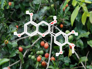
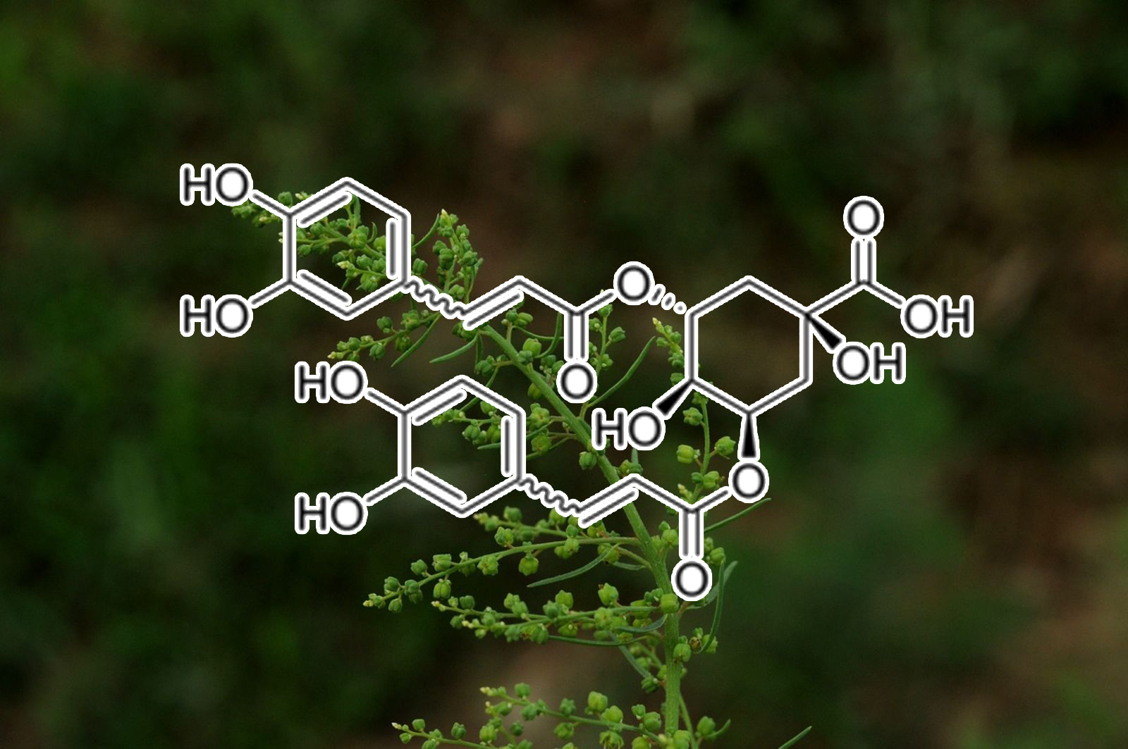
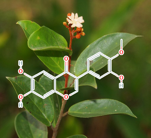


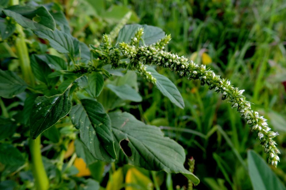
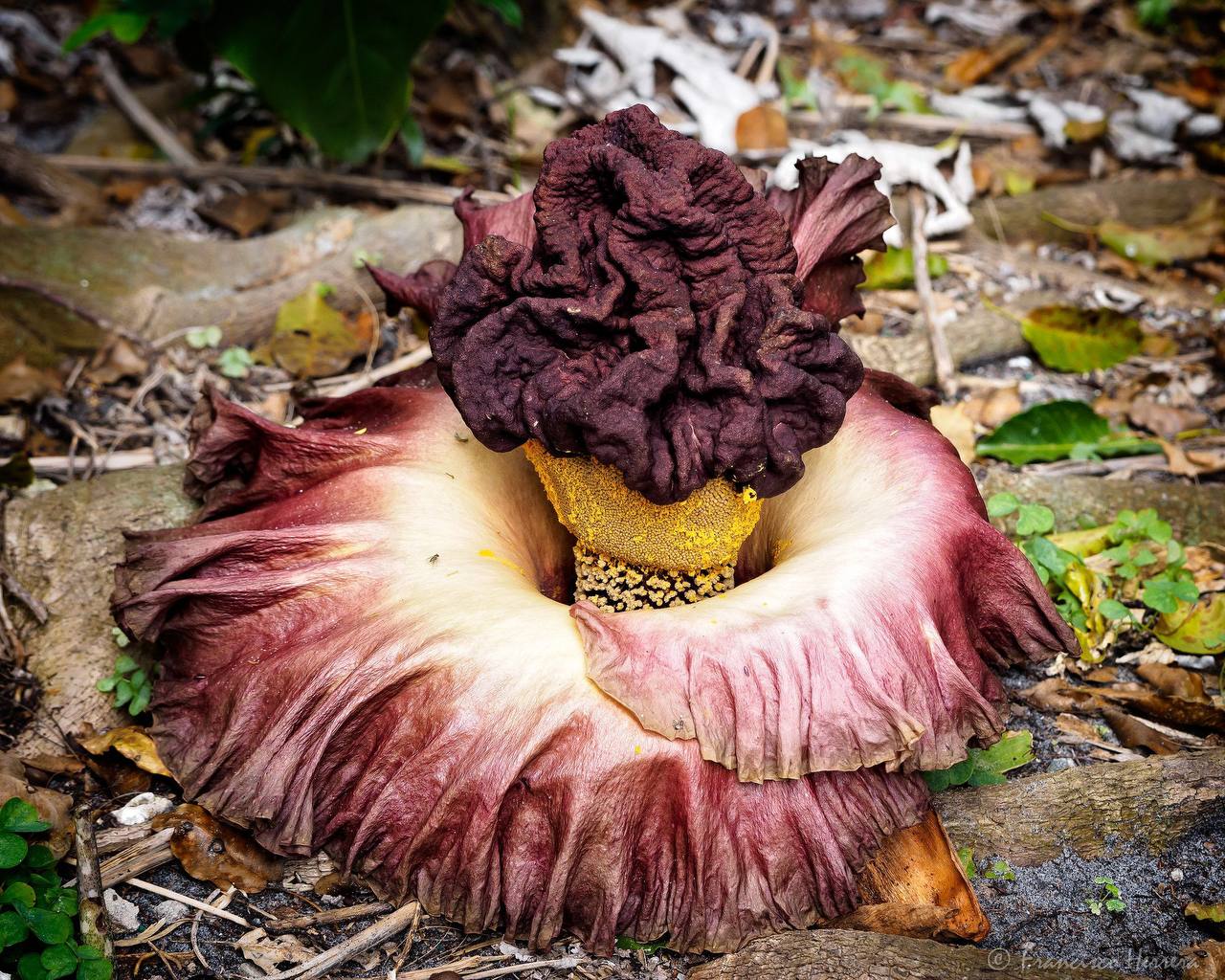


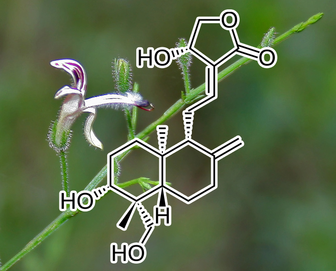
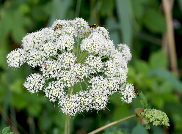

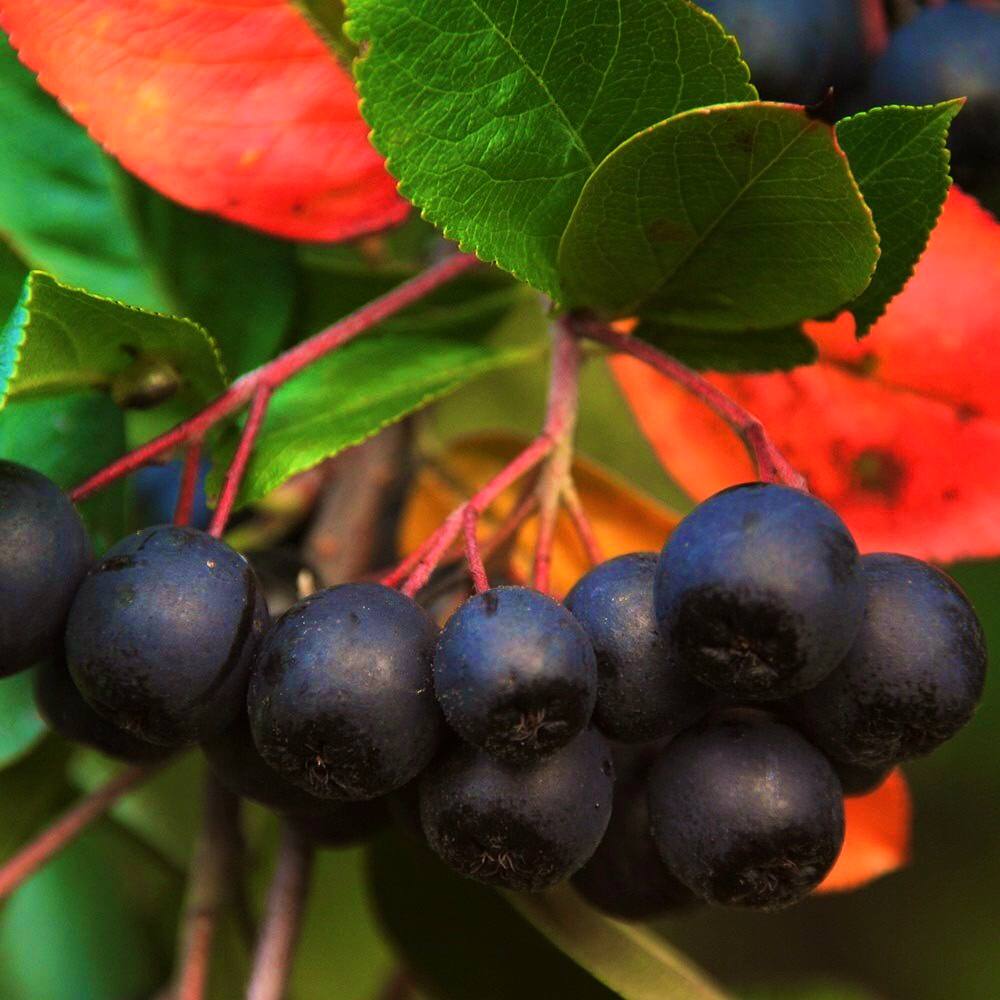
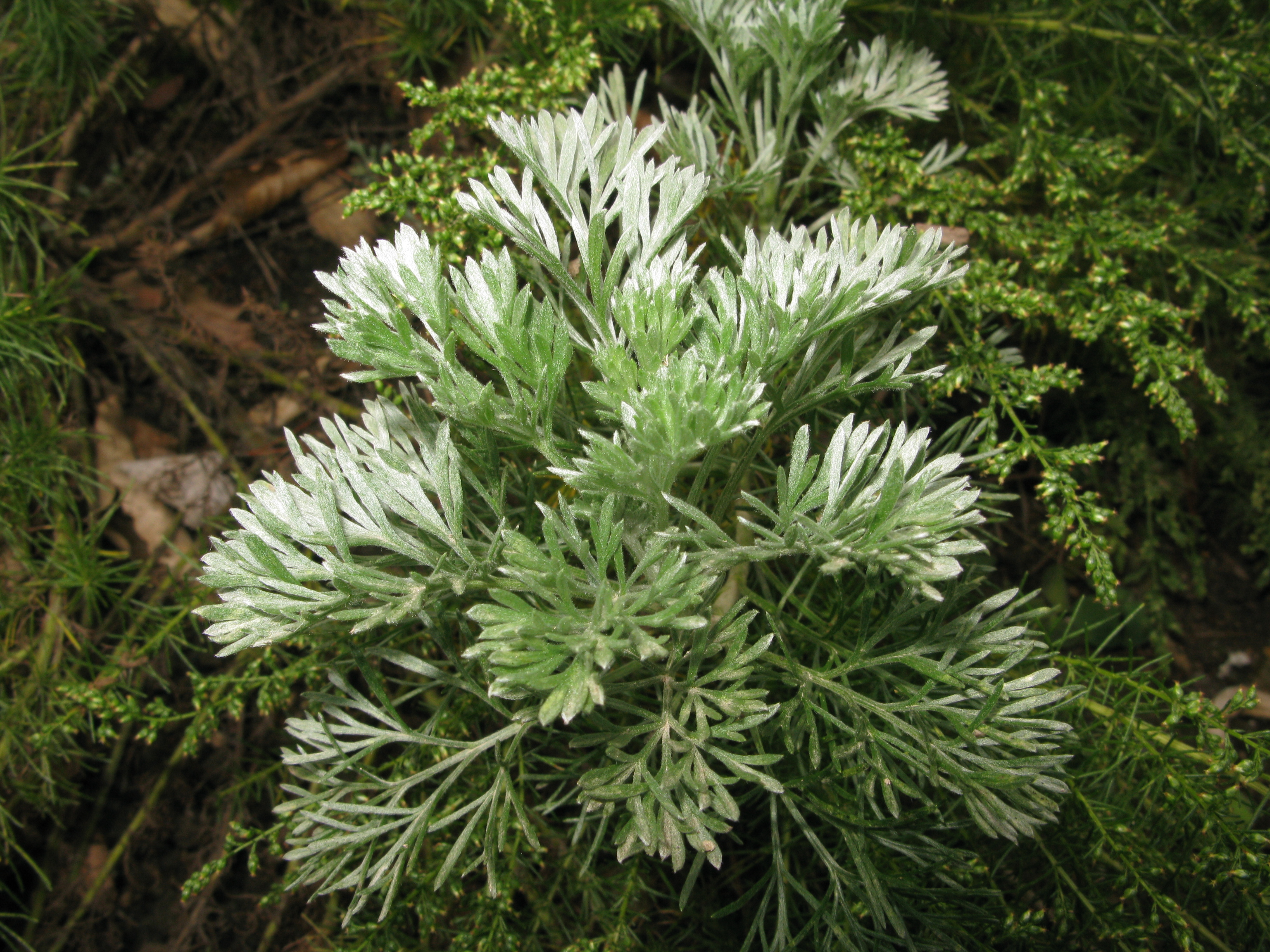

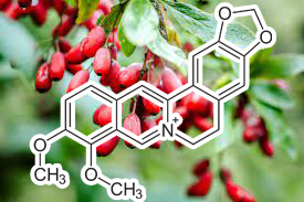

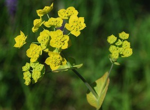

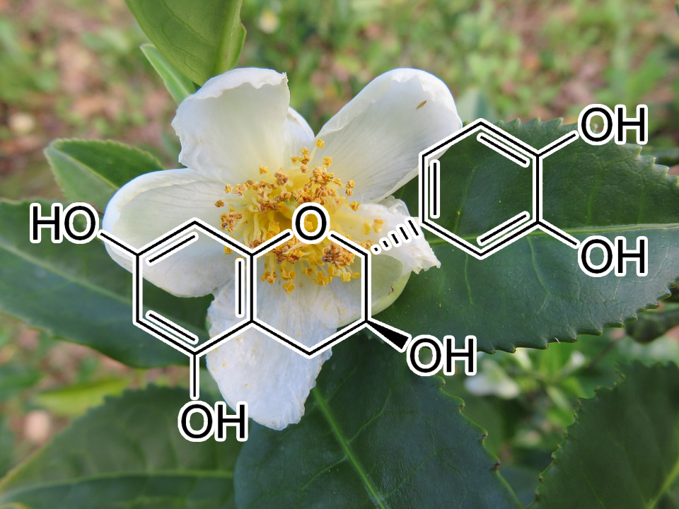
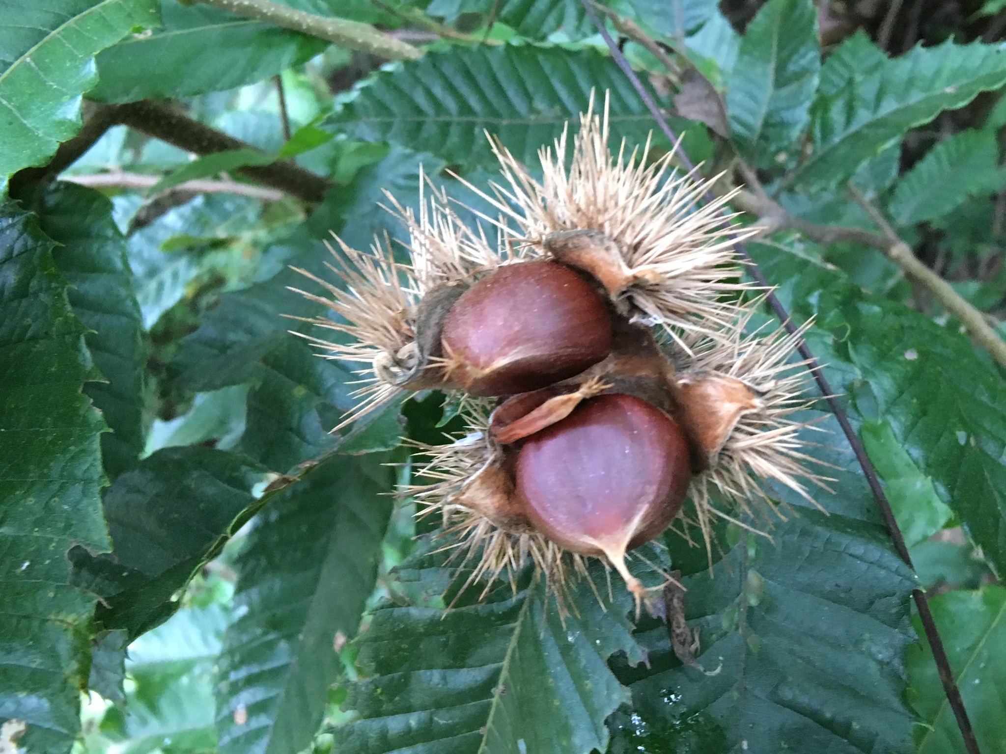


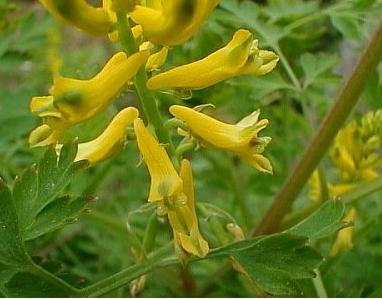


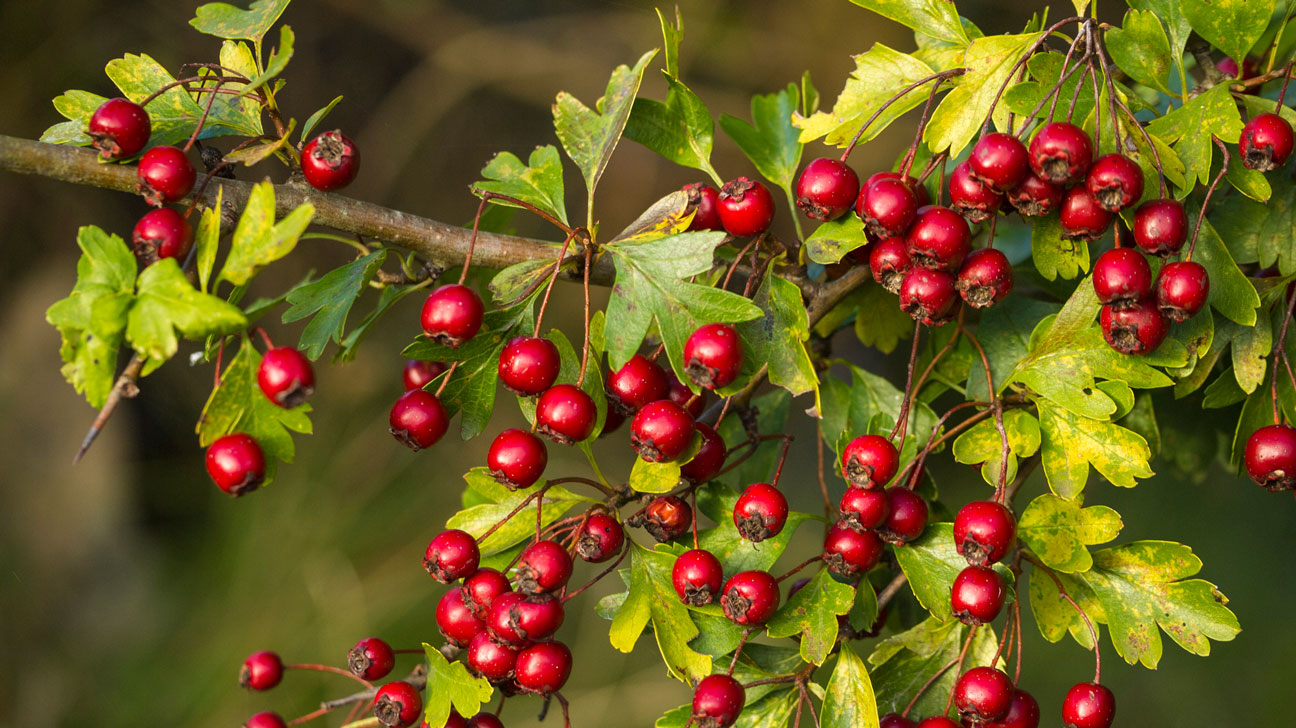
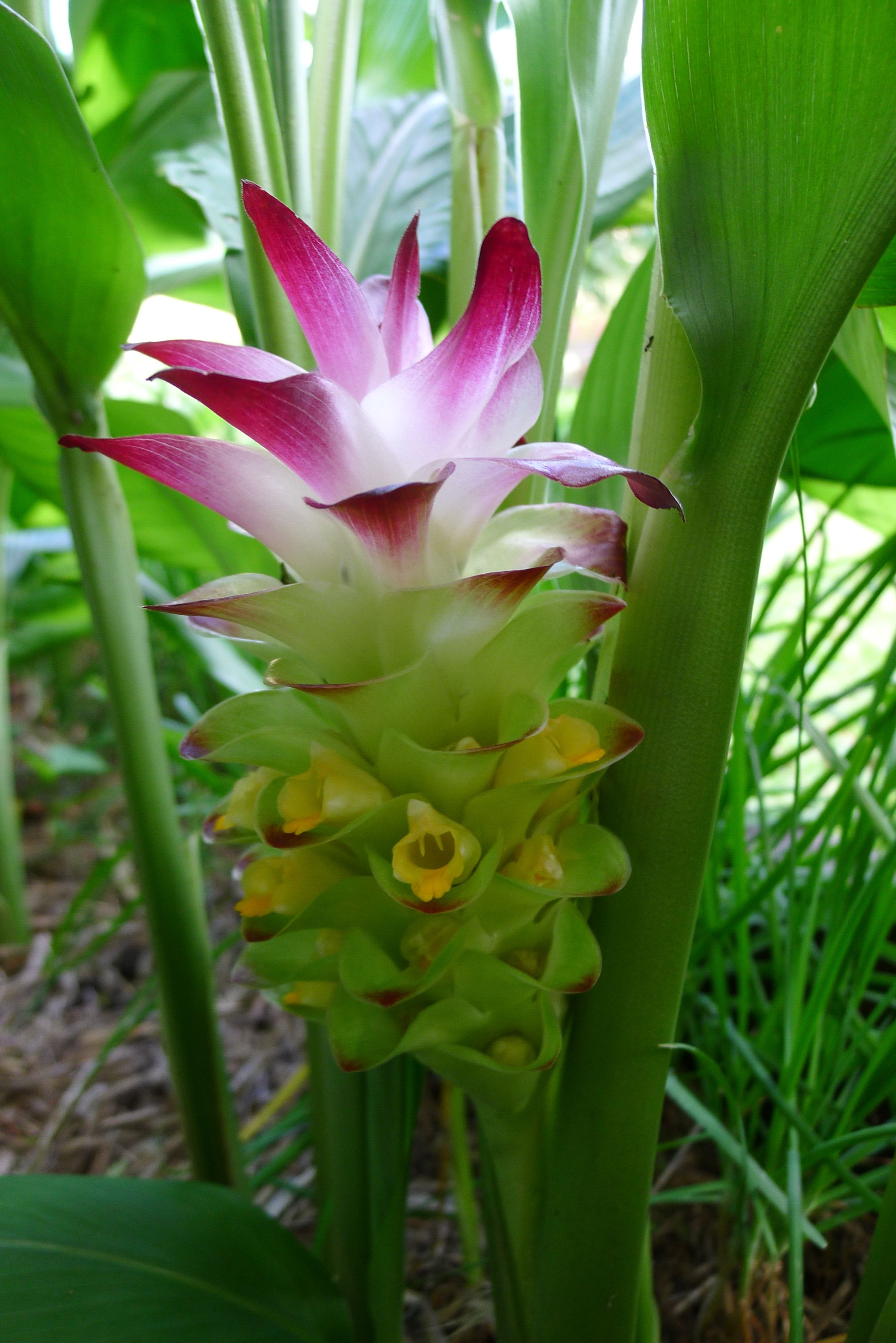


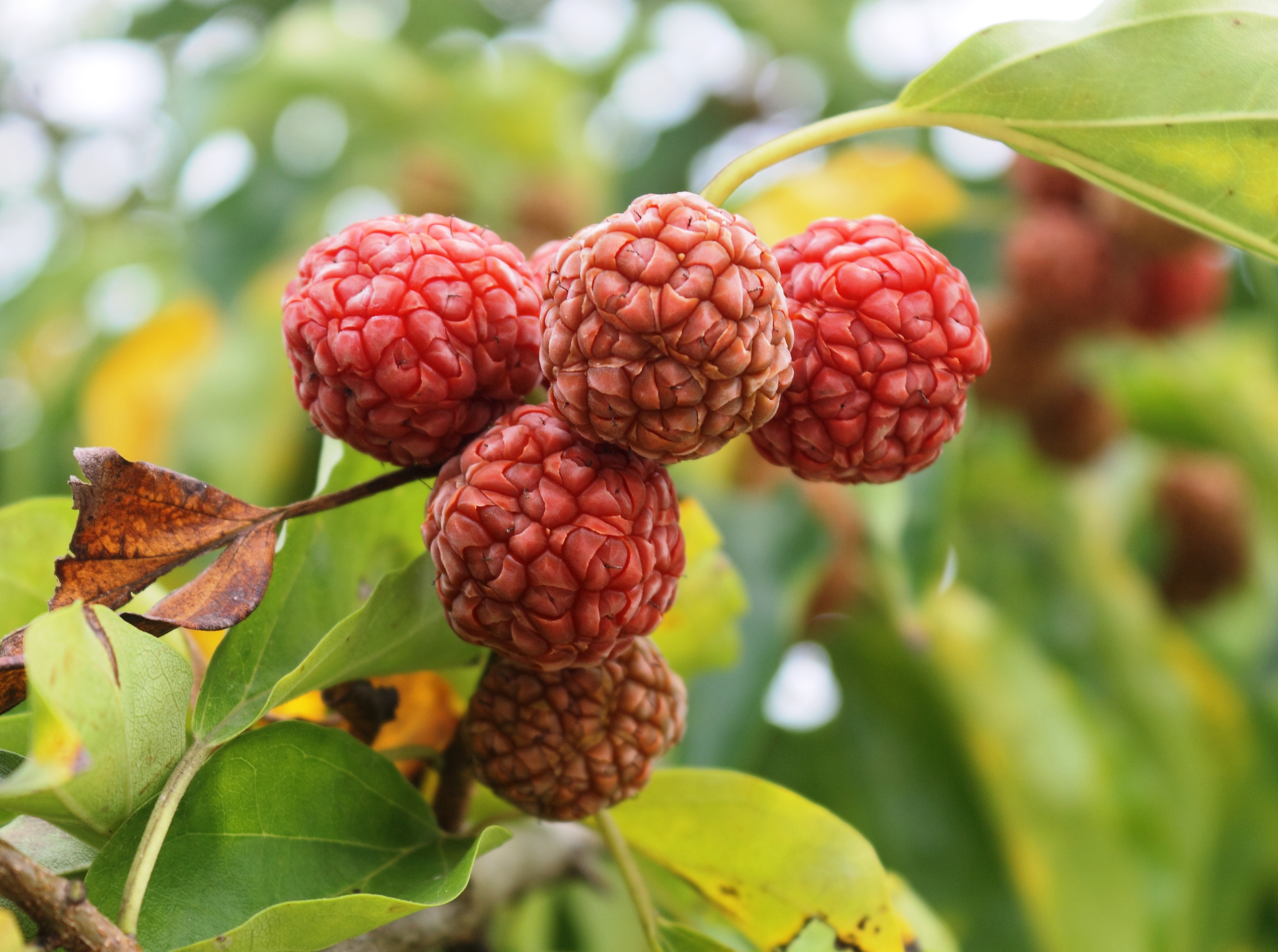


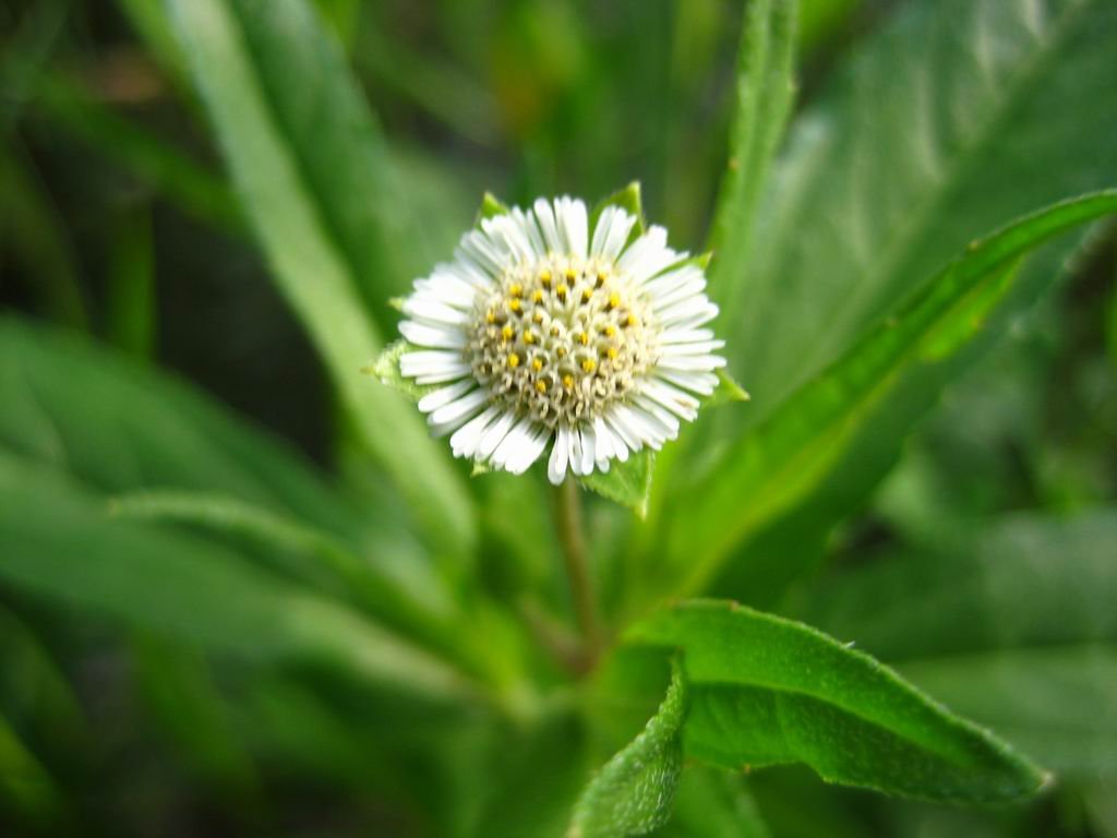

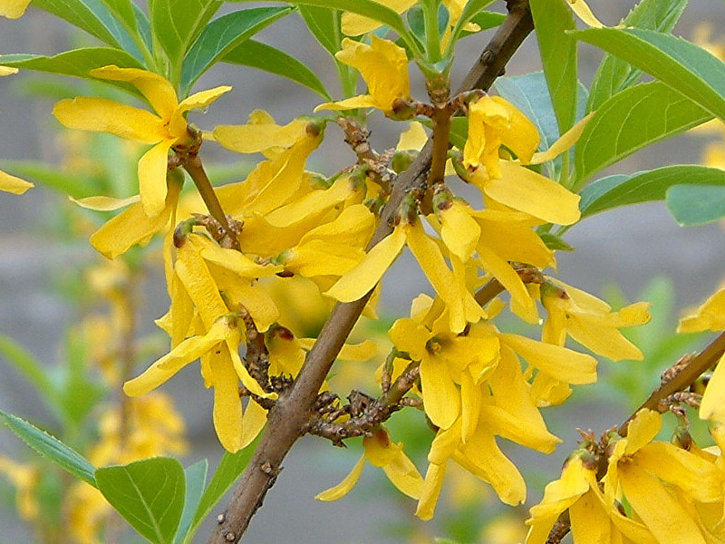
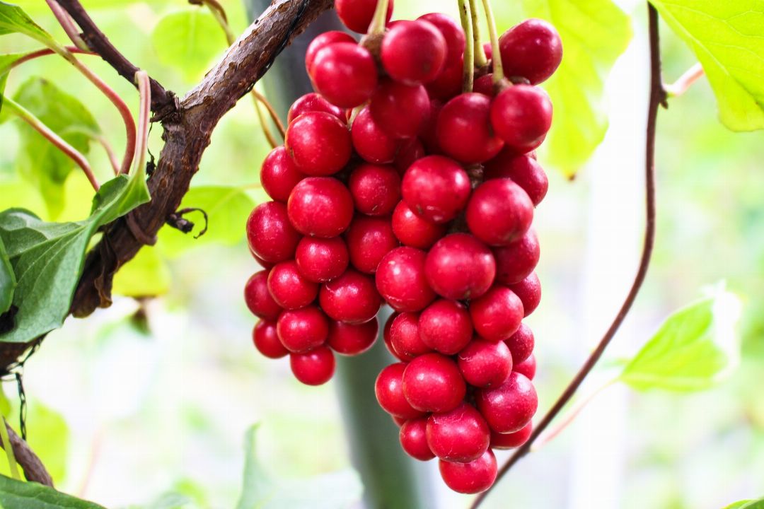
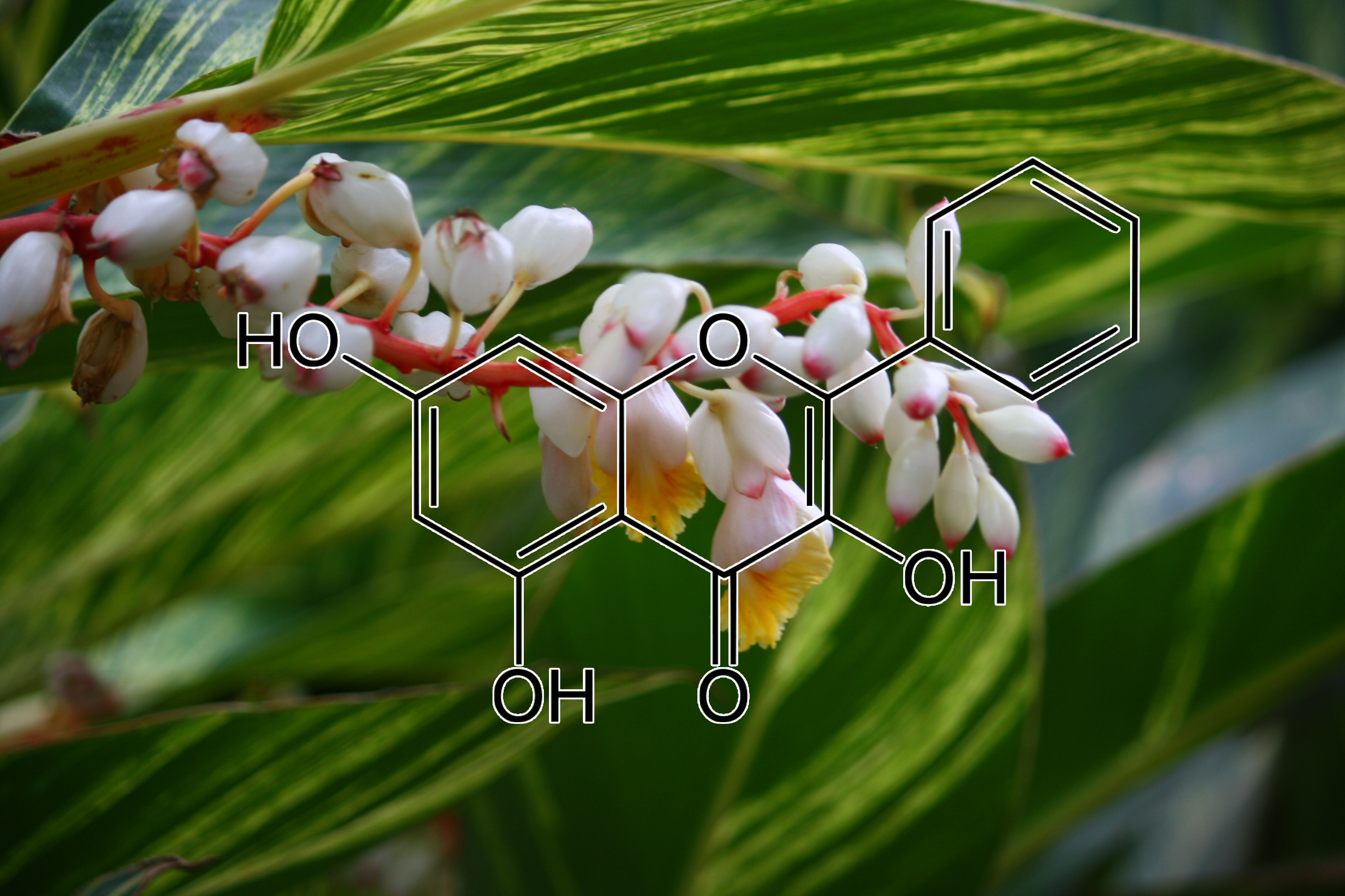
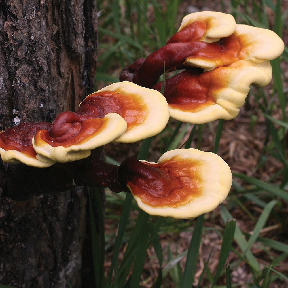

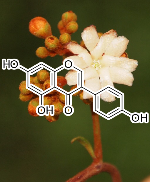


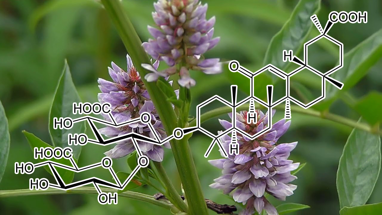
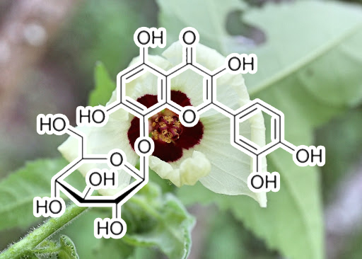
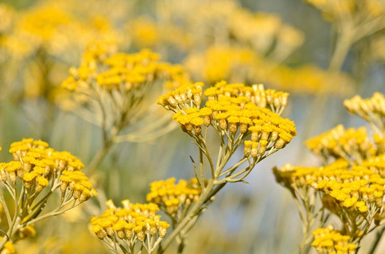
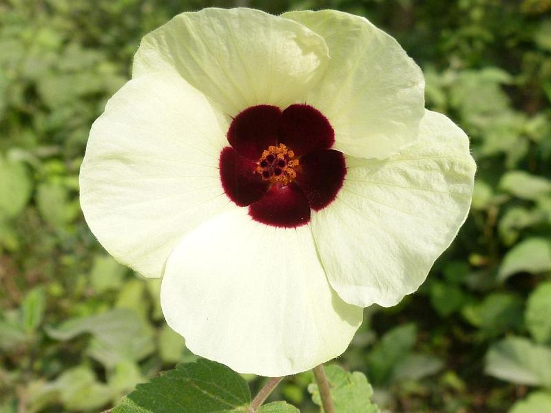

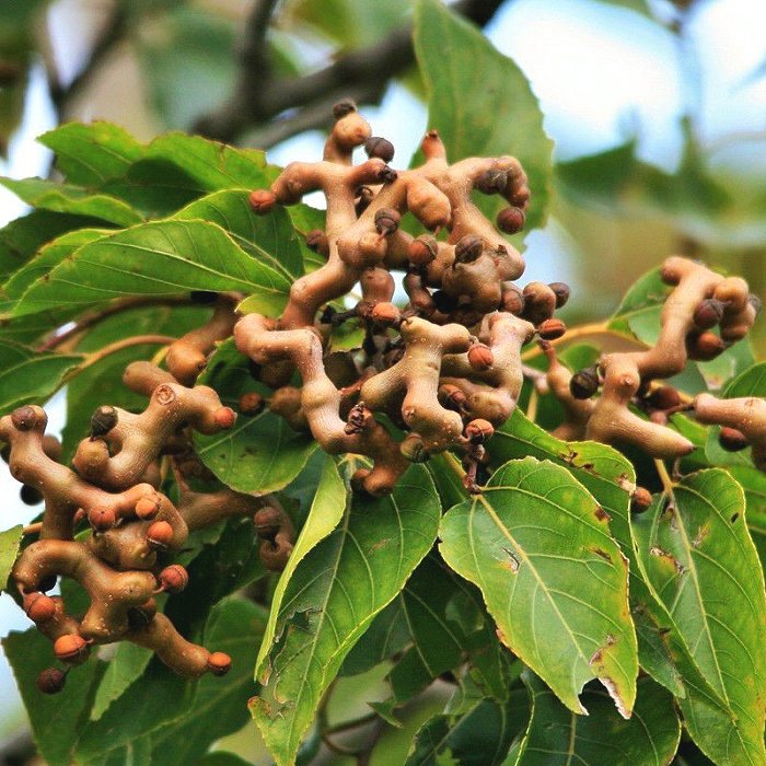
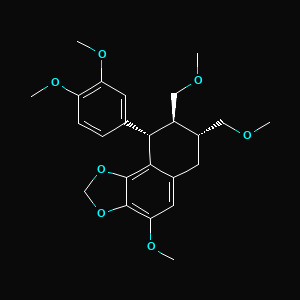
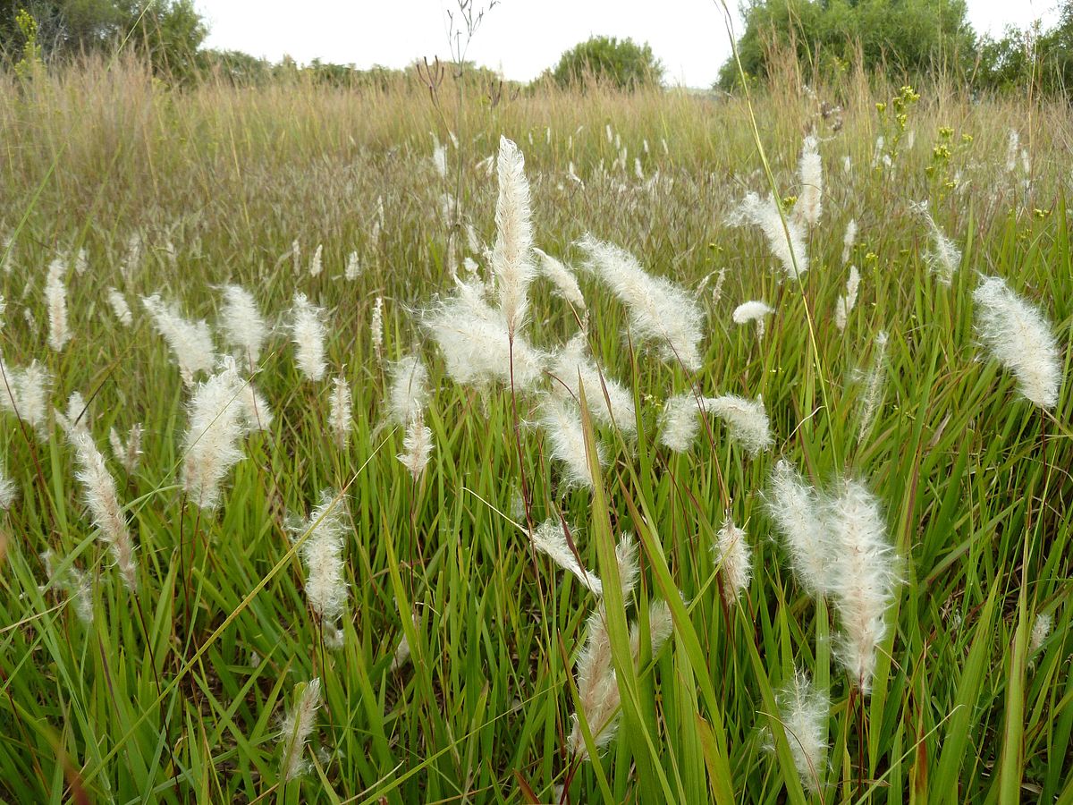
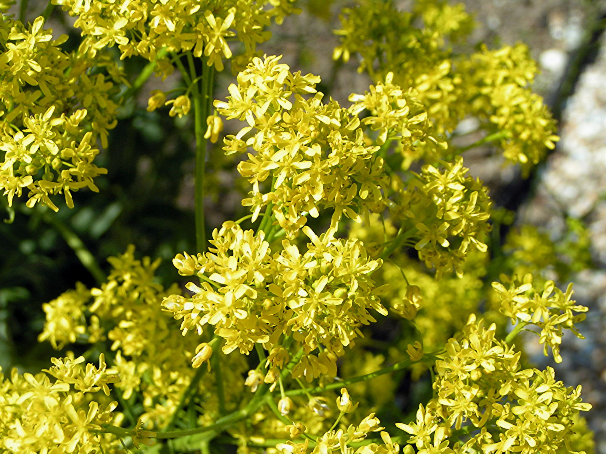
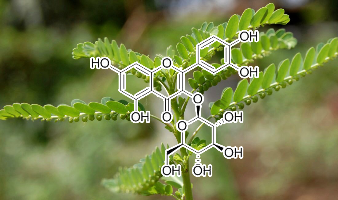
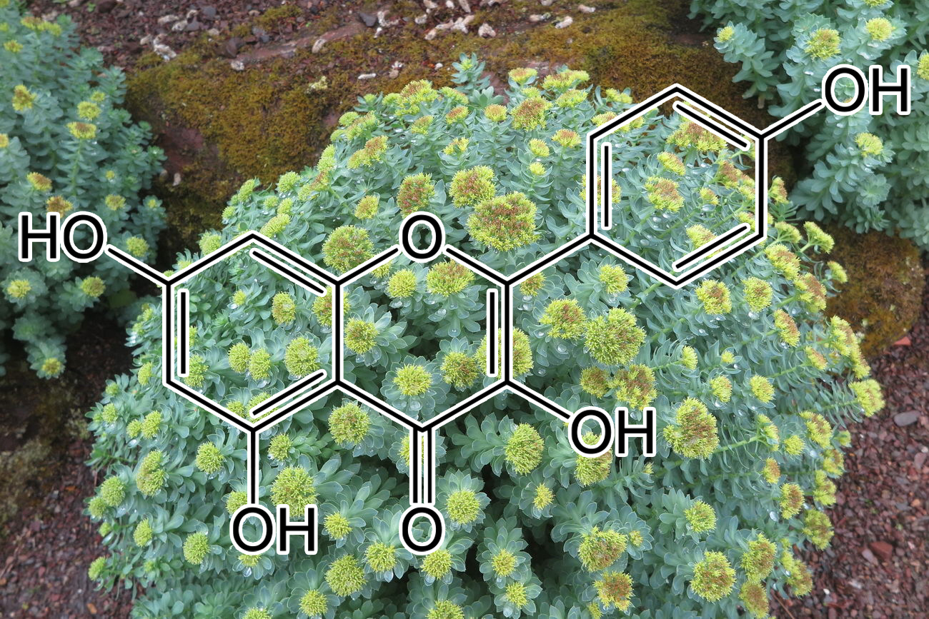

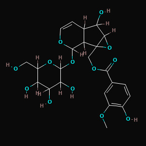


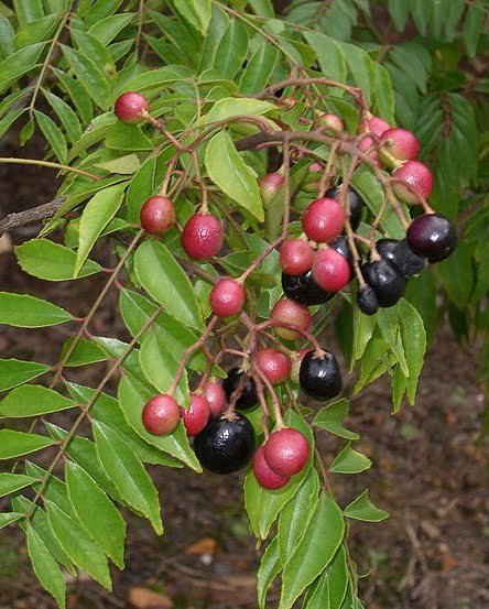
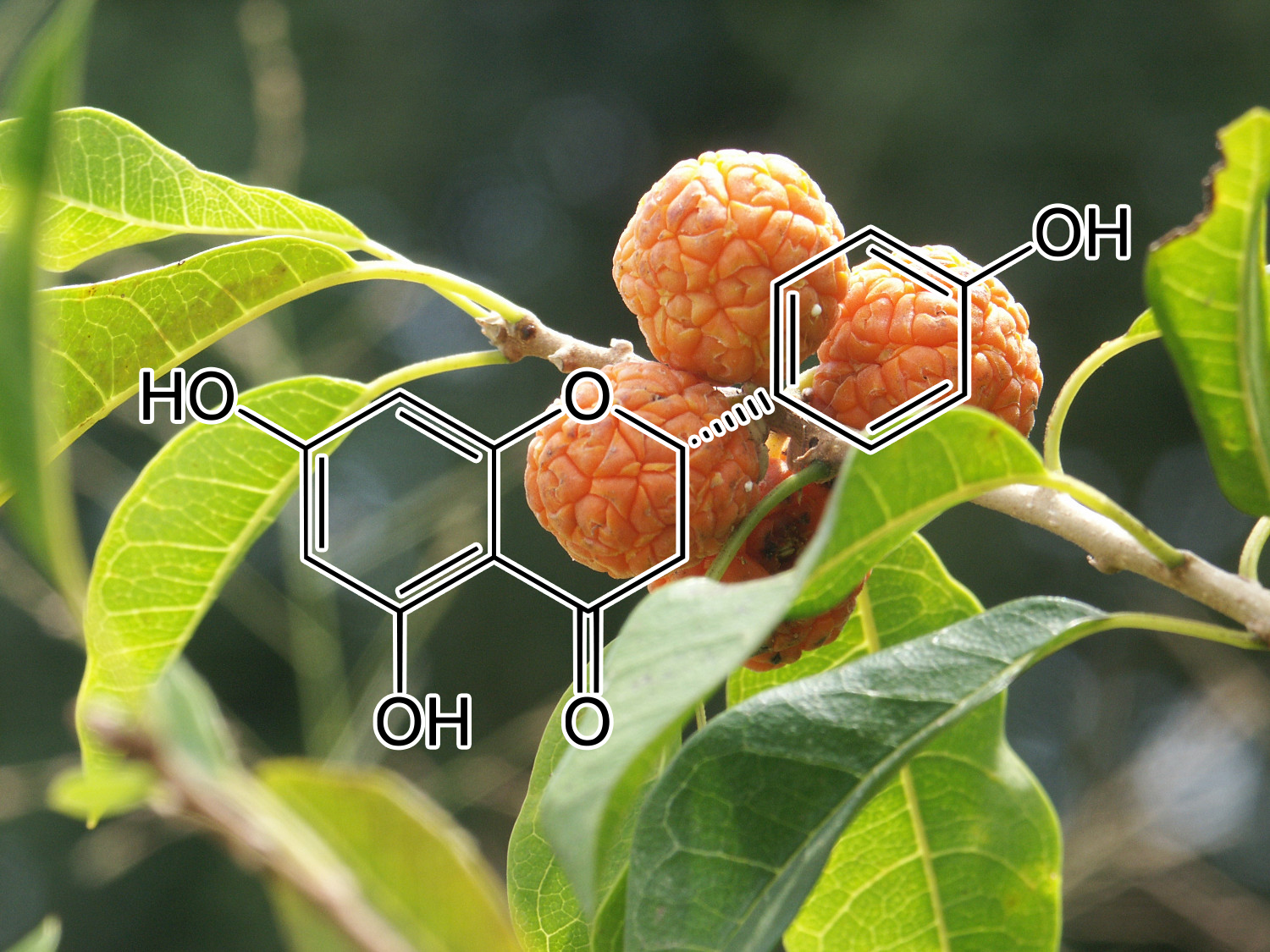

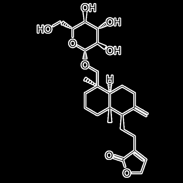


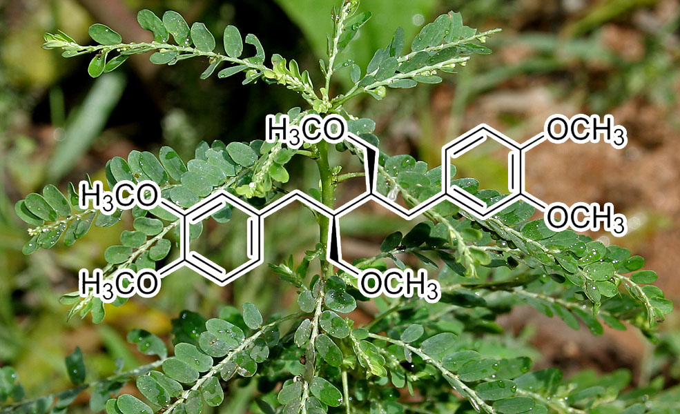
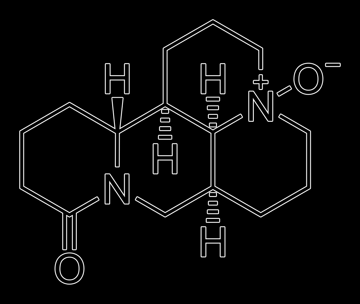

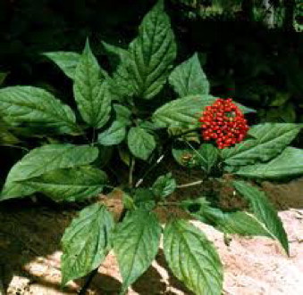
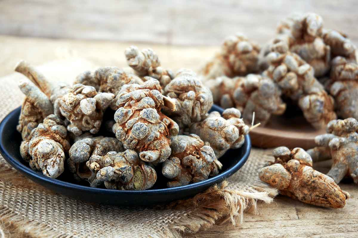

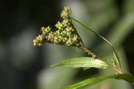
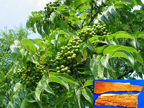


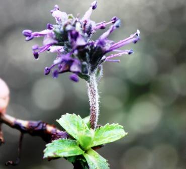
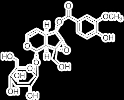
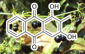
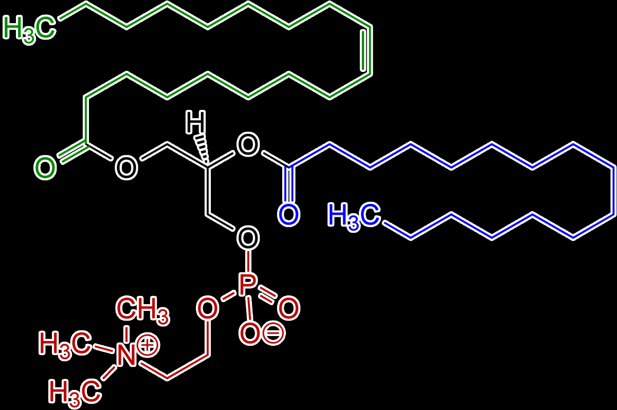


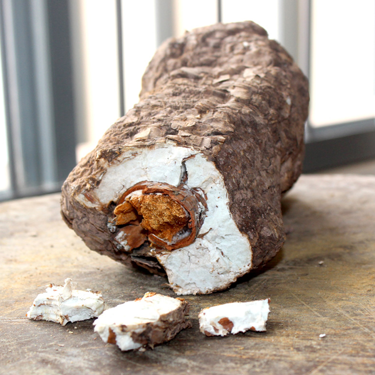
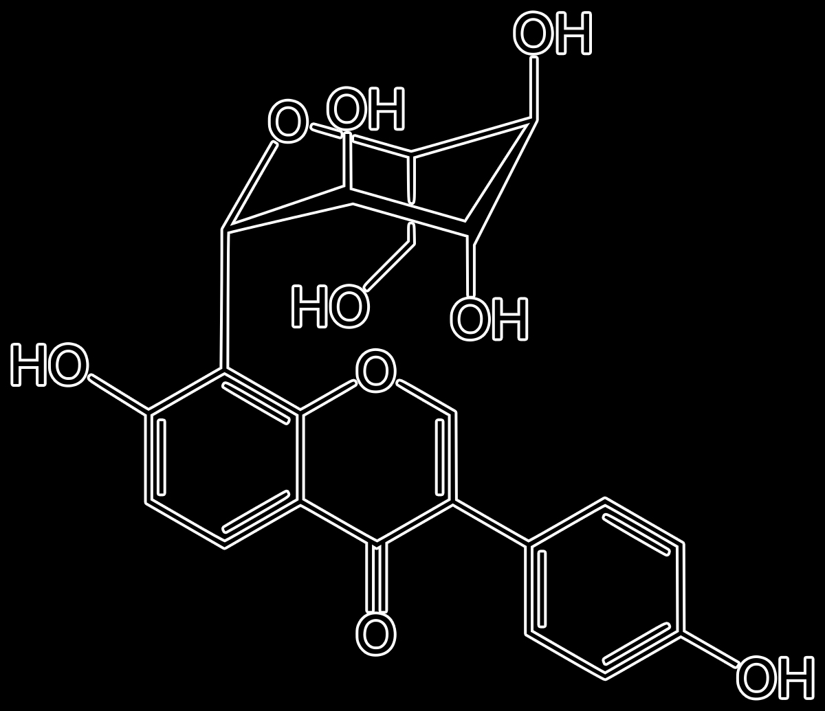
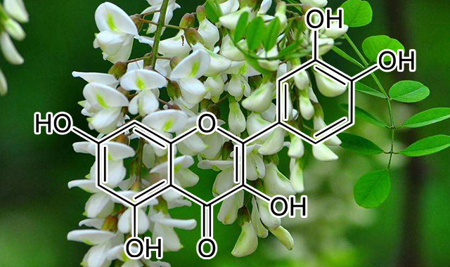


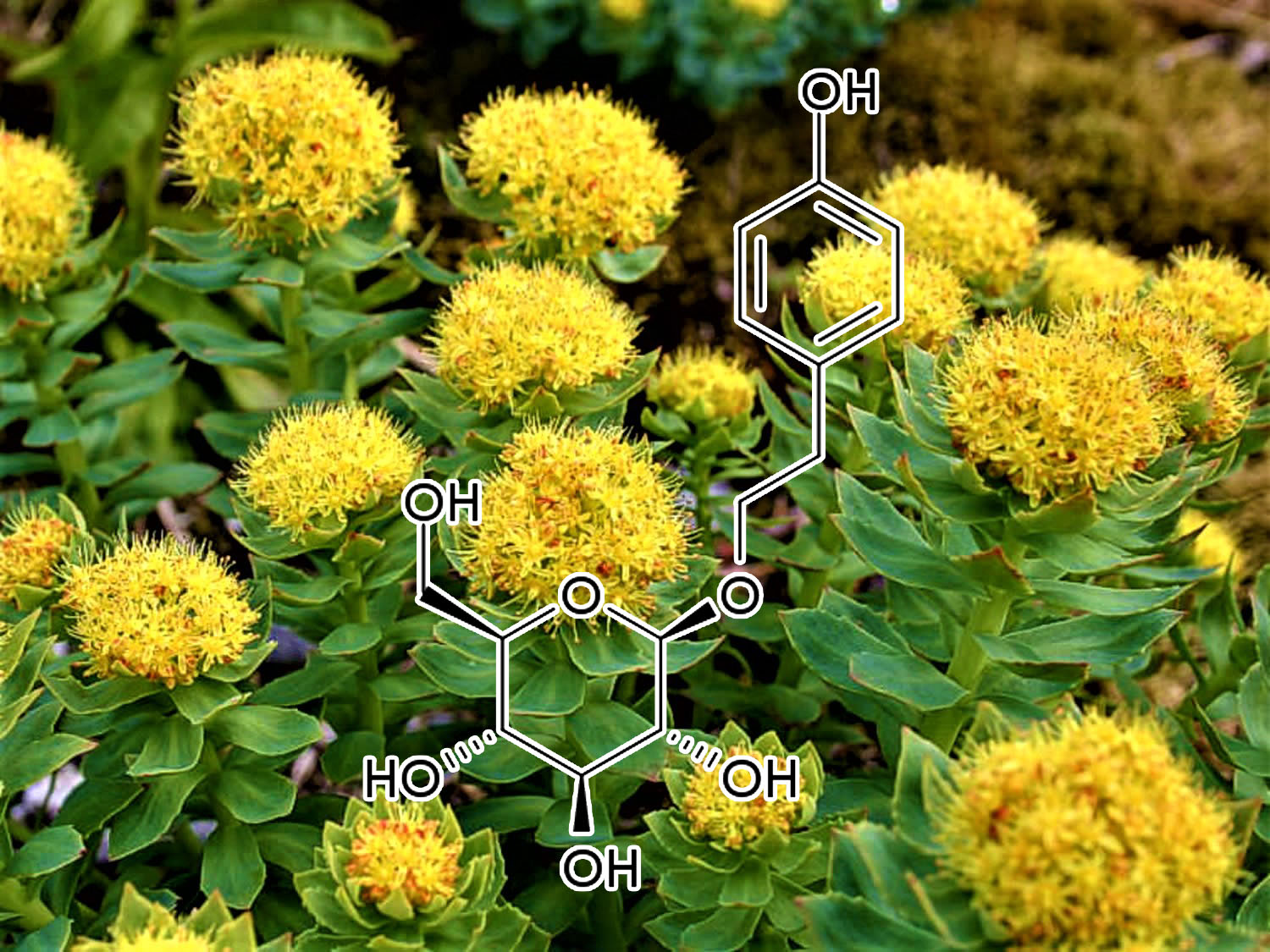
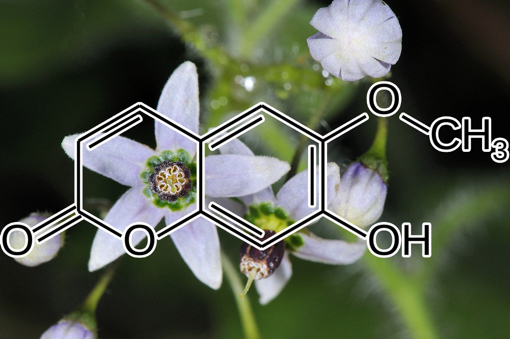
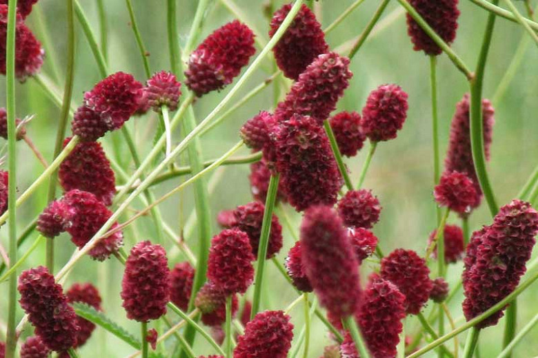
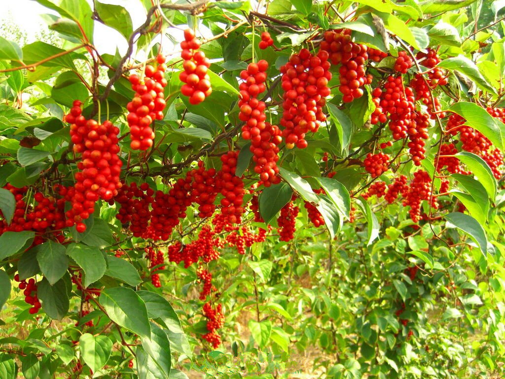

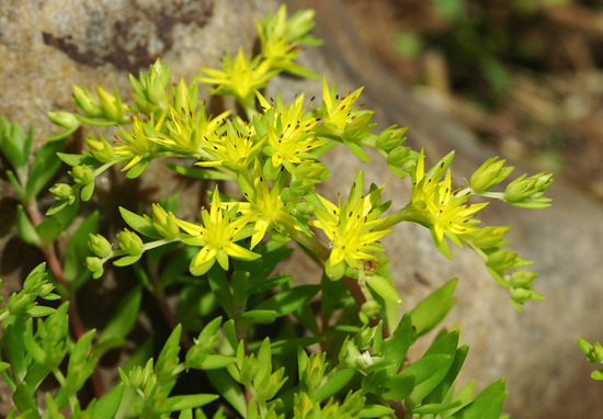
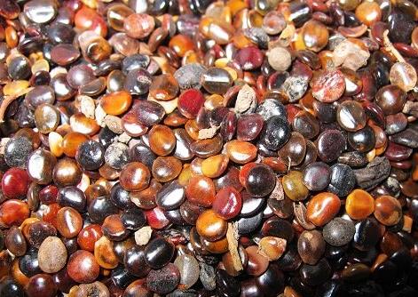
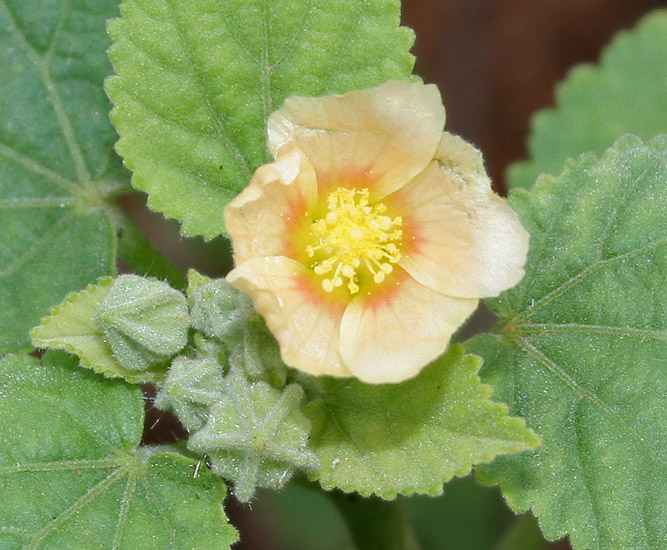
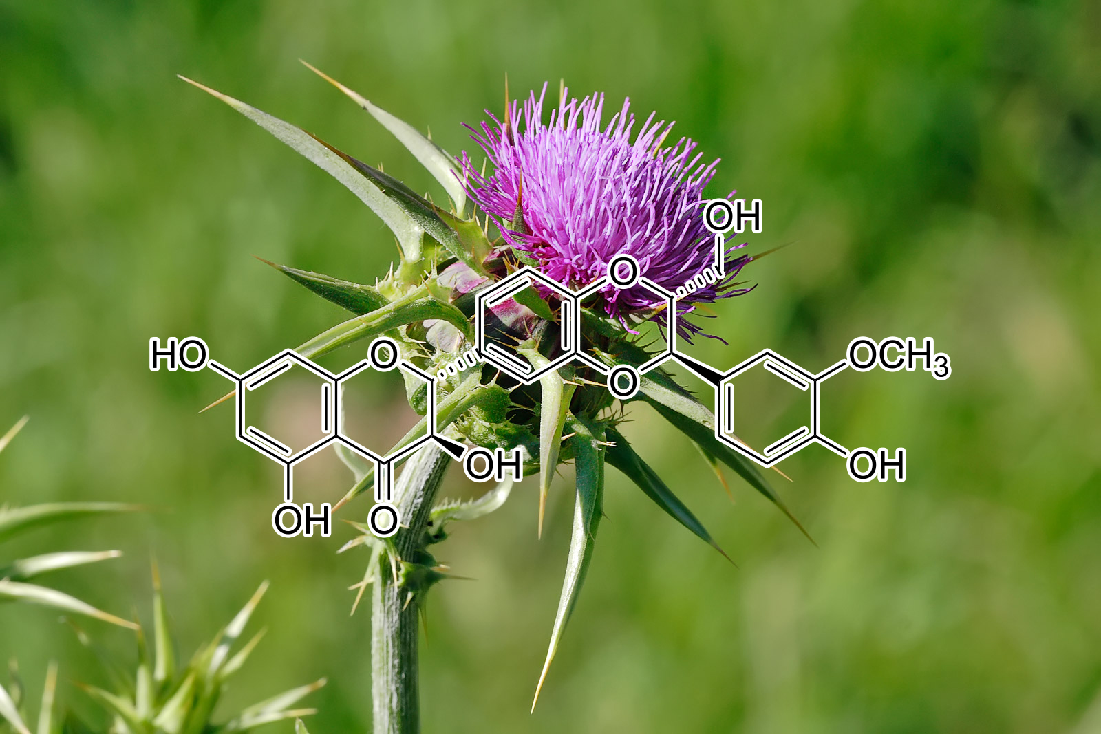
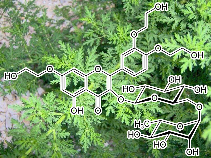
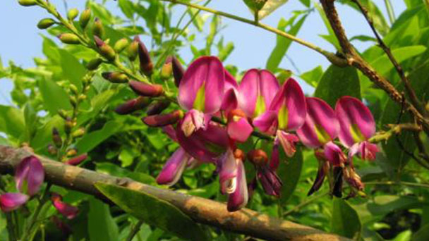

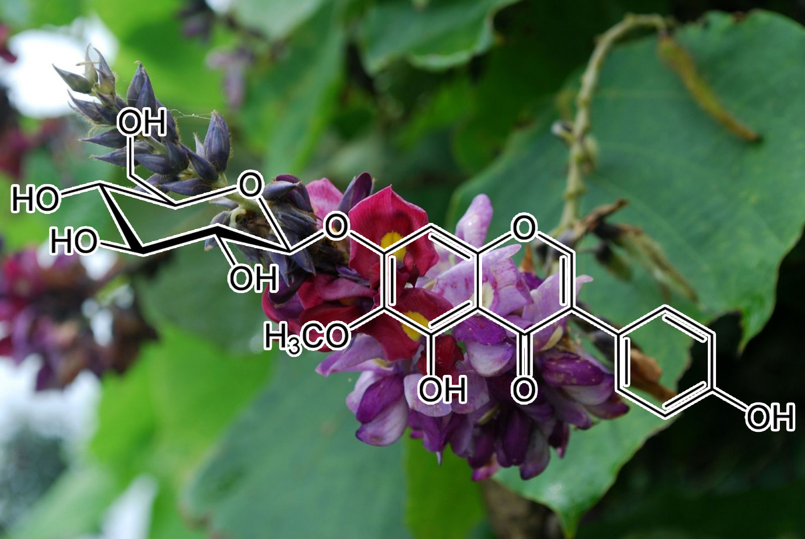
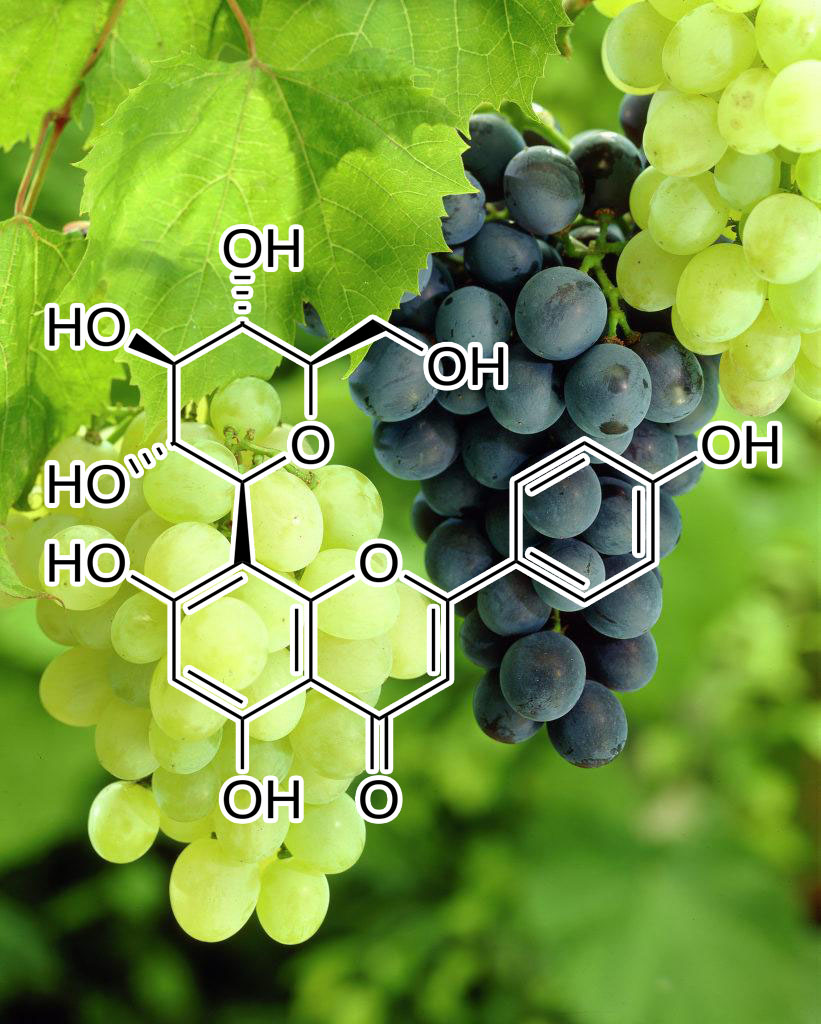


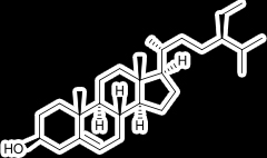


Bernadette Andersen –
i’ve been waiting to post my review on the newer blends. liver regenerator and spaceborn.
i’m a little unique because i only half half of my pancreas and no gallbladder. my liver works twice as hard as the average person. i’m 61 and work long hours very well actually with the help of the blends. i was struggling this last year with being bone chilling cold. i know the fasting makes you cold. This was far beyond what most people would consider feeling cold. i went to a accupucture dr who is very knowledgeable and does extreme healing. He told me my liver was over taxed which was causing the extreme cold. i started very slow with liver regenerator – a salt tsp – i’ve been doing 1/8 tsp 3 times a day and it feels amazing to not feel cold. to actually sweat when i work out. To want to go out and do the things i love outside going into winter here in oregon.
As for the spaceborn. it totally curbs my appetite. it is actually more than that and it’s hard to explain. it takes away the feeling over overwhelm when you to start to eat. My awareness with others and there energy is crystal clear. i higher level of consciousness and taping into what or who feels good without attachment to any outcome.
it’s flow – not force
thank you gavin and the whole team behind the scenes who work to bless our lives. Extremely grateful ♥️
Lynn Montecalvo –
As much as I love Trinity and wiil never be without it , LIVER REGENERATOR has given me my life back. When your liver is overtaxed many negative things can occur….your gut for one never has a chance of healing no matter how many supplements you take or how clean you are eating. It will eventually cause issues with your kidney and pancreas ! Imagine your whole detox organ system shutting down ?!!!
Next your Brain Gut connection is huge ..a positive connection is how you become healthy…a negative connection is how DIS-EASE and inflammation begins…it also regulates your pain receptors and body temp. Liver Regen has restored my brain gut balance ! How I know is my energy, clarity, focus and creativity are back in full force ( Trinity plays a part in this as well )!!
I get up early to see the sunrise which brings tears to my eyes…previously I couldn’t drag myself out of bed without and emotional tug of war…”just ten more minutes” leads to another hour of restless dozing. That is a very negative start to my day causing brain fog, grogginess and zero energy all day!
With Liver Regen in my morning routine my day is full of enegy. My gut balance has improved greatly, I even stopped all those unnecessary expensive supplements that really weren’t working.
I will never be without this combo..Liver Regen and Trinity are pure magic and why I will continue to try other blends for all of my issues. I have added two more blends to my routine this week and look forward to seeing the results. Believe me when I say I am uber aware of my body changes with Interstellar Blends and I thank Gavin for being the great knowledge behind them. I also appreciate his unique attutude toward life…no holds barred !! BAAAAM💥
Rich Ryan –
Wow! Another great blend from my friend Gavin. He just keeps ’em coming!
Liver Regen feels very tonifying for the whole body. Very solidifying. The whole body feels cleaner, like everything is working better in a well-oiled machine..
My mind was clearer and everything felt more solid throughout the day. I noticed an improvement in overall energy too. More sustained energy throughout the day with fewer ups and downs.
I noticed improvements in gut health and digestion. After finishing Liver Regen, there were a few times when I ate too much and experienced some indigestion and bloating. I then realized that I hadn’t experienced that the entire time I was on Liver Regen.
I had less eye strain using the computer for several hours at a time too. After finishing Liver Regen, I was back to limiting my screen time to around 3 hours a day. I then remembered that I hadn’t had to limit myself the entire time being on Liver Regen. Amazing!
I definitely need to take this one for a year or two, to make sure the Liver is fully regenerated.
Great stuff!
Santana (verified owner) –
Gavin, I want to express my heartfelt gratitude for your incredible gift.
After six months of using your blends, my life has transformed in remarkable ways. I was blessed enough to meet an amazing man who introduced me to these blends. I’ve undergone two liver transplants, which made managing my health challenging due to medication changes and complications.
Taking Liver Regen alone yielded incredible results – boosted energy and improved function. Incorporating other blends led to weight loss, enhanced mood, and focus. Niagra regulated my cycle and eliminated any typical mood swings.
Today, I’m thrilled to share that I received a clean bill of health at my annual transplant clinic appointment. My specialist was amazed by my bloodwork. Thanks to your blends, I’ve improved my health and my quality of life.
With immense gratitude,
Santana B.
Lori Lines (verified owner) –
I want to express my heartfelt gratitude to Interstellar Blends for their life-changing Liver Regenerator product. Six months ago, my life took a remarkable turn when I was introduced to these blends by Gavin. Having Type 2 Diabetes and trying to stay off pharmaceutical medications was a daunting task, managing my health had become a confusing maze due to complications. Liver Regenerator has been nothing short of a miracle for me. It has not only boosted my energy but also significantly improved my liver function.
Taking Liver Regenerator has delivered impressive results, making me feel like my entire body was undergoing a revitalization process. With proper diet and the blends, my mind became clearer, and I experienced more sustained energy throughout the day. Not only did my gut health and digestion improve, but I also noticed a reduction in eye strain during long hours in front of the computer. Liver Regenerator has given me my life back, allowing my detox organ system to function optimally and restoring the vital brain-stomach connection that is essential for overall well-being.
What’s truly remarkable about Liver Regenerator is the profound impact it has had on my daily life. I no longer struggle to face the day, as I now greet the day with renewed energy and enthusiasm. Regenerator indeed! The improved gut balance has even allowed me to eliminate expensive supplements that were previously ineffective. I am committed to making Liver Regenerator and other Interstellar Blends a permanent part of my wellness routine, as they have proven to be pure magic in restoring my health and vitality. I eagerly look forward to exploring more blends to address all of my health concerns. Interstellar Blends has indeed been a game-changer for me, and I am immensely grateful for this gift that has transformed my life.
Ronald –
So since i was young growing up in Ecuador we consume a lot of unhealthy fats and back in 2019 i was really sick all the time sonogram revealed that i had some gallstones and inflammation and since i was unaware of what was really going on in my body and how the liver worked i decided to have the surgery and yes i do regret it. The problem was not my gallbladder, the problem is the liver but back then i was unaware of that. See if your liver is backed full of toxins and get inflamed that is the root cause of gallbladder issues.
So my liver was not the healthiest from having too many fatty fried foods over the years and overeating on carbs its just sad to me that my own doctors and surgeons were not aware of this or did not care because at the end of the day its a business so health its not their priority thats why i thank Gavin for these blends. Lately i was feeling more sluggish than usual and my mind was a little foggy so since i dont have a gallbladder i know that my liver needs some help to get rid of all the toxins The liver is such a major organ and it helps w so many things that it is critical in my opinion that you use this blend daily.
At first i fasted then i started taking the liver blend and it was hard the first couple of days because i guess i had a lot of toxins to detox from but now its gettin much better and iam feeling better and my energy level is up and i have less brain fog. Highly recommend this blend to all and blessings !!!!