THROMBOLYTIC : Blood Clot Destroyer 200:1
April 5, 2022
ASCENDANCE : Pain Annihilator / Non Opioid Anti-Inflammatory Analgesic 200:1
July 14, 2022SABRETOOTH : Tooth Decay & Gum Disease Destroyer 200:1
$275.00
Introducing:
SABRE
TOOTH
TOOTH DECAY & GUM DISEASE DESTROYER
200:1 Concentration
benefits:
- Prevent / REVERSE Tooth Decay
- Eliminate Gum Disease / Gingivitis
- Prevent Tarter & Plaque BUILDUP
- NEUTRALIZE SENSITIVITY & PAIN
- REMINERALIZE Enamel, Pulp & Dentin
- RESTORE / REGROW RECEDIng Gums
- KILL PATHOGENIC Oral Bacteria & Biofilm
- Clean & Disinfect ENTIRE Mouth
- FRESH HealthY BREATH
- REMOVe / Prevent STAINS
- ENJOY SPARKLING WHITE YOUTHFUL SMILE
- EXTEND LIFESPAN OF Teeth
- FEWER DENTIST VISITS
- AVOID CAVITIES, ExtractIONS & Root CANALS
- KEEP ALL YOUR Teeth
- Prevent OTHER DiseaseS AND Health PROBLEMS ASSOCIATED WITH Periodontitis (Gum Disease) SUCH AS:
- HYPERTENSION
- DIABETES
- CANCER
- DEMENTIA
- SYSTEMIC INFLAMMATION
- STROKE
- PREMATURE AGING
- DEPRESSION
- ARTHRITIS
- ANXIETY
- HEART ATTACK
- INFECTIONS
- OBESITY
- PSORIASIS
- ERECTILE DYSFUNCTION
- GERD (ACID REFLUX/HEARTBURN)
- ALL CAUSE MORTALITY
SABRETOOTH PROTOCOL :
1. Schedule a proper Cleaning to remove all Plaque and Tarter for a fresh start. Once gone we are going to keep it gone.
2. Brush with baking soda and 1/16th tsp (gold spoon) SABRETOOTH 3 x a day; use soft bristle electric tooth brush. *Brush within 1 hour of all meals.
3. Get waterpik and use daily with 1 tsp baking soda, 1/8 tsp SABRETOOTH, 1 tsp INTERSTELLAR MATCHA & 1 Oz Hydrogen Peroxide in warm water.
4. Take 1/2 tsp SABRETOOTH a day in 4oz fresh grapefruit juice for first week; 1/4-1/2 tsp a day to maintain after. You can also add 1/8 tsp to your coffee or INTERSTELLAR MATCHA with other blends.
5. If you have an active Tooth infection use a baking soda tablet and sleep with it placed between cheek and infected Tooth. *I personally do this nightly regardless as a precautionary measure to maintain alkaline saliva all night long.
VERY IMPORTANT:
It is absolutely critical to understand that
Tooth Decay and Gum Disease BEGINS when saliva pH drops and becomes acidic (read it again)
Sugar rapidly lowers saliva pH and destroys Teeth and Gums (the baking soda raises pH and SABRETOOTH helps restore Gum Health and remineralize Teeth both internally and externally).
You can buy pH strips to test your saliva pH. 7 and above is what you want to maintain as often as possible. DO THIS and you will have a lifetime of perfect Teeth and Gums.
BAKING SODA. BAKING SODA. BAKING SODA. Got it? Good. Brush with BAKING SODA 3 times a day to keep SALIVA pH ALKALINE.
Why???
Because the bacteria behind tooth decay and gum disease can’t survive in an alkaline environment!
WHAMMO!!!!
Featuring:
4-Chromanol, The Major Constituent Of Piper Betle • Acemannan • Aceriphyllum Rossii Engler (Saxifragaceae) • Achyranthes Bidentata • Agastache Rugosa • Allium Sativum Bulb • Angelica Dahurica • Angelica Sinensis • Arctium Lappa • Astilbin, Flavanone From Rhizoma Smilacis Glabrae • Astragalus Longistylus • Azadiratcha Indica (Neem) • Baicalin • Bitter Guard (Momordica Charantia) • Bletilla Striata • Boswellia Carterii Birdw • Caffeic Acid • Carvacrol • Chitosan • Cimicifugae Rhizoma • Citrus Reticulata Blanco Peel • Cocoa Pod Husks • Coptidis Rhizoma • Cortex Phellodendri (Huang Bai) • Cranberry • Cymbopogon Flexuosus • Deoxynojirimycin • Dimethylaminododecyl Methacrylate • Dodonaea Viscosa Var Angustifolia Leaf • Epigallocatechin‐3‐Gallate (EGCG) • Eucalyptus Camaldulensis • Eugenol From Essential Oil Of Syzygium Aromaticum Clove) • Fructus Armeniaca Mume (Wu Mei) • Galla Chinensis • Gallic Acid • Genipin • Ginkgo Bilobal Leaf • Glycyrrhiza Glabra • Glycyrrhiza Uralensis Fisch • Glycyrrhizol A • Grape Seed • Green Tea Tea Polyphenols • Guaijaverin • Hesperidin • Honokiol Isolated From Magnolia Sp Bark • Houttuynia Cordata • Humulus Lupulus • Isopanduratin A • Juniperus Excelsa M Bieb Essential Oil • Kaempferia Galanga • LACTOFERRIN • Lagerstroemia Speciosa (Lythraceae) Leaves • Lippia Sidoides • Macelignan • Magnolol • Marticariarecutitia (German Chamomile) • Matricaria Chamomilla • Melaleuca Alternifolia (Tea Tree Oil) • Mentha Arvensis • Mentha Piperita • Mentha Spicata • Morinda Citrifolia (Indian Mulberry) • MSM • Myricetin • Myrtus Communis • Nano-hydroxyapatite • Nelumbo Nucifera Leaves • Nepeta Cataria • Nigella Stevia • Oleanolic Acid • Ophiopogon Japonicus Kergawl • Origanum Glandulosum • Osmanthus Fragrans Lour Flower • Paeonia Lactiflora Pall • Paeonia Suffruticosa • Panax Ginseng C A Meyer • Panaxnotoginseng • Pearl Powder • Perilla Seed (Perilla Frutescens Britton Var Japonica Hara) • Physalis Angulata • Polygonum Cuspidatum • Propolis • Propolis Ethanol • Psidium Cattleianum Leaf • Pycnogenol • Quercetin • Quercitrin • Radix Et Rhizoma Rhei (Da Huang) • Rheedia Brasiliensis Fruit (Bacupari) • Ricinus Communis • Rosmarinus Officinalis • Salidroside From Rhodiola Rosea • Salvadora Persica Aqueous • Salvia Miltiorrhiza Bge • Satureja Hortensis • Saussurea Lappa • Shikonin From Lithospermum Erythrorhizo • Sophora Japonical Flower • Tamarillo Skin • Tamarindus Indica (Cesalpiniaceae) Seed • Terpinen-4-Ol • Thymbra Capitata • Thymus Zygis • Triphala • Ursolic Acid • Vitamin D • Vitamin K2 Mk4 Mk7 • Xanthorrhizol • Zerumbone From Zingiber Zerumbet • Α-Mangostin •
Calorie restriction and intermittent fasting can play a key role in the cost‐effective resolution of Periodontal Inflammation as a primary Prevention strategy for the management of chronic inflammatory Diseases, including Periodontal Diseases.
Promotion of Natural Tooth Repair by small molecule GSK-3 antagonists:
INGREDIENTS & SCIENCE:
4-chromanol, the major constituent of Piper betle Extract
To evaluate the antiBacterial activity of 12 ethanol Extracts of Thai traditional herb against Oral Pathogens. The antiBacterial activities were assessed by agar well diffusion, broth microdilution, and time-kill methods. AntiBiofilm activity was investigated using a 3-[4,5-dimethyl-2-thiazolyl]-2,5-diphenyl-2H-tetrazolium-bromide (MTT) assay. High performance liquid chromatography (HPLC), thin layer chromatography (TLC) fingerprinting, and TLC-bioautography were used to determine the active antiBacterial compounds. Piper betle showed the best antiBacterial activities against all tested strains in the minimal Inhibitory concentration and minimal bactericidal concentration, ranged from 1.04–5.21 mg/mL and 2.08–8.33 mg/mL, respectively. Killing ability depended on time and concentrations of the Extract. P. betle Extract acts as a Potent antibiofilm agent with dual actions, preventing and eradicating the Biofilm. The major constituent of P. betle Extract was 4-chromanol, which responded for antiBacterial and antibiofilm against Oral Pathogens. It suggests that the ethanol P. betle leaves Extract may be used for Preventing Oral Diseases.
Betelvine (Piper betle L.): A Potential source for Oral care
Piper betle L. (betelvine) is a valuable crop that is widely used as masticatory and with a long past history of varied traditional
uses. Betelvine possesses numerous phytochemicals with important pharmacological attributes. Active molecules such
as Fluoride, Eugenol, Hydroxylchavicol, Chlorogenic acid etc. present in betelvine with Potent antibacterial, antifungal
as well as anti-carcinogenic properties signify tremendous prospective of the Plant for the formulation of Natural Product
based drugs for maintaining hygiene and cure of Diseases in the Oral Cavity.
In this study the effect of crude aqueous Extract of the leaves of Piper betle L. on the virulence properties of Streptococcus Mutans ATCC 25175 was investigated. It was carried out based on the effect of the Extract towards growth, cell surface hydrophobicity, adhering property and glucosyltransferase activity of the S. Mutans. The concentration of crude aqueous Extract of Piper betle L . used in the experiments above was between 0 to 20 mg mL<sup>-1</sup>. Chlorhexidine (0.12%) and sterile deionised water was used as positive and blank control, respectively. The results obtained showed that the crude Extract at a concentration as low as 2.5 mg mL<sup>-1</sup> exhibited reduced effect towards the growth (p<0.01), adhering ability (p<0.01), glucosyltransferase activity (p<0.05) and cell surface hydrophobicity (p<0.05) of S. Mutans when compared with the blank control. This implies that the Piper betle L . Extract may have anti-virulence property towards S. Mutans .
Scanning Electron Microscopic study ofPiper betle
L. leaves Extract effect against Streptococcus
mutans ATCC 25175
Introduction
Previous studies have shown that Piper betle L. leaves Extract Inhibits the adherence of Streptococcus Mutans to glass surface, suggesting its Potential role in controlling Dental Plaque development. Objectives: In this study, the effect of the Piper betle L. Extract towards S. Mutans (with/without sucrose) using scanning electron microscopy (SEM) and on partially purified cell-associated glucosyltransferase activity were determined.
Material and Methods
S. Mutans were allowed to adhere to glass beads suspended in 6 different Brain Heart Infusion broths [without sucrose; with sucrose; without sucrose containing the Extract (2 mg mL-1 and 4 mg mL-1); with sucrose containing the Extract (2 mg mL-1 and 4 mg mL-1)]. Positive control was 0.12% chlorhexidine. The glass beads were later processed for SEM viewing. Cell surface area and appearance and, cell population of S. Mutans adhering to the glass beads were determined upon viewing using the SEM. The glucosyltransferase activity (with/without Extract) was also determined. One- and two-way ANOVA were used accordingly.
Results
It was found that sucrose increased adherence and cell surface area of S. Mutans (p<0.001). S. Mutans adhering to 100 µm2 glass surfaces (with/without sucrose) exhibited reduced cell surface area, fluffy extracellular appearance and cell population in the presence of the Piper betle L. leaves Extract. It was also found that the Extract Inhibited glucosyltransferase activity and its Inhibition at 2.5 mg mL-1 corresponded to that of 0.12% chlorhexidine. At 4 mg mL-1 of the Extract, the glucosyltransferase activity was undetectable and despite that, bacterial cells still demonstrated adherence capacity.
Conclusion
The SEM analysis confirmed the Inhibitory Effects of the Piper betle L. leaves Extract towards cell adherence, cell growth and extracellular polysaccharide formation of S. Mutans visually. In bacterial cell adherence, other factors besides glucosyltransferase are involved.
Acemannan
Herbal Medications in Endodontics and Its Application—A Review of Literature
Herbal Products are gaining popularity in Dental and medical practice nowadays due to
their biocompatibility, higher Antimicrobial activity, antioxidant and anti-inflammatory properties.
Herbal medicine has experienced rapid growth in recent years due to its beneficial properties, ease
of availability, and lack of side Effects. As pathogenic Bacteria become more resistant to antibiotics
and chemotherapeutic agents, researchers are becoming more interested in alternative Products
and treatment choices for Oral Diseases. As a result, Natural phytochemicals separated from Plants
and utilized in traditional medicine are suitable substitutes for synthetic chemicals. The aim of this
review article is to list and understand several herbal alternatives that are currently accessible for
use as efficient endodontic medicaments. The herbal Products used in endodontics have several
advantages, including safety, ease of use, increased storability, low cost, and a lack of microbial
tolerance. However, preclinical and clinical testing and interactions with other materials and adverse
Effects are required for these herbal Products.
Introduction
This study investigated the Effects of acemannan, a polysaccharide from Aloe vera, on human deciduous Pulp cells in vitro and the response after vital Pulp therapy in dog deciduous Teeth.
Methods
Human primary Dental Pulpal cells were treated with acemannan in vitro and evaluated for proliferation, alkaline phosphatase activity, type I collagen, bone morphogenetic protein (BMP-2), BMP-4, vascular endothelial growth factor, and Dentin sialoprotein expression and mineralization. Osteogenesis-related gene expression was analyzed by complementary DNA microarray. Pulpal Inflammation was induced in dog Teeth for 14 days. The inflamed Pulp was removed, retaining the Healthy Pulp. The Teeth were randomly divided into 3 treatment groups: acemannan, mineral trioxide aggregate, and formocresol. Sixty days later, the Teeth were Extracted and evaluated histopathologically.
Results
Acemannan significantly increased Pulp cell proliferation, alkaline phosphatase, type I collagen, BMP-2, BMP-4, vascular endothelial growth factor, and Dentin sialoprotein expression and mineralization approximately 1.4-, 1.6-, 1.6-, 5.5-, 2.6-, 3.8-, 1.8-, and 4.8-fold, respectively, compared with control. In vivo, partial Pulpotomy treatment using acemannan generated outcomes similar to mineral trioxide aggregate treatment, resulting in mineralized bridge formation with normal Pulp tissue without Inflammation or Pulp necrosis. In contrast, the formocresol group demonstrated Pulp Inflammation without mineralized bridge formation.
Conclusions
Acemannan is biocompatible with the Dental Pulp. Furthermore, acemannan stimulated Dentin Regeneration in Teeth with reversible Pulpitis
Aceriphyllum rossii Engler (Saxifragaceae)
Aceriphyllum rossii Engler (Saxifragaceae) have been used as a nutritious food in Korea. We found that the methanol Extract of A. rossii Root and its components, aceriphyllic acid A and 3-oxoolean-12-en-27-oic acid, Potently Inhibited the growth of the key cariogenic Bacteria, Streptococcus Mutans, with MIC of 2 to 4 μg/mL. They also showed antibacterial activity against other cariogenic Bacteria such as S. Oralis, S. sobrinus, and S. salivarius with the similar potency. In the time-kill study, aceriphyllic acid A reduced the viable counts of S. Mutans by 90% in 1 min at 8 μg/mL, indicating that aceriphyllic acid A had the fast bacteriostatic activity. Severe damages of the cell surface of S. Mutans by aceriphyllic acid A were observed by transmission electron microscopy, suggesting with its fast antibacterial activity that its mechanism of action might be membrane disruption. These results suggest that the methanol Extract of A. rossii Root and its components, aceriphyllic acid A and 3-oxoolean-12-en-27-oic acid, could have the great Potential as Natural agents for preventing Dental Caries.
Achyranthes bidentata Bl. Root Extract
Radiation-induced Oral mucositis represents an influential factor in cancer patients’ accepted radiation therapy, especially in head and neck cancer. This research investigates the treatment effect of Ecdysterone (a steroid derived from the dry Root of Achyranthes bidentate) and Paeonol (a compound derived from Cortex Moutan) on radiation-induced Oral mucositis and possible underlying mechanisms. Concisely, 20 Gy of X-rays (single-dose) irradiated the cranial localization in rats for the modeling of Oral mucositis. The therapeutic Effects of Ecdysterone-Paeonol Oral Cavity directly administered on radiation-induced Oral mucositis were investigated by weight changes, direct observations, visual scoring methods, ulcer area/total area, and basic recovery days. Assessments of tumor necrosis factor α and interleukin-6 were performed to evaluate the inflammatory cytokines secretion in the damaged areas of tongues harvested post-treatment, and changes in signaling pathways were investigated by Western blotting. System Drug Target (SysDT) methods revealed the targets of Ecdysterone-Paeonol in order to support compound-target network construction. Four representative targets with different functions were chosen. The binding interactions between the compound and receptor were evaluated by molecular docking to investigate the binding affinity of the ligand to their protein targets. Ecdysterone-Paeonol, administered Orally, effectively improved radiation-induced Oral mucositis in rats, and the therapeutic effect was better than Ecdysterone administered Orally on its own. In this study, calculational chemistry revealed that Ecdysterone-Paeonol affected 19 function targets associated with radiation-induced Oral mucositis, including apoptosis, proliferation, Inflammation, and wound healing. These findings position Ecdysterone-Paeonol as a Potential treatment candidate for Oral mucositis acting on multiple targets in the clinic.
Agastache rugosa (Fisch. et Mey.) O. Ktze. Extract
In the study, we evaluated chemical composition and Antimicrobial, antibiofilm, and antitumor activities of essential oils from dried leaf essential oil of leaf and flower of Agastache rugosa for the first time. Essential oil of leaf and flower was evaluated with GC and GC–MS methods, and the essential oil of flower revealed the presence of 21 components, whose major compounds were pulegone (34.1%), estragole (29.5%), and p-Menthan-3-one (19.2%). 26 components from essential oil of leaf were identified, the major compounds were p-Menthan-3-one (48.8%) and estragole (20.8%). At the same time, essential oil of leaf, there is a very effective Antimicrobial activity with MIC ranging from 9.4 to 42 μg ml−1 and Potential antibiofilm, antitumor activities for essential oils of flower and leaf essential oil of leaf. The study highlighted the diversity in two different parts of A. rugosa grown in Xinjiang region and other places, which have different active constituents. Our results showed that this native Plant may be a good candidate for further biological and pharmacological investigations.
Allium sativum bulb
Biofilm producing clinical bacterial isolates were isolated from Periodontal and Dental Caries samples and identified as, Lactobacillus acidophilus, Streptococcus sanguis, S. salivarius, S. Mutansand Staphylococcus aureus. Among the identified bacterial species, S. aureus and S. Mutansshowed strong Biofilm producing capacity. The other isolated Bacteria, Streptococcus sanguis, S. salivarius showed moderate Biofilm formation. These Pathogens were subjected for the Production of extracellular polysaccharides (EPS) in nutrient broth medium and the strain S. aureus synthesized more amounts of EPS (610 ± 11.2 µg/ml) than S. sanguis (480 ± 5.8 µg/ml).EPS Production was found to be less in S. salivarius (52 ± 3.8 µg/ml).The solvent Extract of A. sativum bulb showed the phytochemicals such as, carbohydrate, total protein, alkaloids, saponins, flavonoids, tannins and sterioids. The solvent Extract of A. sativum bulb showed wide ranges of activity against the selected Dental Pathogens. The difference in antibacterial activity of the solvent Extract revealed differences in solubility of phytochemicals in organic solvents. Ethanol Extract was highly active againstS. aureus (25 ± 2 mm). The Minimum Inhibitory Concentration (MIC) of crude garlic bulb varied widely and this clearly showed that Bacteria exhibits different level of susceptibility to secondary metabolites. MIC value ranged between 20 ± 2 mg/ml and 120 ± 6 mg/ml and Minimum Bactericidal Concentration (MBC) value ranged from 60 ± 5 mg/l to 215 ± 7 mg/ml. To conclude, A. sativum bulb can be effectively used to treat Periodontal and Dental Caries infections.
Background: Allium sativum, commonly known as garlic, exhibits antibacterial Effects against a wide range of Bacteria.
Aim: The objective of this in vitro study was to assess the antibacterial effect of different concentrations of garlic Extract against human Dental Plaque microbiota.
Materials and Methods: antibacterial activities of four different concentrations of garlic Extract (5%, 10%, 20%, and 100%) were evaluated against Streptococcus Mutans, Streptococcus sanguis, Streptococcus salivarius, Pseudomonas aeruginosa, and lactobacillus spp. using the disk diffusion method. Papers soaked in 0.2% concentration chlorhexidine gluconate and saline were used as positive and negative controls, respectively. The data were subjected to one-way ANOVA and the Tukey multiple comparisons test at a 5% significance level.
Results: All bacterial strains were Inhibited by all test materials. The Inhibition zones of the different concentrations of garlic Extract were not significantly different for S. mutans, S. sanguis, and S. salivarius. For P. aeruginosa and lactobacillus spp. the Inhibition zones of 5%, 10% and 20% concentrations were not significantly different from one another, but they were significantly more than that of the 100% Extract.
Conclusion: The 5%, 10%, 20%, and 100% concentrations of garlic Extract had similar Effects, so further studies seem to be indicated on the usefulness of the 5% Extract.
Garlic (Allium sativum L.) Bioactives and Its Role in Alleviating Oral Pathologies
Garlic (Allium sativa L.) is a bulbous flowering Plant belongs to the family of Amaryllidaceae and is a predominant horticultural crop originating from central Asia. Garlic and its Products are chiefly used for culinary and therapeutic purposes in many countries. Bulbs of raw garlic have been investigated for their role in Oral Health, which are ascribed to a myriad of biologically active compounds such as alliin, allicin, methiin, S-allylcysteine (SAC), diallyl sulfide (DAS), S-ally-mercapto cysteine (SAMC), diallyl disulphide (DADS), diallyl trisulfide (DATS) and methyl allyl disulphide. A systematic review was conducted following the PRISMA statement. Scopus, PubMed, Clinicaltrials.gov, and Science direct databases were searched between 12 April 2021 to 4 September 2021. A total of 148 studies were included and the qualitative synthesis phytochemical profile of GE, biological activities, therapeutic applications of garlic Extract (GE) in Oral Health care system, and its mechanism of action in curing various Oral pathologies have been discussed. Furthermore, the safety of incorporation of GE as food supplements is also critically discussed. To conclude, GE could conceivably make a treatment recourse for patients suffering from diverse Oral Diseases.
Introduction: Sodium hypochlorite (NaOCl) has long been the most preferred Root canal irrigant in endodontic treatment, but besides being an effective anti-microbial agent, it is highly cytotoxic. Thus, a search for an alternative herbal irrigant which would be more biocompatible but equally effective led to this study. Aim: To assess the anti-microbial efficacy of garlic Extract (GE) against Enterococcus faecalis Biofilm and its ability to penetrate into Root Dentin. Materials and Methods: E. faecalis was cultured and treated with the test agents – normal saline, 5.25% of NaOCl, and the three different concentrations of GE (10%, 40%, and 70%). The experiment was done in four groups namely, 24-h Co-treatment group, 24-h Biofilm treatment group, 1-week Biofilm group, and 3-week Biofilm group. These groups were subjected to microbial viability assay and fluorescence microscopic analysis. The most effective concentration of garlic (70%) was further tested and compared with 5.25% NaOCl for its Dentin penetration property using 0.2% alizarin red under a fluorescence microscope. Results: The findings revealed that GE was able to disrupt as well as Prevent the formation of Biofilm produced by E. faecalis. All the concentrations of GE displayed considerable anti-microbial efficacy where 70% concentration was most effective and exhibited similar anti-microbial efficacy as 5.25% NaOCl. In terms of Dentin penetration, no significant difference was found between GE and NaOCl. Conclusion: The results indicate that GE has a Potential to serve as an alternative herbal Root canal irrigant being an effective and biocompatible anti-microbial agent with good Dentinal penetration property.
Effectiveness of Allium sativum on bacterial Oral Infection
Garlic or Allium sativum is a species in the Allium genus. Its name is derived from an old English word that means spear and leek. Garlic is found all over the world. It has some important chemical compositions that show various activities. The Health benefits of consuming garlic are very well known. The use of A. sativum as an antibacterial agent and its Effects on Oral flora are currently being studied in vitro and in vivo. The application of garlic in Oral therapy has shown promising results against Porphyromonas Gingivalis and Actinobacillus actinomycetemcomitans and also on the proteases of Porphyromonas Gingivalis that are found in Periodontitis. Furthermore, in vivo studies have reported that Mouthrinse containing garlic Extract is efficient in the treatment of Streptococcus Mutans Bacteria by reducing their complete count in saliva. The ongoing interest in garlic as an Oral antibacterial agent has grown since it appears that the resistance of Bacteria to garlic is much less than conventional antibiotics. It has also been found that garlic has antifungal, anticancer, antiallergic, antiobesity, and antiviral properties. Garlic could conceivably become a treatment option for patients suffering from a variety of Oral Diseases.
Angelica dahurica Extract
Abstract
Ethnopharmacological relevance
Anti-inflammatory Effects of Angelica dahurica (AD) have been reported in previous studies. In this study, we investigated the anti-inflammatory Effects of AD on Periodontitis.
Materials and methods
Male Sprague-Dawley rats aged 7 weeks (n=7) were subjected to ligature around bilateral mandibular first molars. 1 and 100 mg/mL of AD were topically applied to first molars for 14 days. Histological changes were observed in Gingival epithelial layer, and the thickness of the Gingival epithelial layer as well as the number of epithelial cells were quantified. To investigate the mRNA expression of pro-inflammatory cytokines in Gingival tissues, reverse transcriptase polymerase chain reaction was performed. To confirm the anti-inflammatory Effects of AD, pro-inflammatory mediators including cytokines and NF-kB, COX-2, and iNOS were analyzed in LPS-stimulated Raw 264.7 cells.
Results
Topical application of AD attenuated not only the thickness of epithelial layer, also the number of epithelial cells in Gingival tissue. The expressions of IL-1β, IL-6, IL-8, and IFN-γ in gingiva were significantly reduced by AD treatment. Additionally, the expressions of IL-1β, IL-6, IL-8 and IFN-γ mRNA were Inhibited by AD in LPS-treated RAW264.7 macrophage cells. Furthermore, AD treatment decreased LPS-induced elevation of NF-κB, COX-2 and iNOS protein levels in RAW264.7 cells.
Conclusion
Taken together, AD application ameliorated the hyperplasia of Gingival epithelial layer by down-regulating pro-inflammatory mediators. AD might have therapeutic Potentials for Periodontal Diseases.
Abstract
Objects: to assess the effectiveness of Periodontitis treatment using syzygium aromaticum and angelica dahurica.
Methods: descriptive – comparative design.
Results: the conditions of Gum tissue was well improved after treatment. After 1-4 weeks of treatment, the percentages of good, medium and poor effectiveness were respectively 59.9%, 32.7%, 7.4%. OHI-S index was improved significantly after 1-4 weeks after treatment, the percentages of good, medium and poor effectiveness were respectively 68.8%, 22.3% and 8.9%. The decreasing of Periodontal attachment loss after 1 and 4 weeks of treatment were respectively 0.11mm and 0.08mm (p>0.05). The decreasing of the depth of Periodontal pocket after 1 and 4 weeks of treatment were respectively 0.32mm and 0.25mm (p>0.05).
Conclusions: Conservation treatment for Periodontitis using the Mouthwash Extracted from syzygium aromaticum and angelica dahurica was remarkably effective in improving Periodontal indexes, such as decreasing of the depth of Periodontal pocket, improving the Gum indexes and Oral hygiene at the time after treatment.
Angelica sinensis Extract
Abstract
Human Dental Pulp stem cells (hDPSCs) are capable of forming mineralized nodules. The proliferation and osteogenic differentiation of hDPSCs are very important for alleviating Tooth defects caused by related Diseases. Angelica polysaccharide (ASP) is the main bioactive ingredient Extracted from the angelica Root. ASP has a variety of biological functions, including immune regulation, antitumor activity, and hematopoiesis. However, its possible Effects on hDPSCs are still unclear. In this study, we aimed to investigate the role of ASP in Periodontal Diseases. We found that ASP Promoted the proliferation of hDPSCs and osteogenic differentiation of hDPSCs. We further found that it Promoted the expression of osteogenic-related genes, including ALP, RUNX2, Col1a1, and OCN. Mechanically, we found that ASP activated the Wnt/β-catenin pathway. In conclusion, our results suggested that ASP Promoted the proliferation and osteogenic differentiation of hDPSCs via the Wnt/β-catenin pathway.
Periodontitis is a major cause of Tooth motility and loss, resulting in destruction of the supporting structures of the Tooth, including Periodontal ligaments and alveolar bone. Periodontal surgery can slow the progression of the Disease, but is costly, invasive, limited by contraindications and technique-sensitive. Recently, non-invasive pharmacological treatments using proteinaceous biologicals have become available. Here, for the first time, the bone-regenerative capabilities of a non-proteinaceous biological – SBD.4A – a novel, stable multicomponent growth factor isolated from a medicinal Plant Angelica sinensis are reported. SBD.4A was tested in osteoblast proliferation and differentiation systems, as well as in a fibroblast-secreted hyaluronic acid assay. Furthermore, SBD.4A was formulated in a slow release matrix and tested in the rat calvarial defect model. Apart from the previously reported strong stimulation of angiogenesis, fibroblast growth and collagen synthesis – the activities needed for Periodontal Regeneration – SBD.4A enhanced the deposition of hyaluronic acid and proliferation of osteoblasts in vitro, as well as bone Regeneration in the rat calvarial defect model. Together, these results indicate the beneficial effect of SBD.4 on Periodontal ligament and bone Regeneration making the case for further development of this botanical growth factor.
Arctium lappa L.Extract
The discovery of Natural biocomponents from Plants with antibacterial activity on endodontic microbiota may lead to new therapies. This study evaluated the antibacterial activity of a phytotherapeutic agent prepared from an ethyl acetate fraction (AcOEt) Extracted from Arctium lappa. This agent was compared with calcium hydroxide as an intracanal dressing. Twenty-seven maxillary canines were instrumented, sterilized and inoculated with a mixed bacterial suspension of Pseudomonas aeruginosa, Escherichia coli, Lactobacillus acidophilus, Streptococcus Mutans and Candida albicans. The Teeth were divided into three groups and their canals filled with: group 1, calcium hydroxide and propylene glycol; group 2, a paste containing AcOEt fraction of A. lappa and propylene glycol; group 3, propylene glycol (control). At 7, 14 and 30 days, three Teeth from each group were opened and a paper point was placed in the Root canal for 5 min. The paper points were transferred to Petri dishes with Brain Heart Infusion (BHI). The bacterial growth was classified. Mild bacterial growth was found in group 1 at all time intervals; in group 2 there was severe growth at 7 days, but no growth at 14 and 30 days. The phytotherapeutic agent Extracted from an AcOEt fraction of A. lappa Inhibited the growth of all the Microorganisms in this study.
Antimicrobial activity of Arctium lappa constituents against Microorganisms commonly found in endodontic infections
This study evaluated in vitro the Antimicrobial activity of rough Extracts from leaves of Arctium lappa and their phases. The following Microorganisms, commonly found in the Oral Cavity, specifically in endodontic infections, were used: Enterococcus faecalis, Staphylococcus aureus, Pseudomonas aeruginosa, Bacillus subtilis and Candida albicans. The agar-diffusion method allowed detection of the hexanic phase as an Inhibitor of microbial growth. Bioautographic assays identified Antimicrobial substances in the Extract. The results showed the existence, in the rough hexanic phase and in its fractions, of constituents that have retention factors (Rf) in three distinct zones, thereby suggesting the presence of active constituents with chemical structures of different polarities that exhibited specificity against the target Microorganisms. It may be concluded that the Arctium lappa constituents exhibited a great microbial Inhibition Potential against the tested endodontic Pathogens.
Objectives
To evaluate the Antimicrobial activity of Arctium lappa L. Extract on Staphylococcus aureus, S. epidermidis, Streptococcus Mutans, Candida albicans, C. tropicalis and C. glabrata. In addition, the cytotoxicity of this Extract was analyzed on macrophages (RAW 264.7).
Design
By broth microdilution method, different concentrations of the Extract (250–0.4 mg/mL) were used in order to determine the minimum microbicidal concentration (MMC) in planktonic cultures and the most effective concentration was used on Biofilms on discs made of acrylic resin. The cytotoxicity A. lappa L. Extract MMC was evaluated on RAW 264.7 by MTT assay and the quantification of IL-1β and TNF-α by ELISA.
Results
The most effective concentration was 250 mg/mL and also Promoted significant reduction (log10) in the Biofilms of S. aureus (0.438 ± 0.269), S. epidermidis (0.377 ± 0.298), S. Mutans (0.244 ± 0.161) and C. albicans (0.746 ± 0.209). Cell viability was similar to 100%. The Production of IL-1β was similar to the control group (p > 0.05) and there was Inhibition of TNF-α (p < 0.01).
Conclusions
A. lappa L. Extract was microbicidal for all the evaluated strains in planktonic cultures, microbiostatic for Biofilms and not cytotoxic to the macrophages.
astilbin, a flavanone compound Extracted from Rhizoma Smilacis Glabrae
Astilbin Inhibits the Activity of Sortase A from Streptococcus Mutans
Streptococcus Mutans (S. Mutans) is the primary etiological agent of Dental Caries. The S. Mutans enzyme sortase A (SrtA) is responsible for anchoring bacterial cell wall surface proteins involved in host cell attachment and Biofilm formation. Thus, SrtA is an attractive target for Inhibiting Dental Caries caused by S. Mutans-associated acid fermentation. In this study, we observed that astilbin, a flavanone compound Extracted from Rhizoma Smilacis Glabrae, has Potent Inhibitory activity against the S. Mutans SrtA, with an IC50 of 7.5 μg/mL. In addition, astilbin was proven to reduce the formation of Biofilm while without affecting the growth of S. Mutans. The results of a molecular dynamics simulation and a mutation analysis revealed that the Arg213, Leu111, and Leu116 of SrtA are important for the interaction between SrtA and astilbin. The results of this study demonstrate the Potential of using astilbin as a nonbactericidal agent to modulate pathogenicity of S. Mutans by Inhibiting the activity of SrtA.
Astragalus Longistylus
Increasing stem cell proliferation and preventing their Aging is a prerequisite for cell-based therapy. Astragalus Longistylus has been used as a medicinal Plant for centuries and is widely dispersed in Iran. In this study, the effect of aqueous and methanolic Extracts of A. Longistylus on the growth and proliferation of human Dental Pulp stem cells was investigated by MTT, BrdU and flow cytometry over 24 and 48 h. The antioxidant activity of the Extracts was also measured by DPPH method. The expression of BTG1, CCND1, and HTERT genes were also analyzed by RT-qPCR. The results of the DPPH test showed that the aqueous and methanolic Extracts at the concentration of 1000 µg/mL had 29.39% and 82.71% antioxidant activity, respectively. Both the aqueous and methanolic significantly increased the cell proliferation and DNA synthesis after 48 h as revealed by MTT and BrdU assay results. Cell cycle analysis showed that the cells arrested in the G1 phase upon treatment with both Extracts after 24 h. However, after 48 h, the number of G1 cells was reduced while the number of cells in G2/M phase approached that of the control group. Meanwhile, expression of CCND1, TERT and BTG1 was reduced after 48 h by both Extracts and increased following a 72 h treatment. Therefore, our findings demonstrate that the effect of the Extracts on the cell proliferation was somewhat decreasing during 24 h and increasing after 48 h. These Effects could be attributed to compounds such as flavonoids, polysaccharides, astragalosides, and calycosin.
Human Dental Pulp stem cells and hormesis
This paper represents the first assessment of hormetic dose responses by human Dental Pulp stem cells (hDPSCs) with particular emphasis on cell renewal (proliferation) and differentiation. Hormetic dose responses were commonly reported in this model, encompassing a broad range of chemicals, including principally pharmaceuticals (e.g., metformin and artemisinin), dietary supplements/Extracts from medicinal Plants (e.g., berberine, N-acetyl-L-cysteine, and ginsenoside Rg1) and endogenous agents (e.g., ATP, TNF-α). The paper assesses mechanistic foundations of the hDPSCs hormetic dose responses for both cell proliferation and cell differentiation, study design considerations, and therapeutic implications.
Azadiratcha Indica (Neem)
Herbal Medications in Endodontics and Its Application—A Review of Literature
Abstract: Herbal Products are gaining popularity in Dental and medical practice nowadays due to
their biocompatibility, higher Antimicrobial activity, antioxidant and anti-inflammatory properties.
Herbal medicine has experienced rapid growth in recent years due to its beneficial properties, ease
of availability, and lack of side Effects. As pathogenic Bacteria become more resistant to antibiotics
and chemotherapeutic agents, researchers are becoming more interested in alternative Products
and treatment choices for Oral Diseases. As a result, Natural phytochemicals separated from Plants
and utilized in traditional medicine are suitable substitutes for synthetic chemicals. The aim of this
review article is to list and understand several herbal alternatives that are currently accessible for
use as efficient endodontic medicaments. The herbal Products used in endodontics have several
advantages, including safety, ease of use, increased storability, low cost, and a lack of microbial
tolerance. However, preclinical and clinical testing and interactions with other materials and adverse
Effects are required for these herbal Products.
Azadirachta indica: A herbal panacea in dentistry – An update
Azadirachta indica commonly known as Neem, is an evergreen tree. Since time immemorial it has been used by Indian people for treatment of various Diseases due to its medicinal properties. It possesses anti-bacterial, anti-cariogenic, anti-helminthic, anti-diabetic, anti-oxidant, astringent, anti-viral, cytotoxic, and anti-inflammatory activity. Nimbidin, Azadirachtin and nimbinin are active compounds present in Neem which are responsible for antibacterial activity. Neem bark is used as an active ingredient in a number of Toothpastes and Toothpowders. Neem bark has anti-bacterial properties, it is quite useful in dentistry for curing Gingival problems and maintaining Oral Health in a Natural way. Neem twigs are used as Oral deodorant, Toothache reliever and for Cleaning of Teeth. The objective of this article is to focus on the various aspects of Azadirachta indica in dentistry in order to provide a tool for future research.
The Inhibiting effect of Azadirachta indica against Dental Pathogens
The present study was carried out to evaluate the Antimicrobial properties of neem Extract against three bacterial strains causing Dental Caries using disc diffusion method. The pathogenic Bacteria such as Streptococcus Mutans, Streptococcus salivarious and Fusobacterium nucleatum were isolated from Dental Caries. The organic Extracts of neem were prepared using different solvents such as petroleum ether, chloroform, ethanol and distilled water and were screened for its Antimicrobial activity. Among the four Extracts of neem, petroleum ether and chloroform Extract showed strong Antimicrobial activity against S. Mutans with Inhibition zone of 18 mm at 500 µg concentrations. Chloroform Extract of neem showed strong activity against Streptococcus salivarius with Inhibition zone of 18 mm. The third strain Fusobacterium nucleatum was highly sensitive to both ethanol and water Extract of neems with Inhibition zone of 16 mm. The results demonstrate that the chloroform Extracts of neem has a strong Antimicrobial activity and suggest that it can be useful in the treatment of Dental Caries.
Evaluation of antiPlaque activity of Azadirachta indica leaf Extract gel—a 6-week clinical study
Various chemical agents have been evaluated over the years with respect to their Antimicrobial Effects in the Oral Cavity; however, all are associated with side Effects that prohibit regular long-term use. Therefore, the effectiveness of neem (Azadirachta indica A. Juss) leaf Extract against Plaque formation was assessed in males between the age group of 20–30 years over a period of 6 weeks. Present study includes formulation of mucoadhesive Dental gel containing Azadirachta indica leaf Extract (25 mg/g). A 6-week clinical study was conducted to evaluate the efficacy of neem Extract Dental gel with commercially available chlorhexidine gluconate (0.2% w/v) Mouthwash as positive control. Microbial evaluation of Streptococcus Mutans and Lactobacilli species was carried out to determine the total decrease in the salivary bacterial count over a period of treatment using a semi-quantitative four quadrant streaking method. The results of the study suggested that the Dental gel containing neem Extract has significantly (P<0.05) reduced the Plaque index and bacterial count than that of the control group.
Baicalin
Herbal Medications in Endodontics and Its Application—A Review of Literature
Herbal Products are gaining popularity in Dental and medical practice nowadays due to
their biocompatibility, higher Antimicrobial activity, antioxidant and anti-inflammatory properties.
Herbal medicine has experienced rapid growth in recent years due to its beneficial properties, ease
of availability, and lack of side Effects. As pathogenic Bacteria become more resistant to antibiotics
and chemotherapeutic agents, researchers are becoming more interested in alternative Products
and treatment choices for Oral Diseases. As a result, Natural phytochemicals separated from Plants
and utilized in traditional medicine are suitable substitutes for synthetic chemicals. The aim of this
review article is to list and understand several herbal alternatives that are currently accessible for
use as efficient endodontic medicaments. The herbal Products used in endodontics have several
advantages, including safety, ease of use, increased storability, low cost, and a lack of microbial
tolerance. However, preclinical and clinical testing and interactions with other materials and adverse
Effects are required for these herbal Products.
Introduction
Periodontal Disease is characterized by a chronic infection, leading to the irreversible destruction of tissues supporting the Teeth. Bacteria, pro-inflammatory mediators and host immune response play important role in the progress of Periodontal Disease. Baicalin is a bioactive flavone Extracted from the dry raw Root of Scutellaria baicalensis, with pharmaceutical actions of anti-Inflammation, anti-oxidants, anti-tumor, antivirus, and so on. The present review summarizes the efficacy of baicalin in Periodontal treatment.
Methods
A computer-based literature search was carried out using Pubmed, Scopus and Web of Science to identify papers published until 2017. Keywords used in the search were “baicalin”/“baicalein” and various words related to Periodontal Disease (Periodontal, Periodontitis, Periodontal tissue, Gingival, Gingivitis, Gingival tissue, Periodontal Disease, Gingival Disease, gingiva, periodontium).
Results
A total of 28 original studies were found, including 3 bacteriological studies, 7 zoological studies and 18 cytological studies. 15 of them were published in English and 13 of them were published in Chinese. Results from these 28 studies could not be pooled to conduct meta-analysis due to the heterogeneity. The pharmacological properties and mechanisms of baicalin for treating Periodontal Disease is mainly focused on five aspects: antibacterial effect on putative periodontopathic Bacteria, protective effect on Periodontal tissues, regulatory effect on pro-inflammatory mediators and matrix metalloproteinases, and regulatory effect on innate immune response.
Conclusions
Baicalin have been shown to possess multiple pharmacological activities in Periodontal tissues. However, the underlying mechanisms have not been fully defined. Further researches are needed to provide more scientific evidence for the clinical Periodontal treatment.
Response of Human Periodontal Ligament Cells to Baicalin
Background: Periodontitis is the most common cause of Tooth loss in adults. Periodontal ligament cell (PLC)–based therapy is considered one of the most promising methods in Periodontal tissue Regeneration. The traditional Chinese medicine baicalin has been shown to possess Antimicrobial and anti-inflammatory activities and enhance cell proliferation and alkaline phosphatase activity. The aim of this study is to investigate the response of human PLCs (HPLCs) to baicalin.
Methods: The effect of baicalin on cultured HPLC proliferation was measured with a 3-(4,5-dimethylthiazol-2-yl)-2,5-diphenyltetrazolium bromide assay. The effect of baicalin on the expression of osteoprotegerin (OPG), receptor activator of nuclear factor-κB ligand (RANKL), core binding factor α1 (Cbfα1), and osteocalcin (OC) was determined by quantitative real-time polymerase chain reaction and immunodetection.
Results: Baicalin at a concentration of 0.01 μg/mL Promoted HPLC proliferation, upregulated OPG messenger RNA (mRNA) and protein expression, and downregulated RANKL mRNA and protein expression. In addition to reducing the RANKL/OPG expression ratio significantly, it also increased Cbfα1 and OC mRNA and protein expression.
Conclusion: Baicalin showed multifaceted regulation of genes with important roles in tissue growth and differentiation, and thus it has the Potential to be a promising candidate for HPLC-based Periodontal Regeneration therapy.
Protective Effects of baicalin on ligature-induced Periodontitis in rats
Background and Objective: Baicalin is a flavonoid compound purified from the medicinal Plant, Scutellaria baicalensis Georgi, and has been reported to possess anti-inflammatory and antioxidant activities. The purpose of this study was to test the ability of baicalin to influence the progression of experimental Periodontitis in rats, as well as the expression of cyclooxygenase-2 and inducible nitric oxide synthase.
Material and Methods: Adult male Sprague–Dawley rats were subjected to placement of a nylon thread around the bilateral lower first molars and killed after 7 d. Baicalin (50, 100 or 200 mg/kg) was supplied to the animals by Oral gavage, starting 1 d before the induction of Periodontitis. The ligature group consisted of rats subjected to Periodontitis and receiving vehicle (0.5% carboxymethylcellulose) alone. The alveolar bone loss and the area fraction occupied by collagen fibers were assessed. The expression of cyclooxygenase-2 and inducible nitric oxide synthase protein in the gingiva were detected by immunohistochemistry and western blotting.
Results: Baicalin-treated groups presented with lower alveolar bone loss than that of the ligature group, reaching statistical significance at the dose of 200 mg/kg (p = 0.009). The area fraction of collagen fibers was significantly higher in the baicalin (200 mg/kg)-treated group than in the ligature group (p = 0.047). Baicalin treatment significantly down-regulated the protein expression for cyclooxygenase-2 (p = 0.000) and inducible nitric oxide synthase (p = 0.003), compared with the ligature group.
Conclusion: Baicalin protects against tissue damage in ligature-induced Periodontitis in rats, which might be mediated, in part, by its Inhibitory effect on the expression of cyclooxygenase-2 and inducible nitric oxide synthase. These activities could support the continued investigation of baicalin as a Potential therapeutic agent in Periodontal Disease.
Bitter Guard (Momordica charantia)
One of the most important objectives of Root canal treatment is
the elimination of Microorganisms from the Root canal system.
Persistent endodontic infections are mainly due to retention of
microorganism in the Dentinal tubules. Enterococcus faecalis is th e
primary organism detected in persistent asymptomatic infections. The
most effective method for eliminating E. faecalis from the Root canal
space and Dentinal tubules is the use of Sodium hypochlorite and 2%
Chlorhexidine. Due to the disadvantages of thes e irrigants like toxicity
and synthetic concern, consumption of preparations from medicinal
Plants has increased over the last few decades.
Objectives
The purpose of this study is to determine methods of Dental Caries Prevention by investigating the use of compounds of Diospyros kaki (D. kaki) peel, Momordica charantia (M. charantia), and Canavalia gladiata (C. gladiata) Extracts to limit the cariogenic traits of Streptococcus Mutans (S. Mutans), such as their ability to proliferate and adhere to the Tooth surface.
Methods
Broth microdilution and the agar spreading assay were used to determine the Antimicrobial effect and minimum Inhibitory concentration (MIC) of S. Mutans Extracts. In order to identify the adhesive ability of S. Mutans at varying concentrations, culture plates were first stained with 1 ml of 0.01% crystal violet for 15 minutes at room temperature, and then eluted with 1 ml of EtOH:Acetone (8:2) solution for 15 minutes in a 37℃ incubator. Eluted solutions were then evaluated by use of a spectrophotometer at 575 nm.
Results
Experiments were conducted in order to investigate the effectiveness of D. kaki peel, M. charantia, and C. gladiata Extracts on limiting the proliferation of S. Mutans. The MIC was measured as an indication of whether the antibacterial activity of D. kaki peel, M. charantia, and C. gladiata Extracts had a significant bacteriostatic effect on S. Mutans. M. charantia Extract was effective for growth Inhibition on S. Mutans at a minimum concentration of 0.25%. From the adhesion ability assay, M. charantia Extract had an anti-adhesive effect.
Conclusions
These results indicate that M. charantia Extract demonstrates antibacterial activity and has an anti-adhesive effect on S. Mutans. Due to these properties, M. charantia Extract may be used to Prevent Dental Caries.
Bletilla striata
In this study, the bioactivity of the Bletilla striata polysaccharides (BSPS) on platelet aggregation and anti-Inflammation is investigated. For this purpose, a physiological system in vitro is established to evaluate the hemostatic effect of BSPS by measuring the platelet aggregation (PAG) rate, cyclic adenosine monophosphate (cAMP), and thromboxane B2 (TXB2). A quantifiable inflammatory model of human Gingival epithelial cells (HGECs) is established by inducing lipopolysaccharide (LPS) to evaluate the anti-inflammatory effect of BSPS. An experimental Gingivitis rat model in vivo is set up to further verify hemostatic and anti-inflammatory efficacy of BSPS, which is comprehensively evaluated through the following five techniques: X-ray morphological observation, hematoxylin-eosin (HE) staining analysis, probe index (Plaque index, Gingival index, bleeding on probing) diagnostic evaluation, immunoglobulin (IgA, IgM, IgG), and cytokines. Interestingly, the changes of cytokines in vivo are in accordance with those in vitro. The positive Effects of BSPS in hemostasis and anti-Inflammation are also confirmed by X-ray observation and HE staining. The above results demonstrate that BSPS Promotes platelet aggregation with an anti-inflammatory effect, which suggests that the BSPS may be beneficial in the treatment of hemostasis and inflammatory Diseases.
Procyanidins and Their Therapeutic Potential against Oral Diseases
Procyanidins, as a kind of dietary flavonoid, have excellent pharmacological properties, such as antioxidant, antibacterial, anti-inflammatory and anti-tumor properties, and so they can be used to treat various Diseases, including Alzheimer’s Disease, diabetes, rheumatoid arthritis, tumors, and obesity. Given the low bioavailability of procyanidins, great efforts have been made in drug delivery systems to address their limited use. Nowadays, the heavy burden of Oral Diseases such as Dental Caries, Periodontitis, endodontic infections, etc., and their consequences on the patients’ quality of life indicate a strong need for developing effective therapies. Recent years, plenty of efforts are being made to develop more effective treatments. Therefore, this review summarized the latest researches on versatile Effects and enhanced bioavailability of procyanidins resulting from innovative drug delivery systems, particularly focused on its Potential against Oral Diseases.
Boswellia carterii Birdw.Extract
Microencapsulated frankincense essential oil: a Potential remedy and Prevention of Mouth Diseases
The global burden of Disease studies estimated that Oral Diseases affected half of the world’s population (3.58 billion people) with Dental Caries (Tooth Decay) in permanent Teeth being the most prevalent condition assessed. On the other hand, the increasing resistance of Dental Caries towards the available Antimicrobials and extensive use of the controversial synthetic chemicals to overcome these problems have attracted the scientific community’s attention to the search for new cost-effective remedies of Natural Products. Frankincense or Boswellia species are highly import-ant aromatic Plants belonging to the Burseraceae family. The present study will focus on an in-vitro anti-inflamma-tion and anti-bacterial activity of Boswellia carterii (BC) Essential oil (EO) encapsulated into the Gum Arabic (GA) polymer. Thus, certain Mouth pathogenic Bacteria, which are the main contributors to Dental Caries and Gingivitis, namely (Streptococcus Mutans and Lactobacillus species), and their in-vitro responses to the defined micro-particles, will pave the way to introduce a new Potential remedy to the forth mentioned problems.
Effect of Using Boswellia Sacra Extract as Final Irrigant on Removal of Smear Layer
Purpose: To evaluate the impact of 10% Boswellia sacra water Extract (B. sacra) as a final irrigant on smear layer removal consequent to primary irrigation with 2.6% sodium hypochlorite (NaOCl). Material and Methods: Thirty-six palatal &distal Roots from Extracted maxillary & mandibular molars have being instrumented and categorized into 3 experimental groups depending on the final irrigant used: (12 samples each), Group I: irrigated with 10% B. sacra Extract. Group II: irrigated with 17% EDTA. Group III: control group irrigated with sterile saline. Irrigation was performed with 5ml of test substances for 1 minute. Scanning electron microscopic analysis was performed to assess smear layer removal on the coronal, middle, and apical portion for each Root canal. Results: no statistically significant difference between using 10% B. sacra Extract & 17% EDTA for smear layer removal at the entire Root canal levels (P=0.000). However, there was statistically significant difference between tested irrigant (10% B. sacra Extract & 17% EDTA) compared to control group. Comparison of the capability to remove smear layer among different Root canal levels for each group showed a significant difference in smear layer removal on coronal and apical part for all assessed groups. Conclusion: The current in-vitro study demonstrated that 10% B. sacra water Extract have a chelating Potential similar to that of EDTA 17%. Boswellia sacra as Natural Product is a promising chelating agent.
AN ALTERNATIVE THERAPEUTIC STRATEGY FOR Root CANAL DisinfectION
Aim of the study : This study was performed to evaluate the antibacterial effectiveness of
Boswellic acids (BA) as Root canal irrigation solution with Sodium hypochlorite, Chlorhexidine.
Materials and Methods: forty five patients having single Rooted Teeth with single canal
diagnosed as necrotic Pulps with chronic apical Periodontitis were included in the study. bacterial
samples were taken from the Root canal before preparation (S1) , Post instrumentation sample S2
after using the tested irrigants. All samples collected were transferred directly for microbiological
analysis, and cultured on blood agar plates in aerobic and anaerobic conditions, and the bacterial
growth was counted as colony forming units (CFUs) using manual counting technique. The anti-
bacterial effectiveness of the tested materials was evaluated by the decrease in the CFUs from
S1 to S2.
Results: NaOCl, CHX, and BA solutions showed significant reduction in the bacterial count
from S1 to S2 (P < 0.05) with no significant difference between them P=0.136. Cleaning and
shaping resulted in > 99.5% decrease in the count of Bacteria from S1 to S2 samples.
Conclusions: BA could be considered as a promising Root canal irrigant owing to its comparable
antibacterial effect with the most commonly used Root canal irrigants (NaOCl, CHX). BA gel
reduced the possibility of post-operative pain compared to the other medicaments.
Caffeic acid phenethyl ester (CAPE)
Dental Caries is the most common Disease in the human Mouth. Streptococcus Mutans is the primary cariogenic bacterium. Propolis is a nontoxic Natural Product with a strong Inhibitory effect on Oral cariogenic Bacteria. The polyphenol-rich Extract from propolis Inhibits S. Mutans growth and Biofilm formation, as well as the genes involved in virulence and adherence, through the Inhibition of glucosyltransferases (GTF). However, because the chemical composition of propolis is highly variable and complex, the mechanism of its Antimicrobial action and the active compound are controversial and not completely understood. Caffeic acid phenethyl ester (CAPE) is abundant in the polyphenolic compounds from propolis, and it has many pharmacological Effects. In this study, we investigated the antibacterial Effects of CAPE on common Oral cariogenic Bacteria (Streptococcus Mutans, Streptococcus sobrinus, Actinomyces viscosus, and Lactobacillus acidophilus) and its Effects on the Biofilm-forming and cariogenic abilities of S. Mutans. CAPE shows remarkable Antimicrobial activity against cariogenic Bacteria. Moreover, CAPE also Inhibits the formation of S. Mutans Biofilms and their metabolic activity in mature Biofilms. Furthermore, CAPE can Inhibit the key virulence factors of S. Mutans associated with cariogenicity, including acid Production, acid tolerance, and the bacterium’s ability to produce extracellular polysaccharides (EPS), without affecting bacterial viability at subInhibitory levels. In conclusion, CAPE appears to be a new agent with Anticariogenic Potential, not only via Inhibition of the growth of cariogenic Bacteria.
Mineral trioxide aggregate (MTA) is a common biomaterial used in endodontics Regeneration due to its antibacterial properties, good biocompatibility and high bioactivity. Surface modification technology allows us to endow biomaterials with the necessary biological targets for activation of specific downstream functions such as promoting angiogenesis and osteogenesis. In this study, we used caffeic acid (CA)-coated MTA/polycaprolactone (PCL) composites and fabricated 3D scaffolds to evaluate the influence on the physicochemical and biological aspects of CA-coated MTA scaffolds. As seen from the results, modification of CA does not change the original structural characteristics of MTA, thus allowing us to retain the properties of MTA. CA-coated MTA scaffolds were shown to have 25% to 55% higher results than bare scaffold. In addition, CA-coated MTA scaffolds were able to significantly adsorb more vascular endothelial growth factors (p < 0.05) secreted from human Dental Pulp stem cells (hDPSCs). More importantly, CA-coated MTA scaffolds not only Promoted the adhesion and proliferation behaviors of hDPSCs, but also enhanced angiogenesis and osteogenesis. Finally, CA-coated MTA scaffolds led to enhanced subsequent in vivo bone Regeneration of the femur of rabbits, which was confirmed using micro-computed tomography and histological staining. Taken together, CA can be used as a Potently functional bioactive coating for various scaffolds in bone tissue engineering and other biomedical applications in the future.
In Vitro Activity of Caffeic Acid Phenethyl Ester against Different Oral Microorganisms
This was an in vitro study that aimed to evaluate the Antimicrobial effect of the propolis Extract caffeic acid phenethyl ester (CAPE) on four different Oral Microorganisms. Seven different concentrations of CAPE (0.2, 0.5, 1, 1.5, 2, 3, and 4 mg/mL) for use against Staphylococcus aureus, Streptococcus Mutans, Streptococcus Oralis, and Streptococcus salivarius were determined using minimum Inhibition concentration (MIC), minimum bactericidal concentration (MBC), broth microdilution, and well diffusion tests over 1, 3, 6, 12, and 24 h, while NaF at 0.05 percent was used as a positive control. Staphylococcus aureus was most affected by CAPE’s Inhibitory effect on bacterial growth, whereas S. Mutans was the least affected. S. Mutans and S. Oralis had similar CAPE MIC and MBC values of 1 mg/mL and 1.5 mg/mL, respectively. The most resistant Bacteria to CAPE were S. salivarius and S. aureus, with MIC and MBC values of 3 mg/mL and 4 mg/mL, respectively. S. Oralis, followed by S. salivarius, S. Mutans, and S. aureus, had the highest viable count following exposure to CAPE’s MBC values, while S. aureus had the lowest. The current results of the Inhibitory effect of CAPE on bacterial growth are promising, and the values of both CAPE MBC and MIC against the related four cariogenic bacterial organisms are significant. CAPE can be employed as an adjunct Dental hygiene substance for maintaining good Oral hygiene, and has a Potential therapeutic effect in the field of Oral Health care.
Caffeic acid phenethyl ester (CAPE), the main component of propolis, has various biological activities including anti-inflammatory effect and wound healing promotion. Odontoblasts located in the outermost layer of Dental Pulp play crucial roles such as Production of growth factors and formation of hard tissue termed reparative Dentin in host defense against Dental Caries. In this study, we investigated the Effects of CAPE on the upregulation of vascular endothelial growth factor (VEGF) and calcification activities of odontoblasts, leading to development of novel therapy for Dental Pulp Inflammation caused by Dental Caries. CAPE significantly induced mRNA expression and Production of VEGF in rat clonal odontoblast-like KN-3 cells cultured in normal medium or osteogenic induction medium. CAPE treatment enhanced nuclear factor-kappa B (NF-κB) transcription factor activation, and furthermore, the specific Inhibitor of NF-κB significantly reduced VEGF Production. The expression of VEGF receptor- (VEGFR-) 2, not VEGFR-1, was up regulated in KN-3 cells treated with CAPE. In addition, VEGF significantly increased mineralization activity in KN-3 cells. These findings suggest that CAPE might be useful as a novel biological material for the Dental Pulp conservative therapy.
Dental Caries is the most common Disease in the human Mouth. Streptococcus Mutans is the primary cariogenic bacterium. Propolis is a nontoxic Natural Product with a strong Inhibitory effect on Oral cariogenic Bacteria. The polyphenol-rich Extract from propolis Inhibits S. Mutans growth and Biofilm formation, as well as the genes involved in virulence and adherence, through the Inhibition of glucosyltransferases (GTF). However, because the chemical composition of propolis is highly variable and complex, the mechanism of its Antimicrobial action and the active compound are controversial and not completely understood. Caffeic acid phenethyl ester (CAPE) is abundant in the polyphenolic compounds from propolis, and it has many pharmacological Effects. In this study, we investigated the antibacterial Effects of CAPE on common Oral cariogenic Bacteria (Streptococcus Mutans, Streptococcus sobrinus, Actinomyces viscosus, and Lactobacillus acidophilus) and its Effects on the Biofilm-forming and cariogenic abilities of S. Mutans. CAPE shows remarkable Antimicrobial activity against cariogenic Bacteria. Moreover, CAPE also Inhibits the formation of S. Mutans Biofilms and their metabolic activity in mature Biofilms. Furthermore, CAPE can Inhibit the key virulence factors of S. Mutans associated with cariogenicity, including acid Production, acid tolerance, and the bacterium’s ability to produce extracellular polysaccharides (EPS), without affecting bacterial viability at subInhibitory levels. In conclusion, CAPE appears to be a new agent with Anticariogenic Potential, not only via Inhibition of the growth of cariogenic Bacteria.
Carvacrol
Background & Aims: Lactobacillus acidophilus (L. acidophilus) and Lactobacillus casei L. casei) are the primary bacterial
Pathogens involved in Dental Caries and Periodontal Diseases. In this study, we aimed to investigate the Antimicrobial activity of
Carvacrol in Inhibiting the growth of these two microbial species in–vitro.
Materials & Methods: In this study, we prepared standard colonies of L. acidophilus and L. casei, then evaluated disk diffusion and
well diffusion tests on De Man–Rugose and Sharpe (MRS) agar plates to determine the Antimicrobial activity of Carvacrol. We used
30 μg tetracycline disks as control. To evaluate the minimum Inhibitory concentration (MIC), Carvacrol was used in the range of 20
to 0.039 μL in MRS broth medium containing Bacteria. To determine the Minimum Bactericidal Concentration (MBC), the contents
of tubes were subsequently cultured on MRS agar plates.
Results: The MIC and MBC of Carvacrol against L. casei were 0.406 ± 0.143 and 0.813 ± 0.287 μg/mL, and against L. acidophilus
were 0.254 ± 0.072 and 0.406 ± 0.143 μg/mL, respectively. In the disk diffusion test, carvacrol solution (2%) significantly induced
Inhibitory zones against L. casei and L. acidophilus. Although In the well diffusion test, 2% carvacrol solution generated Inhibitory
zones against L. casei. and against L. acidophilus with detectableInhibitory zones, but they werer not statistically significant.. We
noted a significant difference only for the volume of 80 μL of solution (p = 0.03).
Conclusion: The present study indicated that Carvacrol could be used as a natural alternative agent against L. acidophilus and L.
casei generated Dental Caries
The aims of this study were to test a locally applied carvacrol gel and determine its efficacy preventing alveolar bone loss in experimental Periodontitis in rats by regular methodology to validate applicability the atomic force microscopy (AFM) as a novel morphology method on this model. Wistar rats were subjected to ligature around second, upper-left molars. Animals were treated carvacrol gel topically (CAG), immediately after Experimental Periodontitis Disease induction for 1′ three-times/day for 11 days. A vehicle gel was utilized as control. The periodontium and the surrounding gingivae were examined at regular histopathology and by AFM method; the neutrophil influx into the gingivae was also assayed using myeloperoxidase activity. The bacterial flora was assessed through culture of the Gingival tissue. Alveolar bone loss was significantly Inhibited by CAG group compared to the Vehicle (V) group, the carvacrol gel treatment reduced tissue lesion at histopathology, with preservation of the periodontium, coupled to decreased myeloperoxidase activity in Gingival tissue and also Prevented the proliferation of Periodontal Microorganisms and the weight loss. The GAC treatment preserved alveolar bone resorption and showed anti-inflammatory and antibacterial activities in experimental Periodontitis. Topographical changes in histological sections were seen bringing into high relief the Periodontal structures, being a simple and cost-effective method for Periodontal evaluation with ultrastructural resolution.
Carvacrol Ameliorates Ligation-Induced Periodontitis in Rats
Background: This study aims to evaluate the ameliorative effect of carvacrol, an anti-inflammatory monoterpenoid phenol and a major component of Plectranthus amboinicus, on Periodontal damage in an experimental rat model of Periodontitis.
Methods: Forty Sprague-Dawley rats were divided into ligation (Lig), non-ligation (n-Lig), and two ligation plus carvacrol (Lig+C) groups. Carvacrol (17.5 or 35.0 mg/kg body weight/day) was administered intragastrically from 1 day before ligation. On day 8, Dental alveolar bone loss and Gingival Inflammation in Periodontal specimens were examined by Dental radiography, microcomputed tomography, and histology. Expressions of tumor necrosis factor-α, interleukin (IL)-1β, IL-6, and inducible nitric oxide synthase messenger (m)RNAs, and levels of matrix metalloproteinase (MMP)-2 and MMP-9 in gingiva were examined by reverse transcription-polymerase chain reaction and zymography.
Results: Dental radiography revealed Periodontal bone-supporting ratios in Lig and Lig+C groups were lower than the n-Lig group, with Lig group ratios being lowest. Compared with the n-Lig group, the cemento-Enamel junction–bone distance was significantly longer in Lig and Lig+C groups, with Lig+C groups showing shorter distances regardless of radiographic methods used. Histology and histometry showed less inflammatory area and stronger connective tissue attachment in Lig+C groups than in the Lig group. Cytokine/mediator mRNA expression and MMP-9 levels were reduced in the Lig+C groups.
Conclusions: Carvacrol reduced tissue damage and bone loss caused by ligation-induced Periodontitis. The present results indicate that carvacrol might reduce tissue destruction by downregulating expression of proinflammatory mediators and MMP-9.
Organic compounds from Plants are an attractive alternative to conventional Antimicrobial agents. Therefore, two compounds namely M-1 and M-2 were purified from Origanum vulgare L. and were identified as carvacrol and thymol, respectively. Antimicrobial and antibiofilm activities of these compounds along with chlorhexidine digluconate using various assays was determined against Dental Caries causing Bacteria Streptococcus Mutans. The IC50 values of carvacrol (M-1) and thymol (M-2) against S. mutans were 65 and 54 µg/ml, respectively. Live and dead staining and the MTT assays reveal that a concentration of 100 µg/ml of these compounds reduced the viability and the metabolic activity of S. mutans by more than 50%. Biofilm formation on the surface of polystyrene plates was significantly reduced by M-1 and M-2 at 100 µg/ml as observed under scanning electron microscope and by colorimetric assay. These results were in agreement with RT-PCR studies. Wherein exposure to 25 µg/ml of M-1 and M-2 showed a 2.2 and 2.4-fold increase in Autolysin gene (AtlE) expression level, respectively. While an increase of 1.3 and 1.4 fold was observed in the super oxide dismutase gene (sodA) activity with the same concentrations of M-1 and M-2, respectively. An increase in the ymcA gene and a decrease in the gtfB gene expression levels was observed following the treatment with M-1 and M-2. These results strongly suggest that carvacrol and thymol isolated from O. vulgare L. exhibit good bactericidal and antibiofilm activity against S. mutans and can be used as a green alternative to control Dental Caries.
Chitosan
Objective
Regenerating a functional Dental Pulp in the Pulpectomized Root canal has been recently proposed as a novel therapeutic strategy in dentistry. To reach this goal, designing an appropriate scaffold able to Prevent the growth of residual endodontic Bacteria, while supporting Dental Pulp tissue neoformation, is needed. Our aim was to create an innovative cellularized fibrin hydrogel supplemented with chitosan to confer this hydrogel antibacterial property.
Methods
Several fibrin–chitosan formulations were first screened by rheological analyses, and the most appropriate for clinical use was then studied in terms of microstructure (by scanning electron microscopy), Antimicrobial effect (analysis of Enterococcus fæcalis growth), Dental Pulp-mesenchymal stem/stromal cell (DP-MSC) viability and spreading after 7 days of culture (LiveDead® test), DP-MSC ultrastructure and extracellular matrix deposition (transmission electron microscopy), and DP-MSC proliferation and collagen Production (RT-qPCR and immunohistochemistry).
Results
A formulation associating 10 mg/mL fibrinogen and 0.5% (w/w), 40% degree of acetylation, medium molar mass chitosan was found to be relevant in order to forming a fibrin–chitosan hydrogel at cytocompatible pH (# 7.2). Comparative analysis of fibrin-alone and fibrin–chitosan hydrogels revealed a Potent antibacterial effect of the chitosan in the fibrin network, and similar DP-MSC viability, fibroblast-like morphology, proliferation rate and type I/III collagen Production capacity.
Significance
These results indicate that incorporating chitosan within a fibrin hydrogel would be beneficial to Promote human DP tissue neoformation thanks to chitosan antibacterial effect and the absence of significant detrimental effect of chitosan on Dental Pulp cell morphology, viability, proliferation and collagenous matrix Production.
Chitosan effect on Dental Enamel de-remineralization: An in vitro evaluation
Objectives
The aim of this work was to evaluate the in vitro effect of chitosan (concentration and time of action) treatment on Enamel de-remineralization behavior upon a pH cycling assay.
Methods
Different group of human Tooth samples were exposed to de-remineralizing solutions of controlled pH using a random experimental design. Microhardness and phosphorus chemical analysis were employed to evaluate the loss of phosphorus from the samples. Optical coherence tomography (OCT) images were obtained for selected specimens in order to evaluate the degree of penetration of chitosan into Enamel.
Results
Vickers microhardness results were higher for samples treated with chitosan for concentration between 2.5 mg/mL and 5.0 mg/mL and time of action between 60 s and 90 s. A maximum Inhibition of mineral loss of 81% was obtained. Chemical analysis indicated lower net pohosphorus loss (net P loss) for samples treated with chitosan. Best results were obtained in the same conditions found out with microhardness measurements. Chitosan had little effect on the remineralization process. OCT results indicated a correlation of chitosan penetration with chitosan concentration. For chitosan concentrations of 2.5 g/mL and 5.0 g/mL the penetration was up to the Dentin–Enamel junction.
Conclusions
Chitosan interferes with the process of demineralization of the Tooth Enamel Inhibiting the release of phosphorus in this laboratory study. Demineralization is influenced by the concentration and exposure time of the biopolymer to the Enamel. Microhardness measurements may be used as an indication of mineral loss from Tooth Enamel. Additionally, OCT images support the idea that chitosan may act as a barrier against acid penetration, contributing to its demineralization Inhibition.
Dental Caries is still a major Oral Health problem in most industrialized countries. The
development of Dental Caries primarily involves Lactobacilli spp. and Streptococcus
mutans. Although antibacterial ingredients are used against Oral Bacteria to reduce
Dental Caries, some reports that show partial antibacterial ingredients could result in side
Effects. Objectives: The main objective is to test the antibacterial effect of water-soluble
chitosan while the evaluation of the Mouthwash appears as a secondary aim. Material and
Methods: The chitosan was obtained from the Application Chemistry Company (Taiwan).
The authors investigated the antibacterial Effects of water-soluble chitosan against Oral
Bacteria at different temperatures (25-37°C) and pH values (pH 5-8), and evaluated the
antibacterial activities of a self-made water-soluble chitosan-containing Mouthwash by in
vitro and in vivo experiments, and analyzed the acute toxicity of the Mouthwashes. The
acute toxicity was analyzed with the pollen tube growth (PTG) test. The growth Inhibition
values against the logarithmic scale of the test concentrations produced a concentration-
response curve. The IC50 value was calculated by interpolation from the data. Results:
The effect of the pH variation (5-8) on the antibacterial activity of water-soluble chitosan
against tested Oral Bacteria was not significant. The maximal antibacterial activity of
water-soluble chitosan occurred at 37°C. The minimum bactericidal concentration (MBC)
of water-soluble chitosan on Streptococcus Mutans and Lactobacilli brevis were 400 μg/mL
and 500 μg/mL, respectively. Only 5 s of contact between water-soluble chitosan and Oral
Bacteria attained at least 99.60% antibacterial activity at a concentration of 500 μg/mL.
The water-soluble chitosan-containing Mouthwash significantly demonstrated antibacterial
activity that was similar to that of commercial Mouthwashes (>99.91%) in both in vitro
and in vivo experiments. In addition, the alcohol-free Mouthwash exhibited no cytotoxicity
and no Oral stinging. To the best of our knowledge, this was the first study to combine
in vitro and in vivo investigations to analyze the antibacterial properties of water-soluble
chitosan-containing Mouthwash. Conclusions: This study illustrated that water-soluble
chitosan may be a viable alternative to commercial Mouthwashes in the future.
Cimicifugae Rhizoma Extract
Cimicifugae Rhizoma is a traditional herbal medicine used to treat various Diseases in Korea, China and Japan. Cimicifugae Rhizoma is primarily derived from Cimicifuga heracleifolia Komarov or Cimicifuga foetida Linnaeus. Cimicifugae Rhizoma has been used as an anti‑inflammatory, analgesic and antipyretic remedy. The present study was performed to evaluate the Extracts of Cimicifugae Rhizoma on the morphology and viability of human stem cells derived from gingiva. Stem cells derived from gingiva were grown in the presence of Cimicifugae Rhizoma at final concentrations that ranged from 0.001 to 1,000 µg/ml. The morphology of the cells was viewed under an inverted microscope and the analysis of cell proliferation was performed using a Cell Counting kit‑8 (CCK‑8) assay on days 1, 3, 5 and 7. Under an optical microscope, the control cells exhibited a spindle‑shaped, fibroblast‑like morphology. The shapes of the cells in the groups treated with 0.001, 0.01, 0.1, 1 and 10 µg/ml Cimicifugae Rhizoma were similar to the shapes in the control group. Significant alterations in morphology were noted in the 100 and 1,000 µg/ml groups when compared with the control group. The cells in the 100 and 1,000 µg/ml groups were rounder, and fewer cells were present. The cultures that were grown in the presence of Cimicifugae Rhizoma at a concentration of 0.001 µg/ml on day 1 had an increased CCK‑8 value. The cultures grown in the presence of Cimicifugae Rhizoma at a concentration of 10 µg/ml on day 7 had a reduced CCK‑8 value. Within the limits of this study, Cimicifugae Rhizoma influenced the viability of stem cells derived from the gingiva, and its direct application onto Oral tissues may have adverse Effects at high concentrations. The concentration and application time of Cimicifugae Rhizoma should be meticulously controlled to obtain optimal results.
Effects of Cimicifugae Rhizoma on the osteogenic and adipogenic differentiation of stem cells
Cimicifugae Rhizoma, a herb with a long history of use in traditional Oriental medicine is reported to have anti-inflammatory, antioxidant, anti‑complement and anticancer Effects. The aim of the present study was to evaluate the Effects of Cimicifugae Rhizoma Extracts on the osteogenic and adipogenic differentiation of human stem cells derived from gingiva. Stem cells derived from gingiva were grown in the presence of Cimicifugae Rhizoma at final concentrations of 0.1, 1 and 10 µg/ml. Cell proliferation analyses were performed at day 15. For osteogenic differentiation experiments, the stem cells were cultured in osteogenic media containing β‑glycerophosphate, ascorbic acid-2-phosphate and dexamethasone, and osteogenic differentiation was evaluated by analysis of osteocalcin expression at 21 days. For adipogenic differentiation experiments, the stem cells were grown in adipogenic induction medium, and the adipogenic differentiation was evaluated by analysis of adipocyte fatty acid‑binding protein at day 14. The cultures grown in the presence of 0.1 µg/ml Cimicifugae Rhizoma showed a significant increase in cellular proliferation at day 15 compared with the control group. The relative osteogenic differentiation in the presence of Cimicifugae Rhizoma for the 0.1, 1 and 10 µg/ml groups was 171.5±13.7, 125.6±28.7 and 150.5±9.0, respectively, when that of the untreated control group on day 21 was considered to be 100%. The relative adipogenic differentiation at day 14 of the 0.1, 1 and 10 µg/ml groups in the presence of Cimicifugae Rhizoma was 97.5±15.0, 102.9±12.8 and 87.0±6.8%, respectively when that of the untreated control group on day 14 was considered to be 100%. Within the limits of this study, Cimicifugae Rhizoma increased the proliferation of stem cells derived from the gingiva, and low concentrations of Cimicifugae Rhizoma may increase the osteogenic differentiation of stem cells.
Effect of Cimicifuga rhizoma Extract on the odontoblastic differentiation of MDPC-23 cells
Objectives: The purpose of this study was to examine the cell proliferation and expression of alkaline phosphatase (ALP) during the differentiation of murine odontoblast-like cells (MDPC-23) by Cimicifuga rhizoma Extract. Cimicifuga rhizoma Extract was prepared using 70% ethanol. Then, the cells were treated with 25, 50, 100, 150, and 200μg of Cimicifuga rhizoma Extract. Methods: We determined the Cimicifuga rhizoma Effects of MDPC-23 using WST-1 (water soluble tetrazolium salt-1) assay, ALP activity assay and histochemical staining. Results: 25−200μg of Cimicifuga rhizoma Extract did not Inhibit the growth of MDPC-23 cells; 100±0, 100±3.29, 99±4.86, 98±3.80, 98±1.73, 99±5.05 (p<0.794). 50μg of Cimicifuga rhizoma Extract stimulated ALP activity on MDPC-23; 5.1±0.20units/μℓ (p<0.001). Conclusions: It was proven that Cimicifuga rhizoma Promoted differentiation of MDPC- 23 cells.
Citrus reticulata Blanco peel Extract
Abstract
Peel of citrus fruit (Citrus reticulata) has a variety of possible chemical compounds that may serve as a Potential whitening Teeth. This research is conducted on a laboratory scale; therefore, it needs to be developed on an application scale. A quasi-experimental was employed in this study. Citric acid Extraction was carried out on the type of Sweet Orange (Citrus Aurantium L), Tangerine (Citrus Reticulata Blanco or Citrus Nobilis), Pomelo (Citrus Maxima Merr, Citrus grandis Osbeck), and Lemon (Citrus Limon Linn). Citric acid’s ability test as Teeth whitener was performed on premolar Teeth with concentrations of 2.5%, 5%, and 10%. The experiments were replicated in 3 times, and Teeth whiteness level was measured using Shade Guide VITA Classical. The result of this research showed that citric acid in every kind of orange peel with various concentration has different abilities on whitening Teeth. The highest colour level obtained from Tangerine peel’s citric acid concentration of 5%. Orange peel Extract has the best Teeth whitening abilities tested by the method of Gass Chromatography to know the active ingredients.
Background: The use of chewing stick as Tooth Cleanser by Arabs and now by most Muslims all around the globe has long been established. Stems of different trees have been used in this process. Stems of Citrus sinensis (Orange) and Citrus aurantifolia (Lime) are used in Nigeria in Cleansing Teeth. Few attempts were made to screen the Antimicrobial activity of the stems of the trees on Microorganisms isolated from Teeth. Aim of the Study: The aim was to determine the phytoconstituent and the Antimicrobial activity of Citrus sinensis and Citrus aurantifolia on organism’s isolated from human Teeth. Materials and Methods: Phytoconstituents of the aqueous and ethanolic Extract of the stems of Lime and Orange tree were determined using standard methods. The Antimicrobial activity of the Extract against some Microorganisms isolated from Teeth was determined using agar well diffusion method. Minimum Inhibitory concentration (MIC) and Minimum bactericidal concentration (MBC) were determined using standard method. Results: Phytochemical screening of stems of the two Plants revealed the presence of alkaloids, flavonoids, steroids, anthraquinones and carbohydrates. Highest zone of Inhibition of 7 mm and 10 mm was recorded on the ethanolic Extracts of orange and lime tree stems on Staphylococcus aureus respectively. No activity was recorded on both the aqueous and ethanolic Extracts of the trees on Pseudomonas aeruginosa. MIC and MBC of 59 mg/ml and 100 mg/ml for the ethanolic Extracts of lime tree stem on S. aureus and Proteus mirabilis were recorded. For the orange tree, MIC and MBC of 25 mg/ml and 100 mg/ml were recorded for the ethanolic Extracts were recorded on S. aureus. Conclusion: Aqueous and ethanolic Extracts of Citrus sinensis and Citrus aurantifolia were shown to be active against some of the Microorganisms isolated from human Teeth.
Chlorhexidine as a treatment of Periodontal Disease has achieved bactericidal Effects over periodontoPathogens and Oral Biofilm. Its use generates adverse Effects; therefore Natural alternatives are presented with a similar Antimicrobial effect. Essential oils have proved effective in controlling Dental Plaque without the adverse Effects of chlorhexidine. The aim of this study was to determine the bacteriostatic and bactericidal effect of essential oil of tangerine against Fusobacterium nucleatum. The Extraction of the essential oil was performed by expression of tangerine peels (Arrayana and Oneco varieties). Concentrations at 20%, 40%, 60%, 80% and 100% of the essential oil diluted in 0,02% Tween were evaluated. The bacteriostatic and bactericidal effect was determined by Antimicrobial susceptibility testing by disk diffusion. As a positive control 0,2% chlorhexidine and water as negative control were used. Inhibition zone (mm) was measured and presence or absence of bacterial growth was determined from colony forming units. To compare proportions of bacteriostatic and bactericidal activity, Fisher and T student test (95% CI p = 0,05) were performed. The 100% concentration zone of Inhibition showed a similar behavior as chlorhexidine (p <0,05). 100% and 80% concentrations were bactericides, 60%, 40% and 20% showed bacteriostatic behavior. No significant differences between the proportions of Inhibition of the two varieties of tangerine (p> 0,05). The use of essential oils of tangerine could be a complementary alternative to treatment of Periodontal Disease.
Cocoa pod husks
Direct Pulp capping employing calcium hydroxide has been used to maintain the Pulp’s vitality and Health and encourage the Pulp cells to establish reparative Dentin. It has been recommended to use calcium hydroxide as a material of direct Pulp capping due to its beneficial properties. However, calcium hydroxide also has several weaknesses. Cocoa pod husks and green tea contain high polyphenols which are useful for their antibacterial, anti-inflammatory, and antioxidant properties. The Extracts of cocoa pod husk and green tea combined with calcium hydroxide are expected to increase the effectiveness of calcium hydroxide as a Pulp capping material. Objectives to prove the Extracts of cocoa pod husk and green tea’s Effects on the SMAD3 and FGF2 expressions in mice with perforated Dental Pulps. A total of 54 rats were used and divided into three groups: given calcium hydroxide treatment with distilled water, calcium hydroxide with the Extract of cocoa pod husk, and calcium hydroxide with the Extract of green tea. Then, cavities were then restored. The experimental animals from each treatment group were killed on days 3, 7, and 14 to observe their SMAD3 and FGF2 expressions. Data analysis with the Tukey HSD test for SMAD3 and FGF2 expressions on the two test groups suggested no significant difference. The Extracts of cocoa pod husk and green tea have the same ability to increase the SMAD3 and FGF2 expression in exposed Dental Pulp.
To analyze the effect of the combination of calcium hydroxide with cocoa pod husk Extract and
calcium hydroxide with a combination of green tea Extract on fibroblast and Alkaline Phospatase
(ALP) activation in mice perforation Dental Pulp and Immunology.
Sixty upper molars in Wistar rats were perforated mechanically and applied the combination
material of Pulp capping then divided into five groups. The first group was treated with calcium
hydroxide and aqudes, the second group was treated with cocoa pod husk Extract, the third group
was treated with green tea extrac, the Fourth group was treated with calcium hydroxide and cocoa
pod husk Extract, the fifth group was treated with calcium hydroxide and green tea Extract, then the
Cavity was restored. Rats from each group were killed after being treated according to a
predetermined time by peritoneal injection to see the number of fibroblast cells and ALP activation.
The average value of the number of fibroblasts in group I was lower compared to the other test
groups. There were statistically significant differences between groups. between groups I with
groups IV and V on day 7 and day 28 with p < 0.05. In ALP activation, the average value of ALP
activation in group I was lower compared to the other test groups and there were statistically
significant differences between groups group 1 and 4 other groups on day 7 and day 28. The
expression of ALP in the wistar (rattus norvegicus) rat Pulp after administration of calcium hydroxide
mixed with green tea Extract was higher than administration of calcium hydroxide mixed with cocoa
pod Extract.
The use of combination calcium hydroxide with green tea Extract has been proven to activate
more ALP than the combination of calcium hydroxide with cocoa pod husk Extract.
Aim and objective: The aim of this research is to analyze the effect of calcium hydroxide combinations with green tea Extract and the combination
of calcium hydroxide with cocoa pod husk Extract on the activation of p38 MAPK and wide area of reparative Dentin in mice Dental.
Materials and methods: This study used 36 rats that were randomly divided into three treatment groups: positive control group was applied
calcium hydroxide and aquades (group I), the test group was applied calcium hydroxide combined with cocoa pod husk Extract (group II), and
the next test group was applied using calcium hydroxide combined with green tea Extract (group III); all the cavities were restored with RMGIC.
On day 7 and 28, experimental animals from each treatment group were killed by peritoneal injection to see the activation of p38 MAPK, while
reparative Dentin was only seen on day 28.
Results: The result of data analysis using Multiple Comparison Tukey HSD test showed significant difference between the positive control
group and the test groups for the average p38 MAPK activation value on day 7 and 28. But there was no significant difference between two test
groups. The same thing was obtained in the calculation of the average area of reparative Dentin, where group I had the lowest value compared
to groups II and III on day 28 with a significant difference. There was no significant difference between groups II and III.
Conclusion: The use of combination calcium hydroxide with green tea Extract and combination calcium hydroxide with cocoa pod husk Extract
have significant effect on p38 MAPK activation and wide area of reparative Dentin in mice Dental.
Clinical significance: The use of combination calcium hydroxide with green tea Extract and combination calcium hydroxide with cocoa pod
husk Extract have been proven to activate more p38 and form a wider reparative Dentin.
Keywords: Calcium hydroxide, Cocoa pod husk Extract, Green tea Extract, p38 MAPK, Pulp capping direct, Reparative Dentin.
Coptidis Rhizoma Extract
Coptidis Rhizoma Inhibits growth and proteases of Oral Bacteria
OBJECTIVE: The aim of this study was to investigate the antibacterial effect of Coptidis Rhizoma (CR), a traditional medicinal Plant, on Oral Bacteria.
MATERIALS AND METHODS: CR Extract was prepared by boiling CR in water for 2 h. Alkaloids contained in CR Extract were assayed by high performance liquid chromatography (HPLC). antibacterial activity of CR Extract was estimated from the lowest concentration that did not permit bacterial growth (minimum Inhibitory concentration, MIC) and the concentrations that Inhibited 50% of bacterial proteolytic activity (IC50). RESULTS: CR Extract Inhibited the growth of Actinomyces naeslundii, Porphyromonas Gingivalis, Prevotella intermedia, Prevotella nigrescens and Actinobacillus actinomycetemcomitans at MIC of 0.031-0.25 mg ml-1, whereas it had less Inhibitory effect (MIC: 0.5-2 mg ml-1) on the growth of Streptococcus and Lactobacillus. The major active component of CR Extract was berberine (Ber), an alkaloid, and its Inhibiting specificity to bacterial growth was similar to that of CR Extract. CR Extract and Ber were bacteriostatic at the MICs against most of the Bacteria, and bacteriocidal at the concentrations higher than the MICs. Ber Inhibited the activities of collagenase from P. Gingivalis and A. actinomycetemcomitans.
CONCLUSION: CR Extract and Ber had an Inhibitory effect on periodontopathogenic Bacteria. These results suggest the possibility of their clinical application for the treatment of Periodontal Diseases.
Screening of aqueous Extracts of medicinal herbs for Antimicrobial activity against Oral Bacteria
Background
Dental Caries is considered to be a Preventable Disease, and various Antimicrobial agents have been developed for the Prevention of Dental Diseases; however, many Bacteria show resistance to existing agents. In this study, 14 medicinal herbs were evaluated for Antimicrobial activity against five common Oral Bacteria as a screen for Potential candidates for the development of Natural antibiotics.
Methods
Aqueous Extracts of medicinal herbs were tested for activity against Enterococcus faecalis, Actinomyces viscosus, Streptococcus salivarius, Streptococcus Mutans, and Streptococcus sanguis grown in brain heart infusion (BHI) broth. A broth microdilution assay was used to determine the minimum Inhibitory concentration (MIC) and minimum bactericidal concentration (MBC). A disk diffusion assay was performed by inoculating bacterial cultures on BHI agar plates with paper disks soaked in each of the medicinal herb Extracts. Inhibition of the synthesis of water-insoluble glucans by S. Mutans was also investigated.
Results
The aqueous Extracts of many of the 14 medicinal herbs demonstrated Antimicrobial activity against the five types of pathogenic Oral Bacteria. The Extracts of Sappan Lignum, Coptidis Rhizoma, and PsOraleae Semen effectively Inhibited the growth of Oral Bacteria and showed distinct bactericidal activity. The Extracts of Notoginseng Radix, Perillae Herba, and PsOraleae Semen decreased the synthesis of water-insoluble glucans by the S. Mutans enzyme glucosyltransferase (GTase). The present study is the first to confirm the Antimicrobial activity of the Extract of Sappan Lignum against all five species of Oral Bacteria strains.
Conclusion
These results suggest that certain herbal medicines with proven Antimicrobial Effects, such as Sappan Lignum and PsOraleae Semen, may be useful for the treatment of Dental Diseases.
This study was conducted to find the optimum Extraction condition of Gold-Thread for antibacterial activity against Streptococcus Mutans using The evolutionary operation-factorial design technique. Higher antibacterial activity was achieved in a higher Extraction temperature (R2=−0.79) and in a longer Extraction time (R2=−0.71). antibacterial activity was not affected by differentiation of the ethanol concentration in the Extraction solvent (R2=−0.12). The maximum antibacterial activity of clove against S. Mutans determined by the EVOP-factorial technique was obtained at 80∘C Extraction temperature, 26 h Extraction time, and 50% ethanol concentration. The population of S. Mutans decreased from 6.110 logCFU/ml in the initial set to 4.125 logCFU/ml in the third set.
Medicinal Plants in the Treatment of Dental Caries
Cortex phellodendri (huang bai)
Twenty traditional Chinese medicines (TCM) were evaluated for their Antimicrobial activity against four common Oral Bacteria. TCMs were tested for sensitivity against Streptococcus mitis, Streptococcus sanguis, Streptococcus Mutans and Porphyromonas Gingivalis. Aliquots of suspension of each bacterial species were inoculated onto a horse blood agar plate with TCMs soaked separately on 6 mm paper disks. The plates were incubated for 48 h anaerobically and the mean diameters of growth Inhibition of three different areas obtained. 0.2% (w/v) chlorhexidine was used as a positive control. Broth microdilution assay was used to determine minimum Inhibitory concentration and minimum bactericidal concentration. Fructus armeniaca mume was effective against all four Bacteria. Thirteen TCMs demonstrated Antimicrobial activity against Porphyromonas Gingivalis, including Cortex magnoliae officinalis, Cortex phellodendri, Flos caryophylli, Flos lonicerae japonicae, Fructus armeniaca mume, Fructus forsythiae suspensae, Herba cum radice violae yedoensitis, Herba menthae haplocalycis, Pericarpium granati, Radix et rhizoma rhei, Radix gentianae, Ramulus cinnamomi cassia and Rhizoma cimicifugae. Cortex phellodendri showed Antimicrobial activity against Streptococcus Mutans, while Radix et rhizoma rhei was effective against Streptococcus mitis and Streptococcus sanguis. Fructus armeniaca mume had Inhibitory Effects against Streptococcus mitis, Streptococcus sanguis, Streptococcus Mutans and Porphyromonas Gingivalis in vitro.
antibacterial Activity of Phellodendri Cortex on Dental Caries Bacteria Streptococcus sanguis
To develop the Natural antibacterial agents which don’t have any toxicity against man, collected several species of medicinal Plants were tested for their antibacterial activity from Dental Caries Bacteria Streptococcus sanguis. The result of using paper disc method and the result of viable cell counting method, Phellodendri Cortex was selected as antibacterial agent. The high antibacterial activity was acquired at high Extraction temperature and long Extraction temperature. The antibacterial of Phellodendri Cortex was not effected by the concentration of ethanol.
Potent anti-microbial activity of traditional Chinese medicine herbs against Candida species
Anti-candidial activities of eight traditional Chinese medicinal (TCM) herbs were evaluated against six different Candida species. TCM preparations were screened for antifungal activity using a standard agar diffusion assay. Following identification of Potential candidate herbs, their minimum Inhibitory concentration (MIC) values were determined using the standardised NCCLS M-27A broth microdilution assay. Among TCM herbs, Rhizoma Coptidis had Potent antifungal activity against Candida glabrata, Candida krusei and Candida tropicalis, but not against Candida albicans, Candida dubliniensis and Candida parapsolosis. The MIC values of the Rhizoma Coptidis against C. glabrata, C. krusei and C. tropicalis were 50, 50 and 100 μg ml−1 respectively. We report here, for the first time, the Potent antifungal activity of Rhizoma Coptidis and Cortex phellodendri Chinesis on three different non-albicans Candida species, C. glabrata, C. krusei and C. tropicalis and hence their possible use as therapeutic agents.
Cranberry
Potential Oral Health benefits of cranberry
In the past decade, cranberry Extracts have been attracting ever-growing attention by Dental researchers. The Potential benefits of cranberry components in reducing Oral Diseases, including Dental Caries and Periodontitis, are discussed in this review. A non-dialysable cranberry fraction enriched in high molecular weight polyphenols has very promising properties with respect to cariogenic and periodontopathogenic Bacteria, as well as to the host inflammatory response and enzymes that degrade the extracellular matrix. Cranberry components are Potential anti-Caries agents since they Inhibit acid Production, attachment, and Biofilm formation by Streptococcus Mutans. Glucan-binding proteins, extracellular enzymes, carbohydrate Production, and bacterial hydrophobicity, are all affected by cranberry components. Regarding Periodontal Diseases, the same cranberry fraction Inhibits host inflammatory responses, Production, and activity of enzymes that cause the destruction of the extracellular matrix, Biofilm formation, and adherence of Porphyromonas Gingivalis, and proteolytic activities and coaggregation of periodontoPathogens. The above-listed Effects suggest that cranberry components, especially those with high molecular weight, could serve as bioactive molecules for the Prevention and/or treatment of Oral Diseases.
Potential Oral Health Benefits of Cranberry
In the past decade, cranberry Extracts have been attracting ever-growing attention by Dental researchers. The Potential benefits of cranberry components in reducing Oral Diseases, including Dental Caries and Periodontitis, are discussed in this review. A non-dialysable cranberry fraction enriched in high molecular weight polyphenols has very promising properties with respect to cariogenic and periodontopathogenic Bacteria, as well as to the host inflammatory response and enzymes that degrade the extracellular matrix. Cranberry components are Potential anti-Caries agents since they Inhibit acid Production, attachment, and Biofilm formation by Streptococcus Mutans. Glucan-binding proteins, extracellular enzymes, carbohydrate Production, and bacterial hydrophobicity, are all affected by cranberry components. Regarding Periodontal Diseases, the same cranberry fraction Inhibits host inflammatory responses, Production, and activity of enzymes that cause the destruction of the extracellular matrix, Biofilm formation, and adherence of Porphyromonas Gingivalis, and proteolytic activities and coaggregation of periodontoPathogens. The above-listed Effects suggest that cranberry components, especially those with high molecular weight, could serve as bioactive molecules for the Prevention and/or treatment of Oral Diseases.
Introduction:
Dental erosion is defined as the loss of Tooth structure due to chemical process that does not involve Bacteria. The management of such a condition calls for a comprehensive approach to identifying the cause and treating it.
Aim:
The aim of this study is to comparatively evaluate the role of grape seed Extract (GSE) and cranberry Extract (CE) in preventing Dental erosion using optical emission spectrometry.
Materials and Methods:
Prepared Enamel specimens were subjected to the erosive challenge using HCl for 10 s, followed by immersion in experimental Natural groups and control fluoride group for 30 s and artificial saliva for 60 min. This cycle was repeated three times. The amounts of calcium and phosphorous present in the acid solution after 1st, 2nd, and 3rd erosive challenges were determined for each group using induced coupled plasma-optical emission spectrometry.
Results:
The cumulative calcium and phosphorous release after the 1st, 2nd, and 3rd erosive challenges were found to be the least in SnF2 group, followed by GSE group and then in CE group.
Conclusion:
The protective of GSE and CE was inferior to the gold standard control group of stannous fluoride role, against Enamel erosion. GSE showed better remineralizing effect; however, there was no statistically significant difference between the two groups.
Inhibitory Effects of cranberry juice on attachment of Oral streptococci and Biofilm formation
Cranberry juice is known to Inhibit bacterial adhesion. We examined the Inhibitory effect of cranberry juice on the adhesion of Oral streptococci strains labeled with [3H]-thymidine to saliva-coated hydroxyapatite beads (s-HA). When the bacterial cells were momentarily exposed to cranberry juice, their adherence to s-HA decreased significantly compared with the control (P < 0.01). Their hydrophobicity also decreased dependently with the concentration of cranberry juice. We also evaluated the Inhibitory effect of cranberry juice on Biofilm formation. By using a microplate system, we found that the high molecular mass constituents of cranberry juice Inhibited the Biofilm formation of the tested streptococci. The Inhibitory activity was related to the reduction of the hydrophobicity. The present findings suggest that cranberry juice component (s) can Inhibit colonization by Oral streptococci to the Tooth surface and can thus slow development of Dental Plaque.
Inhibitory Effect of Cranberry Polyphenol on Cariogenic Bacteria
The purpose of this study was to investigate the effect of cranberry polyphenol fraction on mutans streptococci. Hydrophobicity is an important factor in the adherence of Bacteria to the Tooth surface. We found that cranberry polyphenol fraction significantly decreased the hydrophobicity of Streptococcus sobrinus 6715, Streptococcus Mutans MT8148R and JC2 in a dose-dependent manner (p<0.05). Biofilm formation by S. sobrinus 6715 and S. Mutans MT8148R was Inhibited by 100 μg/ml cranberry polyphenol fraction (p<0.01). When dosage was increased to 500 μg/ml, Biofilm formation by S. Mutans JC2 was significantly Inhibited (p<0.05). Addition of 500 μg/ml cranberry polyphenol fraction to medium Inhibited growth of S. Mutans MT8148R compared with the control (p<0.05).
Background: Previous investigations showed that a high-molecular-weight, nondialyzable material (NDM) from cranberries Inhibits the adhesion of a number of bacterial species and Prevents the coaggregation of many Oral bacterial pairs. Aim: In the present study, the effect of Mouthrinse containing high-molecular-weight component of cranberry was evaluated on colonization of Streptococcus Mutans in children and compared it with a control Mouthrinse without high-molecular-weight component on Streptococcus Mutans counts. Materials and Methods: A high-molecular-weight NDM was isolated from cranberry juice concentrate after the dialysis of the cranberry concentrate; followed by lyophilization. A Mouthwash was prepared especially for the study having NDM in the concentration of 3 mg/ml. Following 4 weeks of daily usage of cranberry-containing Mouthwash by the children of an experimental group (n = 20), the Streptococcus Mutans counts in Plaque and saliva were compared with that in control group using placebo Mouthwash (n = 20) with the help of Dentocult SM strips. Results: There was a highlysignificant reduction in Streptococcus Mutans counts in saliva and Plaque of children using Mouthwash containing cranberry NDM (P < 0.05) compared to control. Conclusion: The data suggest that the high-molecular-weight cranberry Extract in Mouthwash has a significant Potential in reducing the Streptococcus counts in the Oral environment.
Oral_<span style=”>Oral Health Benefits of Cranberry: A Review
Cranberry has a unique combination of phytochemicals which are used for treatment of various systemic Diseases including Oral Diseases like Caries,Periodontitis and Oral cancer. Many in vitro studies have outlined the Potential Health benefits of cranberry but in vivo studies are still inconclusive. Cranberry Inhibit acid Production, attachment and Biofilm formation by Streptococcus Mutans thereby being an effective antiCaries agent. It also Inhibits host inflammatory response and adherance of Periodontal Pathogens on Tooth surfaces. Proanthocyanidins in cranberries demonstrate significant cancer Prevention. The review aims to well into the Potential benefits of cranberry in improving Oral Health as well as a peep into the still unexplored facets of Natural medicaments in Oral Disease Prevention.
Cranberry crude Extracts, in various vehicles, have shown Inhibitory Effects on the formation of Oral Biofilms in vitro. The presence of proanthocyanidins (PAC) in cranberry Extracts has been linked to biological activities against specific virulence attributes of Streptococcus Mutans, e.g. the Inhibition of glucosyltransferase (Gtf) activity. The aim of the present study was to determine the influence of a highly purified and chemically defined cranberry PAC fraction on S. Mutans Biofilm formation on saliva-coated hydroxyapatite surface, and on Dental Caries development in Sprague-Dawley rats. In addition, we examined the ability of specific PAC (ranging from low-molecular-weight monomers and dimers to high-molecular-weight oligomers/polymers) to Inhibit GtfB activity and glycolytic pH drop by S. Mutans cells, in an attempt to identify specific bioactive compounds. Topical applications (60-second exposure, twice daily) with PAC (1.5 mg/ml) during Biofilm formation resulted in less biomass and fewer insoluble polysaccharides than the Biofilms treated with vehicle control had (10% ethanol, v/v; p < 0.05). The incidence of smooth-surface Caries in rats was significantly reduced by PAC treatment (twice daily), and resulted in less severe carious lesions compared to the vehicle control group (p < 0.05); the animals treated with PAC also showed significantly less Caries severity on sulcal surfaces (p < 0.05). Furthermore, specific A-type PAC oligomers (dimers to dodecamers; 0.1 mg/ml) effectively diminished the synthesis of insoluble glucans by GtfB adsorbed on a saliva-coated hydroxyapatite surface, and also affected bacterial glycolysis. Our data show that cranberry PAC reduced the formation of Biofilms by S. Mutans in vitro and Dental Caries development in vivo, which may be attributed to the presence of specific bioactive A-type dimers and oligomers.
Cymbopogon flexuosus
Adjunctive use of essential oils following scaling and Root planing –a randomized clinical trial
Hitherto no study has been published on the effect of the adjunctive administration of essential oils following scaling and Root planing (SRP). This study describes the effect of a Mouthrinse consisting of essential oils (Cymbopogon flexuosus, Thymus zygis and Rosmarinus officinalis) following SRP by clinical and microbiological variables in patients with generalized moderate chronic Periodontitis.
Methods
Forty-six patients (aged 40–65 years) with moderate chronic Periodontitis were randomized in a double-blind study and rinsed their Oral Cavity following SRP with an essential oil Mouthrinse (n = 23) or placebo (n = 23) for 14 days. Probing depth (PD), attachment level (AL), bleeding on probing (BOP) and modified sulcus bleeding index (SBI) were recorded at baseline and after 3 and 6 months. SubGingival Plaque was taken for assessment of major Bacteria associated with Periodontitis.
Results
AL, PD, BOP and SBI were significantly improved in both groups after three (p < 0.001) and 6 months (p ≤ 0.015). AL improved significantly better in the test than in the control group after 3 and 6 months (p < 0.001), so did PD after three months in the tendency (p = 0.1). BOP improved better in the test group after 3 months (p = 0.065). Numbers of Treponema denticola (p = 0.044) and Fusobacterium nucleatum (p = 0.029) decreased more in the test than in the control group after 3 months, those of Tannerella forsythia after 6 months (p = 0.039). Prevotella micra (p < 0.001, p = 0.035) and Campylobacter rectus (p = 0.002 , p = 0.012) decreased significantly in both groups after 3 months.
Conclusions
The adjunctive use of a Mouthrinse containing essential oils following SRP has a positive effect on clinical variables and on bacterial levels in the subGingival Biofilm.
The objective of this study was to investigate the Antimicrobial activities of essential oil obtained from medicinal and aromatic Plants found in Northeastern region of India namely, Cymbopogon flexuosus, Pogostemon cablin, Curcuma caesia, Cymbopogon winterianus, Clausena heptaphylla, Cinnamomum tamala, Psidium guajava, Ocimum sanctum, Cinnamomum camphora and one rhizome Kaempferia galanga. The oils were analyzed by GC and GC/MS. The Plant species have good oil yielding capacity of more than 0.50%. The Antimicrobial activities were performed against five pathogenic Bacteria and four fungal strains. In disc diffusion method, the highest antibacterial activity for all the tested Bacteria was shown by Ocimum sanctum oil while Cinnamomum camphora and Psidium guajava oil showed no Inhibition except for E. coli and S. aureus respectively. Again the highest antifungal efficiency was shown by Cymbopogon flexuosus oil against the tested Microorganisms while the Inhibition was negligible for Psidium guajava and Cinnamomum camphora oil. MIC test was performed to confirm the results. Till date very scanty information is available on the Plants under study; therefore an effort was made to find out efficacy and effectiveness of different essential oils on different pathogenic strains thereby playing an important role as Disinfectant, preservatives in food industry or substitute for synthetic Antimicrobial drugs in pharmaceuticals.
Aims
To determine the sensitivity of five strains of Staphylococcus aureus to five essential oils (EOs) and to investigate the anti-Biofilm activity of lemongrass and grapefruit EOs.
Methods and Results
Antimicrobial susceptibility screening was carried out using the disk diffusion method. All of the strains tested were susceptible to lemongrass, grapefruit, bergamot and lime EOs with zones of Inhibition varying from 2·85 to 8·60 cm although they were resistant to lemon EO. Lemongrass EO Inhibited Biofilm formation at 0·125% (v/v) as measured by colorimetric assay and at 0·25% (v/v) no metabolic activity was observed as determined by 2,3-bis(2-methoxy-4-nitro-5-sulfophenyl)-2H-tetrazolium-5-carboxanilide (XTT) reduction. Grapefruit EO did not show any anti-Biofilm activity. Following exposure to lemongrass EO extensive disruption to Staph. aureus Biofilms was shown under scanning electron microscopy.
Conclusions
In comparison to the other EOs tested, lemongrass exhibited the most effective Antimicrobial and anti-Biofilm activity.
Significance and Impact of the Study
The effect of lemongrass EO highlights its Potential against antibiotic resistant Staph. aureus in the Healthcare environment.
Deoxynojirimycin
Objectives
To evaluate the synergistic effect of Quercitrin and Deoxynojirimycin (DNJ) together with their individual Inhibitory effect against virulence pathways of Streptococcus Mutans.
Methodology
MICs of both the compounds were determined by the microdilution method, followed by their in vitrosynergy using checkerboard and time kill assay. The nature of interaction was classified as synergistic on the basis of fractional Inhibitory concentration index (FICI) value of ≤0.5. Furthermore, the activity of Quercitrin and DNJ was evaluated individually and in combination against various cariogenic properties of S. Mutans UA159 such as acidogenesis, aciduracity, glucan Production, hydrophobicity, Biofilm and adherence. Moreover, expression of virulent genes in S. Mutans was analysed by quantitative RT- PCR (qRT-PCR) and Inhibition of F1F0-ATPase, lactate dehydrogenase and enolase was also evaluated. Finally, scanning electron microscopy (SEM) was used to investigate structural obliteration of Biofilm.
Results
The in vitro synergism between Quercitrin and DNJ was observed, with a FICI of 0.313. Their MIC values were found to be 64 μg/ml and 16 μg/ml respectively. The synergistic combination consistently showed best activity against all the virulence factors as compared to Quercitrin and DNJ individually. A reduction in glucan synthesis and Biofilm formation was observed at different phases of growth. The qRT-PCR revealed significant downregulation of various virulent genes. Electron micrographs depicted the obliteration of Biofilm as compared to control and the activity of cariogenic enzymes was also Inhibited.
Conclusions
The whole study reflects a prospective role of Quercitrin and DNJ in combination as a Potent Anticariogenic agent against S. Mutans.
The present study focused on isolation, characterization and evaluation of purified compounds from Morus alba against Streptococcus Mutans Biofilm formation.
The effect of crude Extract from M. alba leaves was evaluated against Oral Pathogens, chiefly S. mutans. MICs were determined by the microdilution method. The compound was purified by employing silica gel chromatography and critically analysed with GC–MS, NMR and IR spectroscopy. The S. mutans traits of adherence and Biofilm formation were assessed at sub-MIC concentrations of the crude Extract and purified compound. Both water-soluble and alkali-soluble polysaccharide were estimated to determine the effect of the purified compound on the extracellular polysaccharide secretion of S. mutans. Its effect on Biofilm architecture was also investigated with the help of confocal microscopy.
The purified compound of M. alba showed an 8-fold greater reduction of MIC against S. mutans than the crude Extract (MICs, 15.6 and 125 mg/L, respectively). The Extract strongly Inhibited Biofilm formation of S. mutans at its active accumulation and plateau phases. The purified compound led to a 22% greater reduction in alkali-soluble polysaccharide than in water-soluble polysaccharide. The purified compound was found to be 1-deoxynojirimycin (DNJ). Confocal microscopy revealed that DNJ distorts the Biofilm architecture of S. mutans.
The whole study reflects a prospective role of DNJ as a therapeutic agent by controlling the overgrowth and Biofilm formation of S. mutans.
Acarbose is known to Inhibit glucoamylase, maltase and sucrase. Our aim was to test whether it would also Inhibit glucosyltransferase (GTF), to determine the type of Inhibition and to compare the Inhibitor potency of acarbose with that of nojirimycin and deoxynojirimycin, two other glucosidase Inhibitors. Enzyme Inhibition was measured either by chemical assay or by incorporation of radioactivity into Product. Acarbose effectively Inhibited the synthesis of polysaccharice by GTF from strains of Streptococcus Mutans and Streptococcus sanguis, but not by fructosyltransferase from Streptococcus salivarius. Acarbose and l-deoxynojirimycin were more Potent Inhibitors of GTF than maltose, nojirimycin or various amino sugars. The mechanism of action of these compounds is consistent with competitive Inhibition.
dimethylaminododecyl methacrylate (DMADDM)
Time-kill behavior against eight bacterial species and cytotoxicity of antibacterial monomers
Objectives
The objectives of this study were to investigate: (1) the antibacterial activity of two antibacterial monomers, dimethylaminododecyl methacrylate (DMADDM) and dimethylammoniumethyl dimethacrylate (DMAEDM), against eight different species of Oral Pathogens for the first time; (2) the cytotoxicity of DMAEDM and DMADDM.
Methods
DMAEDM and DMADDM were synthesized by reacting a tertiary amine group with an organo-halide. Minimum Inhibitory concentration (MIC) and minimum bactericidal concentration (MBC) against eight species of Bacteria were tested. Time-kill determinations were performed to examine the bactericidal kinetics. Cytotoxicity of monomers on human Gingival fibroblasts (HGF) was assessed using a methyl thiazolyltetrazolium assay and live/dead viability assay.
Results
DMADDM showed strong bactericidal activity against all Bacteria, with MIC of 1.2 to 9.8μg/mL. DMAEDM had MIC of 20 to 80mg/mL. Time-kill determinations indicated that DMADDM and DMAEDM had rapid killing Effects against eight species of Bacteria, and Eliminated all Bacteria in 30min at the concentration of 4-fold MBC. Median lethal concentration for DMADDM and DMAEDM was between 20 to 40μg/mL, which was 20-fold higher than 1 to 2μg/mL for BisGMA control.
Conclusions
DMAEDM and DMADDM were tested in time-kill assay against eight species of Oral Bacteria for the first time. Both were effective in Bacteria–Inhibition, but DMADDM had a higher potency than DMAEDM. Different killing efficacy was found against different Bacteria species. DMAEDM and DMADDM had much lower cytotoxicity than BisGMA. Therefore, DMADDM and DMAEDM are promising for use in bonding agents and other restorative/Preventive materials to combat a variety of Oral Pathogens.
antibacterial bonding agents and composites containing dimethylaminododecyl methacrylate (DMADDM) have been recently developed. The objectives of this study were to investigate the antibacterial effect of novel adhesives containing different mass fractions of DMADDM on Streptococcus Mutans (S. Mutans) Biofilm at different developmental stages. Different mass fractions of DMADDM were incorporated into adhesives and S. Mutans Biofilm at different developmetal stages were analyzed by MTT assays, lactic acid measurement, confocal laser scanning microscopy and scanning electron microscopy observations. Exopolysaccharides (EPS) staining was used to analyze the Inhibitory effect of DMADDM on the Biofilm extracellular matrix. Dentin microtensile strengths were also measured. Cured adhesives containing DMADDM could greatly reduce metabolic activity and lactic acid Production during the development of S. Mutans Biofilms (p < 0.05). In earlier stages of Biofilm development, there were no significant differences of Inhibitory Effects between the 2.5% DMADDM and 5% DMADDM group. However, after 72 h, the anti-Biofilm Effects of adhesives containing 5% DMADDM were significantly stronger than any other group. Incorporation of DMADDM into adhesive did not adversely affect Dentin bond strength. In conclusion, adhesives containing DMADDM Inhibited the growth, lactic acid Production and EPS metabolism of S. Mutans Biofilm at different stages, with no adverse effect on its Dentin adhesive bond strength. The bonding agents have the Potential to control Dental Biofilms and combat Tooth Decay, and DMADDM is promising for use in a wide range of Dental adhesive systems and restoratives.
Quaternary ammonium compounds constitute a large group of antibacterial chemicals with a Potential for Inhibiting Dental Plaque. The aims of this study were to evaluate short-time antibacterial and regulating Effects of dimethylaminododecyl methacrylate (DMADDM) on multispecies Biofilm viability, reformation, and bacterial composition in vitro. DMADDM, chlorhexidine (CHX), and sodium fluoride (NaF) were chosen in the present study. Streptococcus Mutans, Streptococcus sanguinis, and Streptococcus gordonii were used to form multispecies Biofilm. Cytotoxicity assay was used to determine the optimal tested concentration. 3-(4,5-dimethyl-thiazol-2-yl)-2,5-diphenyltetrazolium bromide (MTT) assay and resazurin test of Biofilm were conducted to study the biomass changes and metabolic changes of controlled multispecies Biofilm. Scanning electron microscopy (SEM) was used to observe Biofilm images. TaqMan real-time polymerase chain reaction was performed to evaluate the proportion change in multispecies Biofilm of different groups. Cytotoxicity assay showed that there existed a certain concentration application range for DMADDM, CHX, and NaF. MTT assay and resazurin test results showed that DMADDM and CHX groups decreased multispecies Biofilm growth and metabolic activity (p < 0.05), no matter after 1 min or 5 min direct contact killing or after 24 h regrowth. The proportion of S. Mutans decreased steadily in DMADDM and CHX groups after 1 min and 5 min direct contact killing and 24 h regrowth, compared to control groups. A novel DMADDM-containing solution was developed, achieving effective short-time antibacterial Effects and regulation ability of Biofilm formation.
Dodonaea viscosa var. angustifolia leaf Extract
Ethnopharmacological relevance
The leaves of Dodonaea viscosa var. angustifolia (DVA) are traditionally used for the treatment of fever, colds, Oral thrush, Toothaches and related problems. Streptococcus Mutans is implicated in many Oral infections. This study investigated the Inhibitory activity of DVA Extract against Streptococcus Mutans and its Biofilm.
Materials and Methods
Crude Extract of the leaves was prepared using methanol. The time-kill curve for Streptococcus Mutans at different concentrations of methanol Extract after 6 and 24 h was determined. Biofilms of Streptococcus Mutans were grown in the presence of subInhibitory concentration of Extract (0.78 mg/ml) for 30 h and the bacterial counts were obtained after 6, 24 and 30 h. The chemical profile of the crude Extract was obtained using gas chromatography-mass spectrometry (GC-MS).
Results
The reduction of Streptococcus Mutans was concentration and exposure time dependent. The crude Extract killed 48% of S. Mutans at a lowest concentration of 0.1 mg/ml and 100% at 25 mg/ml after 6 h. Biofilm formation was reduced by 95, 97 and 99% after 6, 24 and 30 h of exposure to the subInhibitory concentration of crude Extract respectively. GC–MS analyses revealed the presence of polyphenols such as catechin or chromene groups, chalcones with trimethoxyphenyl group and tannin with 4-O-β-D-xylopyranoside. At high concentration the crude Extract was bactericidal to Streptococcus Mutans but subInhibitory concentration significantly reduced the planktonic cells and Biofilm formation.
Conclusions
These results suggest that this Plant has the Potential to be used to control S. Mutans and its Biofilm which are responsible for Oral infections.
Aim. This study investigated the effect of Dodonaea viscosa var. angustifolia (DVA) on the virulence properties of cariogenic Streptococcus Mutans and Porphyromonas Gingivalis implicated in Periodontal Diseases. Methods. S. Mutans was cultured in tryptone broth containing a crude leaf Extract of DVA for 16 hours, and the pH was measured after 10, 12, 14, and 16 h. Biofilms of S. Mutans were grown on glass slides for 48 hours and exposed to Plant Extract for 30 minutes; the adherent cells were reincubated and the pH was measured at various time intervals. Minimum bactericidal concentration of the Extracts against the four Periodontal Pathogens was determined. The effect of the subInhibitory concentration of Plant Extract on the Production of proteinases by P. Gingivalis was also evaluated. Results. DVA had no effect on acid Production by S. Mutans Biofilms; however, it significantly Inhibited acid Production in planktonic cells. Periodontal Pathogens were completely Eliminated at low concentrations ranging from 0.09 to 0.02 mg/mL of crude Plant Extracts. At subInhibitory concentrations, DVA significantly reduced Arg-gingipain (24%) and Lys-gingipain (53%) Production by P. Gingivalis ( ). Conclusions. These results suggest that DVA has the Potential to be used to control Oral infections including Dental Caries and Periodontal Diseases.
Objective
Dental Caries is caused by Plaque associated Oral Bacteria including a pioneer species Streptococcus Mutans. It has ability to form Biofilm and produce acids in the Oral Cavity. Many Oral hygiene Products containing Plant derived compounds have been investigated for their anti-S. Mutans activity. Dodonaea viscosa var. angustifolia (DVA), has been found to have this property. However, beneficial concentrations are difficult to maintain in the Oral Cavity due to continual saliva flow which can be overcome using nanotechnology. The aim of this study was to investigate the anti-acidogenic, anti-Biofilm and slow release properties of DVA derived flavone stabilized polymeric nanoparticles.
Methods
Crude Extract prepared from DVA leaves was fractionated to produce subfractions and the beneficial subfraction (F5.1) was obtained. Polymeric nanoparticles (PLGA-PEG) were prepared, stabilized with the DVA subfraction (F5.1/NPs) and characterized. Anti-S. Mutans, anti-acidogenic and antibiofilm properties were determined. The subfraction release profile (substantivity) and cytotoxicity was determined. Results were analyzed using the Wilcoxon sum test (Mann-Whitney).
Results
F5.1/NPs showed anti-S. Mutans property (MIC 1.56 mg/ml). SubInhibitory concentrations of these nanoparticles significantly reduced the acid Production in S. Mutans (p < 0.01) and also reduced the Biofilm formation by 92%. The retention and slow release of the beneficial compound was detected up to 12 h, reaching 0.1 mg/ml concentration at pH 7.4 after 4 h and at pH 5.5 after 5 h. IC50 of F5.1/NPs was 62.5 µg/ml.
Conclusion
the DVA flavone containing nanoparticles showed Anticariogenic activity with improved substantivity. Therefore, they have Potential for use to control Dental Caries.
Introduction:
The link between Streptococcus Mutans and Dental Caries is well documented. The use of Natural
Plant Products in the treatment of Oral Diseases is gaining popularity. One Plant that has gained
recognition as a source of traditional medicine is Dodonaea viscosa var. angustifolia. The aim of
this study was to analyse the phytochemical constituents of D. viscosa var. angustifolia (DVA)
and establish their beneficial effects against S. Mutans.
Materials and methods
Cultures of S. Mutans ATCC 10923 and SM1 were obtained from the Oral Microbiology
laboratory and the DVA was collected from the Pypeklipberg, Mkhunyane Eco Reserve, South
Africa. Dry DVA leaves were Extracted with methanol. The crude Extract was fractionated into
six fractions (F1–F6) using silica gel column chromatography and thin layer chromatography.
The Minimum Inhibitory Concentrations (MIC) and Minimum Bactericidal Concentration
(MBC) of the crude Extract and six fractions were determined using microtitre plate dilution
technique. The effect of the crude Extract and fractions on Biofilm formation and acid Production
were investigated using standard techniques. The bioautography technique was also used to
identify fractions with bioactive compounds. The most active fraction (F5) was further
fractionated and purified into two subfractions, 5.1 and 5.2. Both subfractions were further
screened to identify the most beneficial subfraction (5.1). Subfraction 5.1 was identified and
elucidated using GC–MS and NMR. The effect of the purified compound on Biofilm formation
and acid Production on S. Mutans was repeated to establish reproducibility of the results.
Cytotoxic effect of the crude Extract and identified subfraction (5.1) was studied using human
v
embryonic kidney cells (HEK). The results were analyzed using Wilcoxon rank–sum test (Mann–
Whitney).
Results
The MIC and MBC of the six fractions and crude Extract ranged from 0.39 to 12.5 mg/ml. On
preliminary screening of 6 fractions, F5 showed lowest MBC of 0.39 mg/ml and highest total
activity value of 2000. In addition, at 0.2 mg/ml, F5 reduced Biofilm formation by 93.3% and
reduced acid Production in S. Mutans. Purification of F5 produced subfraction 5.1 and 5.2.
Subfraction 5.1 showed higher Antimicrobial activity (MIC-0.05 mg/ml) compared to the crude
Extract (MIC-0.78 mg/ml) and subfraction 5.2 (MIC-0.78 mg/ml). At a concentration of 0.05
mg/ml, subfraction 5.1 exhibited an Inhibitory effect on Biofilm formation at both 6 hours (94%
reduction) and 24 hours (99% reduction) which was higher compared to the crude Extract (87%
reduction at 0.78 mg/ml after 6 hours). Subfraction 5.1 also exhibited a higher Inhibitory effect
on acid Production compared to the crude Extract. Subfraction 5.1 was identified as, 5,6,8-
Trihydroxy-7,4l-dimethoxyflavone. Cytotoxicity analysis of the crude Extract and subfraction 5.1
(5,6,8-Trihydroxy-7,4l-dimethoxyflavone) on HEK 293 cells showed IC50 values of 0.09 mg/ml
and 0.03 mg/ml respectively.
Conclusion
Phytochemical analysis of D. viscosa var. angustifolia produced an Anticariogenic constituent,
5,6,8-Trihydroxy-7,4l-dimethoxyflavone. The compound showed improved Antimicrobial and
Anticariogenic activity at lower concentrations than the crude Extract. At subInhibitory
concentrations, the compound significantly Inhibited Biofilm formation and acid Production by S.
mutans. Cytotoxicity analysis established the safe use of this newly isolated compound therefore
it has Potential to be used in the Oral Cavity to Prevent Dental Caries.
Egcg
Objective
This study aimed to investigate the effect of epigallocatechin gallate (EGCG) on the proliferation, mineralization, Inflammation and hypoxia responses of human Dental Pulp stem cells (hDPSCs) in vitro and its effect on inflammatory Pulp tissue in rats in vivo.
Design
The optimum concentration of EGCG was selected by creating a dose response curve. Expression of odontogenic/osteogenic-related genes and inflammatory cytokines after stimulation with Lipopolysaccharide (LPS) was detected by real-time PCR. Under hypoxic conditions, cell proliferation and expression of reactive oxygen species (ROS) and superoxide dismutase (SOD) were detected.In vivo, the maxillary first molars of SD rats were Pulpotomized and stimulated with 5 mg/mL LPS for 30 min. Normal saline and EGCG were used to flush the Pulp chamber. After 2 months, samples were removed for micro-CT scanning and HE staining.
Results
CCK-8 assay revealed that 10 μg/mL EGCG had no significant effect on the proliferation of hDPSCs. EGCG Inhibited expression of IL-1β, IL-6, and TNF-α. Furthermore, EGCG rescued cell proliferation ability, increased SOD activity and reduced ROS expression under hypoxia.In vivo, reduced inflammatory cell accumulation was observed in the coronal Pulp in the EGCG group, while in the control group, diffuse inflammatory cells were observed in the radicular Pulp.
Conclusion
EGCG had no obvious Effects on calcified nodule formation but significantly Inhibited the inflammatory response of hDPSCs and Inhibited apoptosis of hDPSCs caused by hypoxia injury. In vivo, EGCG exerts Inhibitory Effects on Pulp tissue Inflammation.
Inhibitory Potential of EGCG on Streptococcus Mutans Biofilm: A new approach to Prevent Cariogenesis
Dental Caries is a common cause for Tooth loss and Streptococcus Mutans is identified as the etiologic pathogen. This study evaluates the Inhibitory Potential of Epigallocatechin gallate (EGCG) on S.mutans glucansucrase enzyme and its Biofilm. Glucansucrase binding and the Inhibitory Potential of EGCG was validated using AutoDock tool and enzyme Inhibitory assay. Biofilm Inhibitory Potential was also confirmed using Scanning Electron Microscopic (SEM) analysis in human Tooth samples. Molecular docking revealed that EGCG interacted with GLU 515 and TRP 517 amino acids and binds to glucansucrase. SEM analysis revealed Inhibition of S.mutans Biofilm by various concentrations of EGCG on surfaces of Tooth samples. Bioinformatics and biological assays confirmed that EGCG Potentially binds to the S. Mutans glucansucrase and Inhibits its enzymatic activity. Enzymatic Inhibition of glucansucrase attenuated Biofilm formation Potential of S. Mutans on Tooth surface. Thus, we conclude that EGCG Inhibitory Potential of S. Mutans Biofilm on the Tooth surface is a novel approach in Prevention of Dental Caries.
Plaque-forming Microorganisms produce insoluble exopolysaccharide (EPS), which allows the Microorganisms to attach strongly to the Tooth surface. Then, they start forming Biofilm, which is responsible for Plaque formation. The green tea polyphenol epigallocatechin gallate (EGCG) is known to Inhibit Biofilm formation of Oral Bacteria. However, its Inhibitory Effects on Biofilm formation between the Teeth have not been well studied due to the lack of devices that mimic the space between the Teeth. In this study, we used a microfluidic device packed with glass beads (250–300 µm in diameter) to mimic the small cavities between the Teeth in order to test the Inhibitory Effects of EGCG on the Biofilm formation of Streptococcus Mutans, one of the Plaque-forming Microorganisms. EPS Production by S. Mutans was more effectively Inhibited by EGCG in the microfluidic device, compared to on agar plates, suggesting that it is more effective against Biofilm formation by S. Mutans cells under flow conditions than under non-flow conditions, such as conditions achieved by agar plates. These results suggest that our microfluidic device may be highly useful for studying the actual Effects of Antimicrobial compounds against Plaqueforming Microorganisms.
Anti-Biofilm activity of epigallocatechin gallate (Egcg) against Streptococcus Mutans Bacteria
Dental Caries is a Disease caused by Streptococcus Mutans. The use of chlorhexidine to Inhibit bacterial colonization has side Effects such as Tooth staining and can kill the normal flora when used long term. Epigallocatechin gallate (EGCG) is a chemical compound in the form of polyphenols from green tea catechins which have Antimicrobial potency to Inhibit microorganism growth and Biofilm formation. Type Laboratory Experimental Research In-vitro. The group that will be studied are the negative control group in the form of S.mutans + 5% sucrose, the treatment group in the form of S. Mutans + 5% sucrose and EGCG concentration of 0.125mg/ml, 0.25mg/ml, 0.375mg/ml and a positive control group is S.mutans + 5% sucrose and 0.1% chlorhexidine. Data were analyzed using the Kolmogorov-Smirnov test to determine the normality of the data, Levene’s test for homogeneity of data, One Way ANOVA Post Hoc Tukey HSD Multiple Comparison to determine differences between treatments. Results: There were significant differences between the treatment groups and the negative control at test results Post Hoc Tukey HSD and the significant differences in the concentration of EGCG 0.375mg/ml with the positive control given chlorhexidine 0.1% (p <0.05). Epigallocatechin gallate (EGCG) influence on the activity of S. Mutans Biofilm formation and EGCG concentration of 0.375mg/ml are more effective as an antibiofilm of S. Mutans compared with chlorhexidine 0.1%.
Inhibitory Effects of green tea polyphenols on glucan synthesis by glucosyltransferases of Streptococcus mutatis MT8148 and Streptococcus sohrinus 6715DP and on sucrose-dependent adherence of the bacterial cells were examined in vitro. The glucan synthesis by the bacterial glucosyltransf erase was strongly Inhibited by (−)-epicatechin gallate (ECg) and (−)-epigallocatechin gallate (EGCg), the main components of the tea polyphenols. It was also demonstrated that ECg and EGCg interfered with the sucrose-dependent adherence of those bacterial cells at much smaller concentrations than those which were needed to Inhibit the growth of the Bacteria.
Introduction
This study aimed to evaluate the efficacy of epigallocatechin gallate (EGCG), an antibacterial cross-linking agent, on the proliferation and differentiation of human Dental Pulp cells (hDPCs) cultured in hydrogel collagen scaffolds.
Methods
The odontogenic differentiation induced by EGCG was evaluated by alkaline phosphatase (ALP) activity and odontogenic-related gene expression using real-time polymerase chain reaction. The antibacterial effect of EGCG was investigated by a disc diffusion assay in comparison with glutaraldehyde. Proliferation was analyzed by cell number counting under both optical and confocal laser scanning microscopes. To assess the mechanical properties of collagen treated with EGCG, the setting time, surface roughness, and compressive strength were measured.
Results
EGCG itself did not up-regulate the odontogenic-related markers (P > .05) although ALP activity was slightly increased. The proliferation and differentiation of hDPCs cultured in collagen increased significantly in the presence of EGCG (P < .05). The antibacterial activity of EGCG was similar to that of glutaraldehyde. The setting time of collagen was significantly shortened when it was treated with EGCG (P < .05). The surface roughness and compressive strength of the cross-linked collagen were higher than those of collagen without EGCG (P < .05).
Conclusions
Our results showed that EGCG, the antibacterial cross-linking agent, Promoted the proliferation and differentiation of hDPCs cultured in collagen scaffolds. Furthermore, the enhanced mechanical properties of collagen scaffolds induced by EGCG may play important roles in cell behavior. Consequently, the application of EGCG to collagen scaffolds might be beneficial for regenerative endodontic therapy.
Green tea is a popular drink throughout the world, and it contains various components, including the green tea polyphenol (−)-epigallocatechin gallate (EGCG). Tea interacts with saliva upon entering the Mouth, so the interaction between saliva and EGCG interested us, especially with respect to EGCG–protein binding. SDS-PAGE revealed that several salivary proteins were precipitated after adding EGCG to saliva. Matrix-assisted laser desorption/ionization time-of-flight mass spectrometry (MALDI-TOF MS) peptide mass fingerprinting indicated that the major proteins precipitated by EGCG were alpha-amylase, S100, and cystatins. Surface plasmon resonance revealed that EGCG bound to alpha-amylase at dissociation constant (Kd) = 2.74 × 10⁻⁶ M, suggesting that EGCG interacts with salivary proteins with a relatively strong affinity. In addition, EGCG Inhibited the activity of alpha-amylase by non-competitive Inhibition, indicating that EGCG is effective at Inhibiting the formation of fermentable carbohydrates involved in Caries formation. Interestingly, alpha-amylase reduced the Antimicrobial activity of EGCG against the Periodontal bacterium Aggregatibacter actinomycetemcomitans. Therefore, we considered that EGCG–salivary protein interactions might have both protective and detrimental Effects with respect to Oral Health.
Eucalyptus camaldulensis
The effect of Mentha spicata and Eucalyptus camaldulensis essential oils on Dental Biofilm
Abstract: Objectives: To assess the Antimicrobial Effects of Mentha spicata and Eucalyptus camaldulensis essential oils and chlorhexidine against Streptococcus Mutans and Streptococcus pyogenes, with a particular focus on in vitro and in vivo Biofilm formation.
Methods: The essential oils were analysed by gas chromatography (GC) and GC-mass spectrometry. In vitro and in vivo Antimicrobial and Biofilm preventing activities of the oils were studied.
Results: Fifteen and 21 compounds were identified in the essential oils of M. spicata and E. camaldulensis respectively. Minimal bactericidal concentrations (MBC) of the M. spicata and E. camaldulensis oils were found to be 4 and 2 mg ml−1, and those of chlorhexidine (2%) were 8 and 1 mg ml−1 for both S. Mutans and S. pyogenes respectively. Decimal reduction time of S. Mutans by M. spicata and E. camaldulensis oils at their MBC levels was 2.8 min, while that of cholrhexidine was 12.8 min. D-value of S. pyogenes exposed to the MBC levels of M. spicata and E. camaldulensis oils and of chlorhexidine were 4.3, 3.6 and 2.8 min respectively. antibacterial and in vivo Biofilm Preventive efficacies of all the concentrations of eucalyptus oil were significantly (P < 0.001) higher than that of M. spicata oil and chlorhexidine. In conclusion, essential oils are capable of affecting Biofilm formation.
Conclusion: The essential oils from E. camaldulensis and M. spicata significantly retard Biofilm formation that can contribute to the development of novel antiCaries treatments.
Antimicrobial Activity of Tanzanian Chewing Sticks Against Oral Pathogenic Microbes
Methanol Extracts from the bark and wood of ten Plants used as chewing sticks in Morogoro region, in Tanzania, were tested for their ability to Inhibit the growth of cariogenic Bacteria, Streptococcus Mutans , Actinomyces viscosus and a yeast Candida albicans . Screening for Antimicrobial activity was done by the agar-hole diffusion method, and minimum Inhibitory concentrations (MICs) were determined by the agar dilution method. Extracts from seven out of the ten Plants showed varying degrees of growth Inhibitory effect on the Microorganisms, with Acacia senegal var. senegal stem bark being the most active, followed by the stem bark of Eriosema psOraleoides . Their MICs ranged from 0.63 mg/ml to 5 mg/ml. Three Plants Ocimum suave , Opilia celtidifolia and Xerophyta suaveolens did not exhibit any Antimicrobial effect. Actinomyces viscosus was relatively more sensitive to the Extracts than S. Mutans and C. albicans . This study has also demonstrated that most bark Extracts possessed Antimicrobial activity, while many wood Extracts were inactive. It is, therefore, advisable to use, for Toothbrushing, unpeeled, rather than peeled chewing sticks, in order to exploit fully their Antimicrobial effect. However, additional studies are needed to determine their antiPlaque, antiCaries and antimycotic Effects.
Antimicrobial activity of Eucalyptus camaldulensis Dehn. Plant Extracts and essential oils: A review
Eucalyptus has become one of the world’s most widely Planted genera and E. camaldulensis (The River Red Gum) is a Plantation species in many parts of the world. The Plant traditional medical application indicates great Antimicrobial properties, so E. camaldulensis essential oils and Plant Extracts have been widely examined. Essential oil of E. camaldulensis is active against many Gram positive (0.07–1.1%) and Gram negative Bacteria (0.01–3.2%). The antibacterial effect is confirmed for bark and leaf Extracts (conc. from 0.08 μg/mL to 200 mg/mL), with significant variations depending on Extraction procedure. Eucalyptus camaldulensis essential oil and Extracts are among the most active against Bacteria when compared with those from other species of genus Eucalyptus. The most fungal model organisms are sensitive to 0.125–1.0% of E. camaldulensis essential oil. The Extracts are active against C. albicans (0.2–200 mg/mL leaf Extracts and 0.5 mg/mL bark Extracts), and against various dermatophytes. Of particular importance is considerable the Extracts’ antiviral activity against animal and human viruses (0.1–50 μg/mL). Although the antiprotozoal activity of E. camaldulensis essential oil and Extracts is in the order of magnitude of concentration several hundred mg/mL, it is considerable when taking into account current therapy cost, toxicity, and protozoal growing resistance. Some studies show that essential oils’ and Extracts’ Antimicrobial activity can be further Potentiated in combinations with antibiotics (beta-lactams, fluorochinolones, aminoglycosides, polymyxins), antivirals (acyclovir), and Extracts of other Plants (e.g. Annona senegalensis; Psidium guajava). The present data confirm the river red Gum considerable Antimicrobial properties, which should be further examined with particular attention to the mechanisms of Antimicrobial activity.
eugenol from essential oil of Syzygium aromaticum (L.) Merr. & LM Perry (clove) lea
The antibacterial effect and mechanism of eugenol from Syzygium aromaticum (L.) Merr. & L. M. Perry (clove) leaf essential oil (CLEO) against Oral anaerobe Porphyromonas Gingivalis were investigated. The results showed that eugenol, with content of 90.84% in CLEO, exhibited antibacterial activity against P. Gingivalis at a concentration of 31.25 μM. Cell shrink and lysis caused by eugenol were observed with Scanning Electron Microscopy (SEM). The release of macromolecules and uptake of fluorescent dye indicated that the antibacterial activity was due to the ability of eugenol to permeabilize the cell membrane and destroy the integrity of plasmatic membrane irreversibly. In addition, eugenol Inhibited Biofilm formation and reduced preformed Biofilm of P. Gingivalis at different concentrations. The down-regulation of virulence factor genes related to Biofilm (fimA, hagA, hagB, rgpA, rgpB, kgp) explained that eugenol suppressed Biofilm formation at the initial stage. These findings suggest that eugenol and CLEO may be Potential additives in food and personal Healthcare Products as a prophylactic approach to Periodontitis.
Eugenol has been widely used in medicine due to its antibacterial, anti-inflammatory, antioxidant, anticancer and analgesic properties. The present study was designed to investigate the Effects of eugenol on the cariogenic properties of Streptococcus Mutans and Dental Caries development in rats. Eugenol demonstrated significant Inhibitory Effects against acid Production by S. Mutans. The synthesis of water-insoluble glucans by glucosyltransferases was reduced by eugenol. Eugenol also markedly suppressed the adherence of S. Mutans to saliva-coated hydroxyapatite beads. Furthermore, topical application of eugenol reduced the incidence and severity of carious lesions in rats. These results suggest that the Natural compound eugenol may be a useful therapeutic agent for Dental Caries.
Fructus armeniaca mume (wu mei)
Twenty traditional Chinese medicines (TCM) were evaluated for their Antimicrobial activity against four common Oral Bacteria. TCMs were tested for sensitivity against Streptococcus mitis, Streptococcus sanguis, Streptococcus Mutans and Porphyromonas Gingivalis. Aliquots of suspension of each bacterial species were inoculated onto a horse blood agar plate with TCMs soaked separately on 6 mm paper disks. The plates were incubated for 48 h anaerobically and the mean diameters of growth Inhibition of three different areas obtained. 0.2% (w/v) chlorhexidine was used as a positive control. Broth microdilution assay was used to determine minimum Inhibitory concentration and minimum bactericidal concentration. Fructus armeniaca mume was effective against all four Bacteria. Thirteen TCMs demonstrated Antimicrobial activity against Porphyromonas Gingivalis, including Cortex magnoliae officinalis, Cortex phellodendri, Flos caryophylli, Flos lonicerae japonicae, Fructus armeniaca mume, Fructus forsythiae suspensae, Herba cum radice violae yedoensitis, Herba menthae haplocalycis, Pericarpium granati, Radix et rhizoma rhei, Radix gentianae, Ramulus cinnamomi cassia and Rhizoma cimicifugae. Cortex phellodendri showed Antimicrobial activity against Streptococcus Mutans, while Radix et rhizoma rhei was effective against Streptococcus mitis and Streptococcus sanguis. Fructus armeniaca mume had Inhibitory Effects against Streptococcus mitis, Streptococcus sanguis, Streptococcus Mutans and Porphyromonas Gingivalis in vitro.
Antimicrobial Action of A Chinese Medicine Extract On E. Faecalis Biofilm.
This manuscript is to investigate the effectiveness of various irrigants and an aqueous Extract of Fructus mume in combating E. faecalis Biofilm. A mono-species Biofilm of E. faecalis was cultivated for 3 days on Thermanox? plates. Each Biofilm specimen was subjected to 10 seconds of immersion in different irrigants: Fructus mume solution, citric acid, sodium hypochlorite or sterile saline. The amount of viable Bacteria remaining on the substrate was quantified by LIVE/DEAD® BacLight? staining and confocal light scanning microscopy (CLSM). Then, the same Biofilm was retrieved and processed for scanning electron microscopy (SEM). Images were obtained from 12 sites throughout the Biofilm, which were grouped into four regions of concern: Bottom where it would be immersed in the solution for most of the duration of the experiment; Centre where it was struck by the stream of irrigant; Middle and Upper where the effect was due to splashing or vapour of the irrigant. Results of the amount of viable Bacteria residual indicated that Fructus mume showed no significant activity, with an effect similar to physiological saline or citric acid, and significantly inferior to sodium hypochlorite. Sodium hypochlorite (0.5%) solution was superior to citric acid, Fructus mume and physiological saline as an Antimicrobial agent against E. faecalis Biofilm.
Galla chinensis
Galla chinensis, a Natural traditional Chinese medicine with main composition of tannic acid and gallic acid, is formed when the Chinese sumac aphid Baker (Melaphis chinensis bell) parasitizes the levels of Rhus chinensis Mill. Galla chinensis has shown the Potential to enhance the remineralization of initial Enamel carious lesion, but the mechanism is still unknown. This study was to investigate whether the Enamel organic matrix plays a significant role in the Potential of Galla chinensis to Promote the remineralization of initial Enamel Caries. Bovine sound Enamel blocks and non-organic Enamel blocks were demineralized and exposed to a 12 day pH cycling. During the pH cycling, 30 specimens with the Enamel organic matrix were randomly divided into three groups, and treated with 1 g L−1 NaF (group A), 4 g L−1 Galla chinensis Extract (group B1) or double deionized water (group C1). Twenty specimens without the Enamel organic matrix were randomly divided into two groups, and treated with 4 g L−1 Galla chinensis Extract (group B2) or double deionized water (group C2). The integrated mineral loss and lesion depth of all the specimens were analysed by transverse microradiography. The integrated mineral loss and lesion depth of group B1 were less than those of groups B2, C1 and C2, and there were no statistical differences among groups B2, C1 and C2. In conclusion, Galla chinensis can enhance the remineralization of initial Enamel carious lesion, and the Enamel organic matrix plays a significant role in this Potential of Galla chinensis.
To determine the chemical composition of Galla chinensis Extract (GCE) by several analysis techniques and to compare the efficacy of GCE and its main component(s) in Inhibition of Enamel demineralization, for the development of future antiCaries agents, main organic composition of GCE was qualitatively determined by liquid chromatography–time of flight–mass spectrometry (LC–TOF–MS) and quantified by high-performance liquid chromatography–diode array detector (HPLC–DAD). Inorganic ions were tested by inductively coupled plasma–atomic emission spectroscopy and F was especially measured by ion chromatography. Then, bovine Enamel blocks were randomly divided into four treatment groups and were subjected to a pH-cycling regime for 12 times. Each cycle included 5-min applications with one of four treatments: 4 g⋅L−1 GCE solution, 4 g⋅L−1 gallic acid (GA) solution, 1 g⋅L−1 NaF solution (positive control), deionized water (DDW, negative control), and then 60-min application in pH 5.0 acidic buffer and 5-min application in neutral buffer. Acidic buffers were retained for calcium analysis. The main organic composition of GCE were GA and its isomer, and, to a lesser extent, small molecule gallotannins. The content of GA in GCE was 71.3%±0.2% (w/w). Inorganic ions were present in various amounts, of which Ca was (136±2.82) µg⋅g−1, and Zn was (6.8±0.1) µg⋅g−1. No F was detected in GCE. In pH cycling, GA showed an effect similar to GCE in Inhibiting Enamel demineralization (P>0.05). GA was found to be the main effective, demineralization Inhibiting component of GCE and could be a promising agent for the development of antiCaries agents.
Gallic Acid
Phytochemical investigation of the ethyl acetate Extract of the Root of Mezoneuron benthamianum, a shrub commonly used as chewing stick in the southwest of Nigeria, led to the isolation of three compounds characterised as trans-resveratrol, piceatannol and gallic acid by the use of spectroscopic techniques. The antiCaries activity of the polyphenols was evaluated against four Oral Bacteria Pathogens (Streptococcus Mutans, Pseudomonas aeruginosa, Staphylococcus aureus and Escherichia coli), isolated from clinical samples presented by patients having Dental Caries, while their antioxidant and cytotoxic activities were determined using 2,2-diphenyl-1-picrylhydrazyl (DPPH) and brine shrimp (artemia salina nauplii) bioassay techniques. The results showed that the isolated compounds exhibited very strong antiCaries activity with minimum Inhibitory concentration (MIC) ranging from 25 to 300 µg/ml, antioxidant activity with IC50 of 35.81, 30.35 and 11.73 µg/ml, when compared with ascorbic acid (IC50 = 38.20 µg/ml) and cytotoxic activity with LC50 of 7.15, 99.99 and 98.71 µg/ml for resveratrol, piceatannol and gallic acid respectively The isolated compounds demonstrated very strong antiCaries, antioxidant and cytotoxic activities. This study further justifies the ethno-medicinal use of M. benthamianum as an effective tool in maintaining Oral hygiene.
Inhibitory effect of methyl gallate and gallic acid on Oral Bacteria
This study examined the ability of methyl gallate (MG) and gallic acid (GA), the main compounds of gallo-tannins in Galla Rhois, to Inhibit the proliferation of Oral bacterial and the in vitro formation of Streptococcus Mutans Biofilms. The Antimicrobial activities of these compounds were evaluated in vitro using the broth microdilution method and a beaker-wire test. Both MG and GA had Inhibitory Effects on the growth of cariogenic (MIC<8 mg/ml) and periodontopathic Bacteria (MIC=1 mg/ml). Moreover, these compounds significantly Inhibited the in vitro formation of S. Mutans Biofilms (MG, 1 mg/ml; GA, 4 mg/ml; P<0.05). MG was more effective in Inhibiting bacterial growth and the formation of S. Mutans Biofilm than GA. In conclusion, MG and GA can Inhibit the growth of Oral Pathogens and S. Mutans Biofilm formation, and may be used to Prevent the formation of Oral Biofilms.
Exploring biological agents to control Biofilm is a vital alternative in combating pathogenic Bacteria that cause Dental Plaque. This study was focused on Antimicrobial, Biofilm formation and Biofilm dispersal efficacy of Gallic acid (GA) against Bacteria, including Proteus spp., Escherichia coli, Pseudomonas spp., Salmonella spp., Streptococcus Mutans, and Staphylococcus aureus and multispecies Bacteria. Biofilm was qualitatively and quantitatively assessed by crystal violet assay, florescence microscopy (bacterial biomass (µm2), surface coverage (%)) and extracellular polymeric substances (EPS). It was exhibited that GA (1–200 mg/L) can reduce bacterial growth. However, higher concentrations (100–200 mg/L) markedly reduced (86%) bacterial growth and Biofilm formation (85.5%), while GA did not exhibit any substantial dispersal Effects on pre-formed Biofilm. Further, GA (20–200 mg/L) exhibited 93.43% biomass reduction and 88.6% (p < 0.05) EPS (polysaccharide) reduction. Microscopic images were processed with BioImageL software. It was revealed that biomass surface coverage was reduced to 2% at 200 mg/L of GA and that 13,612 (µm2) biomass was present for control, while it was reduced to 894 (µm2) at 200 mg/L of GA. Thus, this data suggest that GA have Antimicrobial and Biofilm control Potential against single and multispecies Bacteria causing Dental Plaque.
Phytochemical investigation of the ethyl acetate Extract of the Root of Mezoneuron benthamianum, a shrub commonly used as chewing stick in the southwest of Nigeria, led to the isolation of three compounds characterised as trans-resveratrol, piceatannol and gallic acid by the use of spectroscopic techniques. The antiCaries activity of the polyphenols was evaluated against four Oral Bacteria Pathogens (Streptococcus Mutans, Pseudomonas aeruginosa, Staphylococcus aureus and Escherichia coli), isolated from clinical samples presented by patients having Dental Caries, while their antioxidant and cytotoxic activities were determined using 2,2-diphenyl-1-picrylhydrazyl (DPPH) and brine shrimp (artemia salina nauplii) bioassay techniques. The results showed that the isolated compounds exhibited very strong antiCaries activity with minimum Inhibitory concentration (MIC) ranging from 25 to 300 µg/ml, antioxidant activity with IC50 of 35.81, 30.35 and 11.73 µg/ml, when compared with ascorbic acid (IC50 = 38.20 µg/ml) and cytotoxic activity with LC50 of 7.15, 99.99 and 98.71 µg/ml for resveratrol, piceatannol and gallic acid respectively The isolated compounds demonstrated very strong antiCaries, antioxidant and cytotoxic activities. This study further justifies the ethno-medicinal use of M. benthamianum as an effective tool in maintaining Oral hygiene.
Background. Gallic acid (GA) has been shown to Inhibit demineralization and enhance remineralization of Enamel; however, GA solution is highly acidic. This study was to investigate the stability of GA solutions at various pH and to examine the resultant Effects on Enamel demineralization. Methods. The stability of GA in H2O or in phosphate buffer at pH 5.5, pH 7.0 and pH 10.0 was evaluated qualitatively by ultraviolet absorption spectra and quantified by high performance liquid chromatography with diode array detection (HPLC-DAD). Then, bovine Enamel blocks were subjected to a pH-cycling regime of 12 cycles. Each cycle included 5 min applications with one of the following treatments: 1 g/L NaF (positive control), 4 g/L GA in H2O or buffered at pH 5.5, pH 7.0 and pH 10.0 and buffers without GA at the same pH (negative control), followed by a 60 min application with pH 5.0 acidic buffers and a 5 min application with neutral buffers. The acidic buffers were analysed for dissolved calcium. Results. GA was stable in pure water and acidic condition, but was unstable in neutral and alkaline conditions, in which ultraviolet spectra changed and HPLC-DAD analysis revealed that most of the GA was degraded. All the GA groups significantly Inhibited demineralization (p < 0.05) and there was no significant difference of the Inhibition efficacy among different GA groups (p > 0.05). Conclusions. GA could Inhibit Enamel demineralization and the Inhibition effect is not influenced by pH. GA could be a useful source as an anti-cariogenic agent for broad practical application.
In vitro Anticariogenic effect of gallic acid against Streptococcus Mutans
Involvement of Streptococcus Mutans in the pathogenesis of Dental Caries among human populations is well established. Here, we studied the effect of gallic acid, a Naturally occurring polyphenol on certain cariogenic activities of S. Mutans. Gallic acid Inhibited the glycosyltransferase activity, a key enzyme of sucrose metabolism by 27-36% in S. Mutans. Minimal Inhibitory concentration (MIC) of gallic acid (136 μg/mL) Inhibited the growth of S.mutans by 50%. About 0.4 mM of the polyphenol reduced Biofilm formation by 40%, hydrophobicity 60% and acid Production 36% by the organism under in vitro growth conditions. Fluorescence microscopy revealed that in absence of gallic acid, the cells were present as clumps, however in the presence of gallic acid (68 µg/mL), they were well segregated due to the Inhibition of Biofilm formation. The present findings suggest that gallic acid has cariostatic activity against S. Mutans, which may have Potential application in Prevention of Dental Caries.
New strategies for Biofilm Inhibition are becoming highly necessary because of the concerns to synthetic additives. As gallic acid (GA) is a hydrolysated Natural Product of tannin in Chinese gall, this research studied the Effects of GA on the growth and Biofilm formation of Bacteria (Escherichia coli [Gram-negative] and Streptococcus Mutans [Gram-positive]) under different conditions, such as nutrient levels, temperatures (25 and 37 °C) and incubation times (24 and 48 h). The minimum Antimicrobial concentration of GA against the two pathogenic organisms was determined as 8 mg/mL. GA significantly affected the growth curves of both test strains at 25 and 37 °C. The nutrient level, temperature, and treatment time influenced the Inhibition activity of GA on both growth and biofim formation of tested Pathogens. The Inhibition effect of GA on Biofilm could be due to other factors in addition to the antibacterial effect. Overall, GA was most effective against cultures incubated at 37 °C for 24 h and at 25 °C for 48 h in various concentrations of nutrients and in vegetable wash waters, which indicated the Potential of GA as emergent sources of Biofilm control Products.
Genipin
Herbal Medications in Endodontics and Its Application—A Review of Literature
Abstract: Herbal Products are gaining popularity in Dental and medical practice nowadays due to
their biocompatibility, higher Antimicrobial activity, antioxidant and anti-inflammatory properties.
Herbal medicine has experienced rapid growth in recent years due to its beneficial properties, ease
of availability, and lack of side Effects. As pathogenic Bacteria become more resistant to antibiotics
and chemotherapeutic agents, researchers are becoming more interested in alternative Products
and treatment choices for Oral Diseases. As a result, Natural phytochemicals separated from Plants
and utilized in traditional medicine are suitable substitutes for synthetic chemicals. The aim of this
review article is to list and understand several herbal alternatives that are currently accessible for
use as efficient endodontic medicaments. The herbal Products used in endodontics have several
advantages, including safety, ease of use, increased storability, low cost, and a lack of microbial
tolerance. However, preclinical and clinical testing and interactions with other materials and adverse
Effects are required for these herbal Products
Genipin, a Cross-linking Agent, Promotes Odontogenic Differentiation of Human Dental Pulp Cells
Introduction
The aim of this study was to investigate the Effects of genipin, a Natural collagen cross-linking agent, on odontogenic differentiation of human Dental Pulp Dental Pulp cells (hDPCs) because the mechanical properties of collagen allow it to serve as a scaffold for engineering of Pulp–Dentin complex. Furthermore, the role of extracellular signal–regulated kinase (ERK) was investigated as a mediator of the differentiation.
Methods
The odontogenic differentiation was analyzed by alkaline phosphatase activity, real time-polymerase chain reaction, Western blotting, and alizarin red S staining. The morphologic features of hDPCs cultured in genipin-treated collagen were evaluated by scanning electron microscopy. For the assessment of mechanical properties of collagen treated with genipin, the surface roughness and compressive strength were measured.
Results
Alkaline phosphatase activity, the expression of odontogenic markers, and mineralized nodule formation increased in the genipin-treated group. Genipin also activated ERK, and treatment with ERK Inhibitor blocked the expression of the markers. The cells cultured in genipin-treated collagen spread across the substrate and attached in close proximity to one another. The proliferation and differentiation of hDPCs cultured in genipin-treated collagen were facilitated. The mechanical properties of collagen, such as surface roughness and compressive strength, were increased by treatment with genipin.
Conclusions
Our results show that genipin Promotes odontogenic differentiation of hDPCs via the ERK signaling pathway. Furthermore, the enhanced mechanical properties of the collagen scaffold induced by genipin may play important roles in cell fate. Consequently, the application of genipin might be a new strategy for Dentin–Pulp complex Regeneration.
Preparation and evaluation of ketorolac tromethamine gel containing genipin for Periodontal Diseases
Ketorolac tromethamine gel (KT gel) and ketorolac tromethamine gel containing genipin (KTG gel) were prepared and their therapeutic Effects on Periodontitis were evaluated. The skin permeation rate of ketorolac from the KT gel and KTG gel was 5.75±0.53 and 5.82 ± 0.74 (ig/cm2/ h, respectively. The skin permeation rate of genipin from the KTG gel was 10.13 ± 1.47 [μl cm2/h. The tensile strength of the KTG gel was larger than the KT gel. After 4 weeks, the Periodontal pocket depth of the KTG gel group (3.22 ± 0.20 mm) significantly decreased compared with the non-treated group (4.50 ± 0.25 mm) and the KT group (3.84 ± 00.26 mm). The KTG gel did not induce separation of the stratum corneum and subcutaneous tissue, and the collagen layers of the corium were closer, more fibrous, and showed longer connections than in the other groups. The KTG gel appears to be effective against Gingivitis in the Periodontal pocket through its increased anti-inflammatory activity and the crosslinking of genipin with the biological tissue.
Objective
An ideal scaffold for endodontic Regeneration should allow the predictableness of the new tissue organization and limit the negative impact of residual Bacteria. Therefore, composition and functionalization of the scaffold play an important role in tissue bioengineering. The objective of this study was to assess the morphological, physicochemical, biological and Antimicrobial properties of a new solid chitosan-based scaffold associated with gelatin, microparticulate Dentin and genipin.
Methods
Scaffolds based on chitosan (Ch); chitosan associated with gelatin and genipin (ChGG); and chitosan associated with gelatin, microparticulate Dentin and genipin (ChGDG) were prepared by using the freeze-drying method. The morphology of the scaffolds was analyzed by scanning electron microscopy (SEM). The physicochemical properties were assessed for biodegradation, swelling and total released proteins. The biological aspects of the scaffolds were assessed using human cells from the apical papilla (hCAPs). Cell morphology and adhesion to the scaffolds were evaluated by SEM, cytotoxicity and cell proliferation by MTT reduction-assay. Cell differentiation in scaffolds was assessed by using alizarin red assay. The Antimicrobial effect of the scaffolds was evaluated by using the bacterial culture method, and bacterial adhesion to the scaffolds was observed by SEM.
Results
All the scaffolds presented porous structures. The ChCDG had more protein release, adhesion, proliferation and differentiation of hCAPs, and bacteriostatic effect on Enterococcus faecalis than Ch and ChGG (p < 0.05).
Significance
The chitosan associated with gelatin, microparticulate Dentin and genipin has morphological, physicochemical, biological and antibacterial characteristics suitable for their Potential use as scaffold in regenerative endodontics.
Ginkgo bilobaL.leaf Extract
Protective Effects of ginkgo biloba Extract on ligature-induced Periodontitis in rats
Objective. The aim of this study was to test the hypothesis that the systemic administration of Extract of Ginkgo biloba (EGb) would Prevent excessive tissue destruction in ligature-induced Periodontitis in a rat model. Materials and methods. Thirty-two male Wistar albino rats were used in the current study. The rats were randomly divided into four groups of eight rats each: (1) non-ligated treatment (NL) group, (2) ligature-only (LO) group, (3) ligature plus GB28 (28 mg/kg, daily for 11 days) group and (4) ligature plus GB56 (56 mg/kg, daily for 11 days) group. Results. Measurement of alveolar bone loss in the mandibular molar Tooth revealed significantly lower bone loss values in the LO group compared to groups NL, GB28 and GB56 (p < 0.05). Conclusion. The present results are the first data which suggests that host response in Periodontitis can be modified by EGb administration. EGb minimized progression of Periodontal Disease.
Abstract
Chronic exposure to fluoride causes Dental and skeletal fluorosis. Fluoride exposure is also detrimental to soft tissues and organs. The present study aimed at evaluation of the effect of Ginkgo biloba and ascorbic acid on learning and memory deficits caused by fluoride exposure. Male Wistar rats were divided into five groups (n=6). Group 1 control. Groups 2 to 5 received 100 ppm of sodium fluoride over 30 days. Groups 3, 4 and 5 were further treated for 15 days receiving respectively 1% Gum acacia solution, 100 mg/kg body weight ascorbic acid, and 100mg/kg body weight Ginkgo biloba Extract. After 45 days, all animals were subjected to behavioural tests. The results showed that fluoride affected learning and memory. Fluoride causes oxidative stress and neurodegeneration, thereby affecting learning and memory. Ascorbic acid and Ginkgo biloba were found to augment the reversal of learning and memory deficits caused by fluoride ingestion.
Glycyrrhiza glabra
Anticariogenic activity of some tropical medicinal Plants against Streptococcus Mutans
The methanol Extracts of five tropical Plants, Baeckea frutescens, Glycyrrhiza glabra, Kaempferia pandurata, Physalis angulata and Quercus infectoria, exhibited Potent antibacterial activity against the cariogenic bacterium Streptococcus Mutans. In particular, G. glabra, K. pandurata and P. angulata conferred fast killing bactericidal effect against S. Mutans in 2 min at 50 μg/ml of Extract concentration.
antibacterial activity of Glycyrrhiza glabra against Oral Pathogens: an in vitro study
Objectives: Oral infections and Dental Caries are still considered as serious public Health problems and inflict a costly burden to Health care services around the world and especially in developing countries.
Materials and Methods: In the present study, we evaluated the antibacterial activity of Glycyrrhiza glabra (G. glabra) against Oral Pathogens by diffusion methods and determined the minimum Inhibitory concentration (MIC) by both broth and Agar dilution methods and minimum bactericidal concentration (MBC) by broth dilution methods.
Results: In this study, G. glabra Extract showed good antibacterial activity against six Bacteria. No strain in this study showed resistance against this Extract.
Conclusion: G. glabrais suggested as an appropriate candidate to help us in order to control Dental Caries and endodontic infections.
Glycyrrhiza glabra: Its role in dentistry
Glycyrrhiza glabra has been used as medicine in Ayurveda and flavouring herb for more than 4000 years. It is reported to have antiviral, anticancer, anti-ulcer, anti-diabetic, anti-oxidant,anti-malarial, anti-fungal, anti-bacterial, immune-stimulant, antithrombotic, anticonvulsant, anti-allergenic and expectorant activities. The presence of alkaloids, tannins, essential oils and flavonoids Prevent the adherence of Bacteria to the Tooth surfaces, Inhibit glucan Production and have Inhibitory effect on amylases. This paper presents a review of role of Glycyrrhiza glabra in dentistry.
Aim: The present study was done to determine the activity of licorice Root Extract on Streptococcus Mutans (S. Mutans) in comparison to
chlorhexidine and fluoride Mouthwash.
Materials and methods: In the current study, the different concentrations of aqueous and ethanolic licorice Root Extract were subjected to
microbiological assay and zone of Inhibition was determined against S. Mutans by agar ditch method. Minimum Inhibitory concentration
(MIC) of aqueous and ethanolic solution was obtained by using broth dilution method and agar dilution method. Chlorhexidine and fluoride
Mouthwash were kept as a positive control in the present study. One-way ANOVA along with Tukey post hoc test were used at 5% level of
significance to analyze data.
Results: Mean zone of Inhibition of chlorhexidine Mouthwash, fluoride Mouthwash, aqueous and ethanolic licorice Root Extracts againstS. Mutans
at 24 hours were 23 mm, 14.2 mm, 15.8 mm and 22.4 mm, respectively. Minimum Inhibitory concentration of aqueous and ethanolic licorice
Root Extract on S. Mutans was 20 mg/mL and 12.5 mg/mL, respectively by both broth dilution method and agar dilution method.
Conclusion: The antibacterial effect produced by ethanolic licorice Root Extract on S. Mutans was comparable to chlorhexidine Mouthwash
while significantly higher in comparison with aqueous form and fluoride Mouthwash.
Clinical significance: The interest in the Plants with antibacterial and anti-inflammatory activity has increased now days to treat various Dental
Diseases as consequences of current problems associated with the conventional agents. Licorice Root is easily available, economically feasible
and culturally acceptable and may possess minimal side Effects as compared to conventional means of chemicotherapeutic agents used for
reduction of S. Mutans in Oral Cavity and hence can be recommended for Prevention of Dental Caries.
Licorice and its Potential beneficial Effects in common oro-Dental Diseases
Licorice, the name given to the Roots and stolons of Glycyrrhiza species, has been used since ancient times as a traditional herbal remedy. Licorice contains several classes of secondary metabolites with which numerous human Health benefits have been associated. Recent research suggests that licorice and its bioactive ingredients such as glycyrrhizin, glabridin, licochalcone A, licoricidin, and licorisoflavan A possess Potential beneficial Effects in Oral Diseases. This paper reviews the Effects of licorice and licorice constituents on both the Oral microbial Pathogens and the host immune response involved in common ora-Dental Diseases (Dental Caries, Periodontitis, candidiasis, and recurrent aphthous ulcers). It also summarizes results of clinical trials that investigated the Potential beneficial Effects of licorice and its constituents for preventing/treating oro-Dental Diseases.
Glycyrrhiza glabra-an Ayurvedic Medicine in Dentistry
Dental Caries, Oral infections and other related endodontic problems pose a serious threat to Dental Health care. This has lead to the development of various Mouthwash made from chemicals to Prevent such infections and Caries. This is seen as a major threat in developing countries and the major cause for these infections being life style changes, lack of awareness and food habits. Since time immemorial Ayurvedic medicines have been used in the treatment of various ailments. Due to the rise in antibiotic resistance, Ayurvedic medicine has been developed recently and it’s usage has also showed great results. This article throws light on the antibacterial and Antimicrobial activity of the herb Glycyrrhiza glabra in dentistry.
Background:
Herbal agents are used for treating different forms of Diseases since decades. In the current study, the antiadhesive property of herbal Extracts has been evaluated using Glycyrrhiza glabra (GG) and Terminalia chebula (TC) herbal Extracts on Streptococcus Mutans.
Materials and Methods:
The Plant Extracts (GG and TC) were powdered in mechanical grinder. Ten gram of each Plant Extract in powder form was placed in porous bag or thimble. The Extract was placed in a round-bottom flask and was transferred into Clean preweighed universal tubes. The yield strength of the Extract was calculated. The antiadherence property of the herbal Extract was evaluated using glass surface adherence test.
Statistical Analysis:
The statistical analysis was done using one-way analysis of variance followed by post hoc Tukey’s test.
Results:
Both herbal Extracts have significant antiadhesive and Antimicrobial activity against S. Mutans, however, high antiadherence property was seen with TC than GG.
Conclusion:
Both the Plant Extracts exhibit Inhibitory activity against S. Mutans. However, TC had more clinically significant results than GG, but it was found statistically insignificant.
An in silico Analysis of Protein Targeted by Glycyrrhizin in Common Dental Pathogens
Glycyrrhizin is a phytocompound which is derived from Glycyrrhiza glabra. It is used in treating the upper respiratory tract Disease like cough, bronchitis, laryngitis, sore throat, etc. It has various medicinal uses in rheumatism, peptic ulcers, asthma, allergies, and Inflammation. Glycyrrhizin has been reported to possess antibacterial, antiviral, antioxidant, anti inflammatory properties. In view of the above facts, the present in silicostudy was designed to demonstrate the molecular mechanism underlying the Antimicrobial activity of glycyrrhizin against common Dental Pathogens such as Streptococcus Mutans, Porphyromonas Gingivalis, Treponema denticola, Enterococcus faecalisandTannerella forsythia.The STITCH tool was used to identify the drug-protein interaction. The functional class of the protein was deduced using VICMPred, followed by the identification of epitopes on the virulence factors using BepiPred. Further, the subcellular location of the virulence factors were also studied using PSORTb software. The computational analysis performed identified several virulence factors viz., short chain dehydrogenase/reductase family oxidoreductase of Treponema denticola and D-mannonate oxidoreductase of Tannerella forsythiawhich were found to interact with glycyrrhizin. Interestingly, phosphopyruvate hydratase was found to be the protein present in all the five genera was shown to interact with glycyrrhizin. Thus the present study reveals the target proteins on the Dental Pathogens which were shown to interact with glycyrrhizin. Furthermore,experimental validation of the resultsare warranted to provide substantial details on the anti-microbial activity of glycyrrhizin against common Dental Pathogens
Glycyrrhiza uralensis Fisch Extract
This study sought to confirm the effect of using a Mouthwash containing Glycyrrhiza uralensis Extract for Oral Health management by investigating changes in the pH of Dental Plaque and Bacteria that cause Dental Caries. A randomized, double-blind, placebo-controlled study was conducted on 60 subjects categorized in either the Glycyrrhiza uralensis Extract gargle group (n = 30) or the saline gargle group (n = 30). Scaling was conducted in order to ensure the homogeneity of the Oral environment, while gargling was performed once daily before the subjects went to bed for 5 days based on the group. Caries activity was assessed using the Cariview test, while detection of the Bacteria that cause Dental Caries was confirmed using microbiological analysis. All clinical measurements and evaluations were conducted by two trained Dental hygienists under the supervision of a dentist. Based on the analysis of Dental Caries activity and Dental Caries-causing Bacteria, the Glycyrrhiza uralensis Extract gargle group showed a clear decrease in Bacteria compared to the saline gargle group. Glycyrrhiza uralensis Extract demonstrated no risk of Tooth demineralization. It also showed excellent antibacterial activity through Inhibition and effective reduction of Bacteria that cause Dental Caries. Therefore, the Mouthwash containing Glycyrrhiza uralensis Extract is an effective Oral care Product suitable for use as an effective Dental Caries Prevention agent.
Antimicrobial Effects against Oral Pathogens and Cytotoxicity of Glycyrrhiza uralensis Extract
We aimed to evaluate the Antimicrobial Effects of Glycyrrhiza uralensis Extract on Streptococcus Mutans and Candida albicans and its biocompatibility for Dental applications. The Antimicrobial activity of the G. uralensis Extracts at concentrations of 50, 100, 150, and 200 µg/mL was assessed using agar disk diffusion tests, counting the total number of colony-forming units (CFUs), spectrophotometric growth Inhibitory assays, and microbial morphology observations using scanning electron microscopy (SEM; Merin, Carl Zeiss, Oberkochen, Germany). We measured the polyphenol and flavonoid contents of G. uralensis Extracts using ultraviolet–visible spectrometry and the cytotoxicity of these Extracts using an MTT (3-(4,5-Dimethylthiazol-2-yl)-2,5-diphenyltetrazolium bromide) assay. We identified that G. uralensis Extracts had significant Antimicrobial Effects against S. Mutans and C. albicans. The optical density of the experimental groups significantly decreased compared with that of the control group. SEM images revealed that the G. uralensis Extract affected the morphology and density of S. Mutans and C. albicans. The Extract concentration of flavonoids, but not polyphenols, increased with increasing concentrations of the G. uralensis Extract. Furthermore, cell viabilities were more than 70% for G. uralensis Extracts with concentrations of 50 and 100 μg/mL. Naturally derived G. uralensis is biocompatible and exhibits an excellent Antimicrobial effect against Oral Pathogens such as S. Mutans and C. albicans. Thus, G. uralensis Extracts can be used for the development of Oral Products that treat and Prevent Oral Diseases.
As one of the most ancient traditional Chinese medicines, Glycyrrhiza uralensis Fisch shows definite curative impact on many Diseases. This study aims to evaluate the influence of Glycyrrhiza uralensis Fisch Extract on cariogenic factors of S. Mutans. These data suggested that MIC of Glycyrrhiza uralensis Fisch Extract against planktonic cells was 8 mg/mL.The Extract at sub-MIC levels had a repressive effect on acid Production of S. Mutans. Glycyrrhiza uralensis Fisch Extract Inhibited the initial adhesion stage of Biofilm formation. Although Glycyrrhiza uralensis Fisch Extract had no effect on mature Biofilm removal, it could partly penetrate through the Biofilm and kill the bacterial cells. These results indicate that Glycyrrhiza uralensis Fisch Extract has a suppressive impact on cariogenic factors of S. Mutans and it represents a promising anti-Caries agent.
Phytochemical investigation of a supercritical fluid Extract of Glycyrrhiza uralensis has led to the isolation of 20 known isoflavonoids and coumarins, and glycycarpan (7), a new pterocarpan. The presence of two isoflavan-quinones, licoriquinone A (8) and licoriquinone B (9), in a fraction subjected to gel filtration on Sephadex LH-20 is due to suspected metal-catalyzed oxidative degradation of licoricidin (1) and licorisoflavan A (2). The major compounds in the Extract, as well as 8, were evaluated for their ability to Inhibit the growth of several major Oral Pathogens. Compounds 1 and 2 showed the most Potent antibacterial activities, causing a marked growth Inhibition of the cariogenic species Streptococcus Mutans and Streptococcus sobrinus at 10 μg/mL and the periodontopathogenic species Porphyromonas Gingivalis (at 5 μg/mL) and Prevotella intermedia (at 5 μg/mL for 1 and 2.5 μg/mL for 2). Only 1 moderately Inhibited growth of Fusobacterium nucleatum at the highest concentration tested (10 μg/mL).
Glycyrrhizol A
Dental Caries (Tooth Decay) is caused by a specific group of cariogenic Bacteria, like Streptococcus Mutans,
which convert dietary sugars into acids that dissolve the mineral in Tooth structure. Killing cariogenic Bacteria
is an effective way to control or Prevent Tooth Decay. In a previous study, we discovered a novel compound
(Glycyrrhizol A), from the Extraction of licorice Roots, with strong Antimicrobial activity against cariogenic
Bacteria. In the current study, we developed a method to produce these specific herbal Extracts in large
quantities, and then used these Extracts to develop a sugar-free lollipop that effectively kills cariogenic Bacteria
like Streptococcus Mutans. Further studies showed that these sugar-free lollipops are safe and their Antimicrobial
activity is stable. Two pilot human studies indicate that a brief application of these lollipops (twice a day for ten
days) led to a marked reduction of cariogenic Bacteria in Oral Cavity among most human subjects tested. This
herbal lollipop could be a novel tool to Promote Oral Health through functional foods.
Grape Seed Extract
Introduction
Streptococcus Mutans has been implicated as primary Microorganisms which cause Dental Caries in humans. There has been an increased interest in the therapeutic properties of some medicinal Plants and Natural compounds which have demonstrated antibacterial activities. Grape is one of the Plants of this group which contains tannin and polyphenolic compound.
Aim
To evaluate and compare antibacterial activity of grape seed Extract at different concentrations with chlorhexidine gluconate against S. Mutans.
Materials and methods
Grape seeds were Extracted with ethanol/water ratio of 70:30 volume/volume. The Extracts were filtered through Whatman No. 1 filter paper until it becomes colorless. Streptococcus Mutans strains were taken. To check the Antimicrobial properties of grape seed Extract at different concentration and chlorhexidine gluconate, they were added to S. Mutans strain and incubated for 48 hours than colony-forming units/mL were checked.
Results
Grape seed Extract at higher concentration were found to be more Potent against S. Mutans. Chlorhexidine gluconate was found to have most Potent antibacterial action compared to all different concentrations of grape seed Extract.
Conclusion
Grape seed Extract as a Natural Antimicrobial compound has Inhibitory effect against S. Mutans.
Grape Seed Extract as a Potential Remineralizing Agent: A Comparative in vitro Study
Objective: Remineralization is an effective treatment that may
stop or reverse early Tooth Decay. Grape seed Extract (GSE) is
the Potential remineralizing agent under investigation.
Materials and methods: Sound human Tooth sections were
obtained from the cervical portion of the Root and stored in
demineralizing solution at 37°C for 96 hours to induce artificial
Root Caries lesions. The sections were divided into four treatment
groups including 6.5% grape seed Extract, sodium monofluoro-
phosphate (220 ppm) with 0.05% calcium glycerophosphate,
0.5% calcium glycerophosphate and control (no treatment). An
in vitro pH cycling model was used to cycle the demineralized
specimens through treatment solutions, acidic buffer and neutral
buffer for 8 days at 6 cycles per day. Subsequently, they were
evaluated using confocal laser scanning microscope. Data were
analyzed using analysis of variance (p < 0.05).
Results: GSE revealed less demineralization and more
remineralization compared with other groups.
Conclusion: GSE Promotes remineralization of artificial Root
Caries lesions.
Prevention of Dental Caries by grape seed Extract supplementation: A systematic review
Background:
Dental Caries are the most prominent chronic Disease of children and adults worldwide, and facilitating evidence-based, Preventative care for their Prevention is critical. Caries are traditionally and successfully Prevented by regular fluoride use, but there are opportunities to halt and restore Caries with alternative agents in addition to fluoride use. Grape seed Extract (GSE) is a readily available Plant-based supplement that, due to its concentrated levels of proanthocyanidins, has promising characteristics that may assist in Dental Caries Prevention.
Aim:
The goal of this review was to investigate whether current research supports use of grape seed Extract to Prevent Dental Caries formation.
Methods:
A systematic review of articles related to grape seed Extract, Prevention of Dental Caries, Inhibition of Streptococcus Mutans, and remineralization was conducted. Articles were first chosen by inclusion of Dental models that used grape seed Extract as an intervention, and then by strength of study design.
Results:
Twenty articles were reviewed. Studies overall supported three unique grape seed Extract properties facilitating Dental Caries Prevention. In the first articles reviewed, grape seed Extract Inhibited proliferation of bacterial Biofilms on Tooth surfaces. In addition, studies reviewed indicated that grape seed Extract Promoted Dental remineralization.
Conclusions:
Caries Prevention by grape seed Extract may be unique compared with fluoride, and is linked to grape seed Extract’s bacteriostatic and collagen crosslinking properties. Future research should investigate Potential delivery methods, and benefits of combined grape seed Extract use with known Caries Preventative agents, in human participants.
Grape seed Extract: An innovation in remineralization
Aim: The aim of this study was to determine remineralizing Potential of grape seed Extract (GSE) compared to casein phosphopeptide-amorphous calcium phosphate (CPP-ACP) and calcium glycerophosphate (CaGP) through pH-cycling model and subsequent evaluation using polarized light microscope (PLM).
Subjects and methods: Twenty sound human Teeth fragments of ten Teeth were obtained from the cervical portion of the Roots and were stored in demineralizing solution for 96 h at 37°C to induce artificial Root carious lesion. The sections then were divided into four treatment groups including: 6.5% GSE, CPP-ACP, 0.5% CaGP, and control group (no treatment). The demineralized samples were then pH cycled through treatment solutions, acidic buffer, and neutral buffer for 8 days at six cycles per day. The samples were subsequently evaluated using PLM.
Statistical analysis used: Data were analyzed using ANOVA and Scheffe post hoc comparison test (P < 0.001).
Results: PLM data revealed a significantly thicker mineral precipitation band on the surface layer of the GSE-treated lesions compared to the other groups (P < 0.001).
Conclusion: GSE positively affects the demineralization and/or remineralization processes of artificial Root Caries lesions.
Objectives
The aim of this study was to evaluate the effect of grape seed Extract (GSE) on Enamel Caries lesion formation in an in vitro Streptococcus Mutans Biofilm model.
Methods
Enamel fragments were prepared from bovine incisors and divided into six treatment groups (n = 12): inoculated Brain Heart Infusion with 1% sucrose (BHIS), 1 mg/mL GSE, 2 mg/mL GSE, 3 mg/mL GSE, 10 ppm fluoride as NaF, and uninoculated BHIS. For Biofilm formation, Tooth fragments were incubated anaerobically in polystyrene 6-well tissue culture plates containing BHIS, the respective agents, and S. Mutans (1 × 10⁵ CFU/mL) for 24 h at 37 °C. Culture medium was replaced with fresh BHIS and respective agents daily over a 7-day period. Following Caries lesion formation, lesion depth (LD) and relative optical density (ROD) were determined by polarized light microscopy (PLM) and confocal laser scanning microscopy (CLSM), respectively, to evaluate lesion progression.
Results
LDs of the 2 mg/mL GSE group (122.86 ± 13.41 μm) and the 3 mg/mL GSE group (111.92 ± 11.39 μm) were significantly smaller than those of the 1 mg/mL GSE (198.33 ± 17.70 μm) and control groups (210.86 ± 15.50 μm) (p < 0.05). Compared with the 2 mg/mL and 3 mg/mL groups, the control and 1 mg/mL GSE groups showed significantly lower ROD values when depth was less than 200 μm, indicating greater mineral loss.
Conclusions
Dose-dependent GSE Inhibits in vitro Enamel Caries formation due to its ability to suppress growth of S. Mutans and the formation of Biofilm.
Introduction:
Dental erosion is defined as the loss of Tooth structure due to chemical process that does not involve Bacteria. The management of such a condition calls for a comprehensive approach to identifying the cause and treating it.
Aim:
The aim of this study is to comparatively evaluate the role of grape seed Extract (GSE) and cranberry Extract (CE) in preventing Dental erosion using optical emission spectrometry.
Materials and Methods:
Prepared Enamel specimens were subjected to the erosive challenge using HCl for 10 s, followed by immersion in experimental Natural groups and control fluoride group for 30 s and artificial saliva for 60 min. This cycle was repeated three times. The amounts of calcium and phosphorous present in the acid solution after 1st, 2nd, and 3rd erosive challenges were determined for each group using induced coupled plasma-optical emission spectrometry.
Results:
The cumulative calcium and phosphorous release after the 1st, 2nd, and 3rd erosive challenges were found to be the least in SnF2 group, followed by GSE group and then in CE group.
Conclusion:
The protective of GSE and CE was inferior to the gold standard control group of stannous fluoride role, against Enamel erosion. GSE showed better remineralizing effect; however, there was no statistically significant difference between the two groups.
Effects of grape seed Extract on Periodontal Disease: an experimental study in rats
Abstract
Natural compounds capable of modulating the host response have received considerable attention, and herbal Products are suggested as adjunctive agents in Periodontal Disease treatment.
Objective
This study aimed to demonstrate the effect of grape seed Extract (GSE) on Periodontitis.
Material and Methods
Ligature induced Periodontitis was created in 40 rats and they were assigned to four equal groups. One group was fed laboratory diet (group A) while three groups received GSE additionally. Silk ligatures were placed around the cervical area of the mandibular first molars for four weeks to induce Periodontitis. The GSE groups were reallocated regarding GSE consumption as: for two weeks before ligation (group B; totally eight weeks), from ligation to two weeks after removal of the ligature (group C; totally six weeks), and for two weeks from ligature removal (group D; totally two weeks). Sections were assessed histologically and immunohistochemically. Inflammatory cell number (ICN), connective tissue attachment level (CAL), osteoclast density (OD), IL-10 and TGF-β stainings in Gingival epithelium (GE), connective tissue (GC), and Periodontal ligament (PL) were used as the study parameters.
Results
Lower ICN, higher CAL, and lower OD were observed in the GSE groups (p<0.05). IL-10 was more intensive in the GSE groups and in the GEs (p<0.05). Group B showed the highest IL-10 for PL (p<0.05). TGF-ß was higher in the GEs of all groups (p<0.017).
Conclusions
The results suggest anti-inflammatory activities of GSE, but further investigations are needed for clarification of these activities.
Grape seed Extract as a Potential remineralizing agent – A structured review.
Background: Dental Caries is the most common chronic Disease of mankind. Dental Caries development is considered to involve a triad of indispensable factors that can be concluded as Bacteria in Dental Plaque, carbohydrates in the diet and susceptible Teeth. Grape seed Extract (GSE) is a rich source of proanthocyanidin (PA), mainly composed of monomeric catechin and epicatechin, gallic acid and polymeric, and oligomeric procyanidins. It verified that GSE, composed mainly of PA, can positively affect the Tooth structure, thus offering a new therapy for carious lesions. Aim: The aim of this systematic review was to analyze the existing literature on the remineralizing effect of GSE on Caries-like lesion compared to other remineralizing agent. Materials and Methods: Search strategy – The Data Bases of PubMed, Cochrane, Science direct, Lilacs, and Google Scholar were searched for the period from January 2000 to October 2017. References of selected articles and relevant reviews were searched for any missed publications. Selection criteria – a in vitro study evaluating the remineralizing Potential of GSE on Enamel, Dentin, and Root Caries on human permanent, primary Teeth, and bovine Teeth. Results: The systematic search revealed a total of 340 publications from PubMed, Cochrane, Science direct, Lilacs, and Google Scholar. The articles were scrutinized based on present inclusion and exclusion criteria. 11 publications fulfilled all the inclusion criteria, and 329 publications were excluded from the review. Conclusion: With the available evidence, GSE was found to have remineralizing effect on Dental Caries. In the future, GSE with its Antimicrobial, antiGingivitis, and antiCaries effect can be utilized in Preventive and restorative materials to preserve the Dental Health.
Green tea Extract tea polyphenols
Antimicrobial activity and Biofilm formation Inhibition of green tea polyphenols on human Teeth
The Antimicrobial Effects and Biofilm formation Inhibition of tea polyphenols (TPP) Extracted from Korean green tea (Camellia sinensis L) were evaluated against 12 Oral Microorganisms. Effective Antimicrobial activity against all Microorganisms tested, including Lactobacillus spp. (Lactobacillus acidophilus and Lactobacillus Plantarum), Streptococcus spp. (Streptococcus Mutans, Streptococcus sanguis, Streptococcus sobrinus, Streptococcus mitis, and Streptococcus salivarius), Staphylococcus aureus, Neisseria meningitidis, Escherichia coli, Enterobacter cloacae, Enterococcus faecalis, and Candida albicans, was shown at 2,000 μg/mL TPP within 5 min of incubation. Scanning electron microscopy (SEM) analysis revealed various morphological changes, such as the presence of perforations, the formation of cell aggregates, and the leakage of cytoplasmic materials from cells treated with TPP, depending on the Bacteria. The Potential role of TPP in Biofilm formation Inhibition on human Teeth was evaluated in BHI broth with 2 mixed strains of S. Mutans and S. sanguis. SEM analysis showed Biofilm formation on the surface of a Tooth shaken only in saline solution, whereas almost no Biofilm was observed on a Tooth incubated in TPP solution. This result suggests that TPP is effective against adherent cells of S. Mutans and S. sanguis. Thus, TPP would be useful for development as an Antimicrobial agent against Oral Microorganisms, and has great Potential for use in Mouthwash solutions for the Prevention and treatment of Dental Caries.
Preventive Effect of Green Tea Polyphenols against Dental Caries in Conventional Rats
The Effects of green tea polyphenols, Inhibitors of various biological activities of cariogenic Bacteria in vitro, on Caries development were examined using conventional rats. A total of 96 male rats were divided into 8 groups and the rats in the test groups were given tea polyphenols ranging from 0.1% to 0.5% in their cariogenic diet or drinking water for 40 days. Total fissure Caries lesions was significantly reduced by the addition of tea polyphenols to the diet or in the drinking water. Diet containing 0.1% tea polyphenols demonstrated about 40% reduction of total fissure Caries lesions. No toxic effect of tea polyphenols on rats were observed under these experimental conditions.
Cariostatic Effect of Green Tea in Comparison with Common Anticariogenic Agents: An in Vitro Study
Background and aims. Anticariogenic Effects of different Mouthrinses have been shown previously. In this in vitro study the Anticariogenic Effects of polyphenol Extract of green tea with 0.05% fluoride, 0.2% chlorhexidine and fluoride-chlorhexidine were compared.
Materials and methods. This in vitro study was performed on 50 maxillary premolars in 5 groups: 1) normal saline; 2) a 10% solution of green tea polyphenol Extract; 3) 0.05% fluoride; 4) 0.2% chlorhexidine; and 5) fluoride-chlorhexidine. Each Tooth was placed in a tube which contained a cariogenic solution. Every day the Teeth were washed (depending on the experimental groups) with 5 mL of Mouthrinse solution. The depth of the Caries was measured under a polarized light microscope. Data were analyzed using SPSS 13.0 with Kolmogorov-Smirnov, one-way ANOVA and Tukey tests.
Results. The mean and standard deviation (in µm) of Caries depth were 194±16.43, 175±17.94, 142±9.34, 155±13.27, and 144±8.57 in groups 1 to 5, respectively, with significant differences between the groups (P<0.001). Tukey test showed that although there was no significant difference in the depth of Caries in groups 1 and 2 (P>0.001), they were significantlyless than those in groups 3 to 5 (P<0.001). There was no significant difference between Decay depth of groups 3, 4 and 5 (P>0.001).
Conclusion. The Anticariogenic effect of fluoride-chlorhexidine was the highest among the groups. Although green tea showed higher cariostatic Effects than normal saline, in comparison with other Mouthrinses, it is less effective. More re-search is strongly recommended for clinical use of green tea as an Anticariogenic agent.
Green tea and its medical advantages assume a job in Oral Cavity. High atomic weight polyphenols are separated from green tea have cell reinforcement, antibacterial cariostatic, antitumor exercises. It also helps to treat Dental Caries, Periodontal Diseases, and also for treating cancers.
An Extract of oolong tea (semifermented tea leaves of Camellia sinensis) and its chromatographically isolated polyphenolic compound was examined for in vitro Inhibitory Effects on glucosyltransferases (GTases) of mutans streptococci and on Caries development in Sprague-Dawley rats infected with mutans streptococci. The samples showed no detectable effect on the growth of mutans streptococci. However, insoluble glucan synthesis from sucrose by the GTases of Streptococcus Mutans MT8148R and Streptococcus sobrinus 6715 was markedly Inhibited, as was sucrose-dependent cell adherence of these mutans streptococci. The administration of the oolong tea Extract and the isolated polyphenol compound into diet 2000 and drinking water resulted in significant reductions in Caries development and Plaque accumulation in the rats infected with mutans streptococci. The active components in the oolong tea Extract were presumptively identified as polymeric polyphenols which were specific for oolong tea leaves. These results indicate that the oolong tea polyphenolic compounds could be useful for controlling Dental Caries.
Guaijaverin
Guaijaverin – a Plant flavonoid as Potential antiPlaque agent against Streptococcus Mutans
Aims: The aim of the present study was to investigate the anti-Streptococcus Mutans activity and the in vitro Effects of subminimal Inhibitory concentrations of guaijaverin isolated from Psidium guajava Linn. on cariogenic properties of Strep. mutans.
Methods and Results: Bioautography-directed chromatographic fractionation, yield biologically active compound, quercetin-3-O-α-l-arabinopyranoside (guaijaverin), from crude methanol Extract of P. guajava. Growth-Inhibitory activity of the compound against Strep. mutans of both clinical and type strain cultures was evaluated. The anti-Strep. mutans activity of the guaijaverin was found to be bacteriostatic, both heat and acid stable and alkali labile with the minimum Inhibitory concentration (MIC) of 4 mg ml−1 for MTCC 1943 and 2 mg ml−1 for CLSM 001. The sub-MIC concentrations (0·0078–2 mg ml−1) of the guaijaverin were evaluated for its cariogenic properties such as acid Production, cell-surface hydrophobicity, sucrose-dependent adherence to glass surface and sucrose-induced aggregation of Strep. mutans.
Conclusions: The active flavonoid compound, quercetin-3-O-α-l-arabinopyranoside (guaijaverin) demonstrated high Potential antiPlaque agent by Inhibiting the growth of the Strep. mutans.
Significance and Impact of the Study: This study demonstrated the new growth-Inhibitory compound guaijaverin against Strep. mutans and led to the acceptance of traditional medicine and Natural Products as an alternative form of Health care.
Purpose: This study investigated whether selected Natural Products could speci fically target the growth o f a Caries–
associated bacterial species (Streptococcus Mutans) without affecting the viability of a Health-associated Oral comme n-
sal bacterial species (Streptococcus sanguinis).
Materials and Methods: Agar diffusion assays were used to screen the Natural Products for bacterial-growth Inhibitory
Effects and the diameters of the Inhibitory zones for the two bacterial species compared. The minimum Inhibitory co n-
centrations (MIC) of the Natural Products that showed growth Inhibitory Effects were determined using the broth micro-
dilution method.
Results: Except for the berr y Extracts (cranberry, wild blueberr y, and strawberr y), all the other selected Natural pro d-
ucts (peppermint, ginger, cinnamon, rosemary, liquorice, xanthorrrhizol, tt-farnesol, guaijaverin, and maceli gnan)
exhibited varying degrees of bacterial growth Inhibition. The MIC values ranged from as low as 4 μg/ml for xanthorrrhizol
to 1000 μg/ml for guaijaverin. All the growth Inhibitory Natural agents tested showed similar Inhibition for both
S. mutans and S. sanguinis.
Conclusions: Although several Natural Products exerted signi ficant antibacterial Effects, none had selective Inhibitor y
action on the growth of S. mutans.
Hesperidin
Hesperidin reduces Dentin wear after erosion and erosion/abrasion cycling in vitro
Objective
To evaluate the action of hesperidin (HPN) at different concentrations to Prevent Dentin erosive wear, associated or not to abrasion.
Methods
A study with 6 experimental groups (n = 10) for erosion (experiment 1) and another 6 for erosion + abrasion (experiment 2). The treatments were: distilled water (DW), DW with collagenase (DW + Col), 0.46% epigallocatechin-3-gallate (EGCG) and 0.1%, 0.5% or 1% HPN. The specimens were submitted to a cycle (3x/day) for 5 days that consisted of immersion on 1% citric acid (5 min), artificial saliva (60 min), treatment (5 min), brushing (150 movements only in experiment 2), and artificial saliva (60 min / overnight). Collagenase was added in artificial saliva for all groups except DW-group. Dentin changes were assessed with optical profilometry and scanning electron microscopy. Data were submitted to one-way analysis of variance and Tukey tests (α = 0.05).
Results
For experiment 1, DW showed the lowest wear and did not significantly differ from EGCG. DW + Col showed the highest wear, being significantly different from HPN at 1%. In experiment 2, DW showed the lowest wear and DW + Col the highest. EGCG showed less wear than the three groups treated with HPN. In addition, for both cycling models, there were no significant differences among the three concentrations of HPN analyzed. In micrographs of HPN-treated groups, it could be observed the formation of a barrier on the Dentin that Promoted the obliteration of the tubules.
Conclusions
HPN was able to preserve the demineralized organic matrix layer but did not overcome the effect of EGCG.
In vitro effect of hesperidin on Root Dentin collagen and de/re-mineralization
The aims of this study were to investigate the Effects of hesperidin, a citrus flavonoid, on human Root Dentin demineralization and collagen preservation, and compare it with chlorhexidine and grape seed Extract. Specimens were assigned to different treatment groups: hesperidin, chlorhexidine and grape seed Extract. Specimens were subjected to pH cycling by demineralization for 14 h, incubation in testing solutions for 2 h and remineralization in presence of bacterial-derived collagenase for 8 h, for 8 days. Calcium release was measured by means of an atomic absorption spectrophotometer, and degraded collagen matrix was investigated by hydroxyproline assay. Specimens were assessed longitudinally with transverse micro-radiography to investigate lesion depth and mineral loss. In hesperidin and grape seed Extract groups, demineralization was reduced when the collagen matrix was preserved. The hesperidin group showed the lowest value in lesion depth and mineral loss, indicating that hesperidin Inhibited demineralization and probably enhanced remineralization even under fluoride-free conditions.
Objectives: To evaluate the Antimicrobial activity and adhesive properties of a simplified total-
etch adhesive system containing different proportions of Hesperidin (HPN).
Materials and Methods: Hesperidin was added to Dental adhesive in three different ratios
producing four experimental adhesive groups (0 [control], 0.2, 0.5, and 1%). The antibacterial
activity of the prepared adhesive groups was studied using agar disc-diffusion test against
Streptococcus Mutans. The viscosity of Dental adhesives was evaluated using a cone and plate
viscometer. Microtensile bond strength was tested immediately and after thermocycling.
The fracture patterns were examined using a stereomicroscope. Data were statistically analyzed
using ANOVA and Tukey HSD tests (α = 0.05).
Results: The Antimicrobial activity of HPN-incorporated experimental adhesives exhibited
a significant Inhibitory effect against Streptococcus Mutans compared with the control (P < 0.05).
The viscosity of the experimental adhesives increased with increasing the concentrations of HPN
incorporation into the adhesive. The incorporation of 0.2 wt% and 0.5 wt% HPN into the Dental
adhesive significantly increased the immediate μTBS (P < 0.05). However, experimental adhesives
incorporating 1 wt% HPN showed no significant differences in the μTBS values compared with the
unmodified adhesive resin (P > 0.05). After thermocycling, all studied adhesive groups revealed
significant reduction in μTBS (p < 0.001).
Conclusions: 0.5 wt% HPN incorporated Dental adhesives could achieve a promising
antibacterial effect without adversely affect the adhesive characteristics; however, thermocycling
significantly reduced the μTBS.
Ameliorative effect of hesperidin on ligation-induced Periodontitis in rats
Background
This study evaluated the ameliorative effect of hesperidin (HES), an anti-inflammatory flavanone, in rats with ligation (Lig)-induced Periodontitis.
Methods
A total of 48 rats were randomly divided into non-ligation group (NL), Lig group, and two ligation-plus-HES groups (L+H). HES was administered immediately after ligature placement at a dose of 75 or 150 mg/kg by intragastric feeding. Destruction of the ligated maxillary second and mandibular first molars were evaluated by Dental radiography, microcomputed tomography (micro-CT), and histometry performed after sacrificing the rats on the seventh day. The expression levels of interleukin (IL)-1β, IL-6, and inducible nitric oxide synthase (iNOS) messenger (m)RNAs in the gingiva were determined by reverse-transcription polymerase chain reaction. The expression of iNOS was examined by immunohistochemistry.
Results
The Dental radiography and micro-CT findings revealed significantly increased alveolar bone loss in the Lig group, which was significantly Prevented by HES. The histometry results revealed less Gingival Inflammation and connective tissue loss in the L+H groups compared with that in the Lig group. The mRNA expression levels of IL-6, IL-1 β, and iNOS were significantly increased in the Lig group but were reduced in the L+H groups. The immunostaining results showed that the ligation-induced iNOS expression was also decreased by HES.
Conclusions
Oral administration of HES Promotes an ameliorative effect against the ligation-induced alveolar bone loss and effectively Inhibits the Production of proinflammatory mediators in rats with experimentally induced Periodontitis. Therefore, HES may be a good candidate for modulating Oral inflammatory Diseases.
Effect of hesperidin in vitro on Root Dentine collagen and demineralization
Objectives
Caries progress might be controlled when collagen matrix could be preserved after demineralization. The aim of this pH cycling study was to investigate the effect of hesperidin, a citrus flavonoid antioxidant, on Dentine collagen and remineralization in Dentine lesion, and compared with that of chlorhexidine.
Methods
The pH cycling was employed on bovine Root Dentine by demineralization for 14 h, incubation in testing solutions (hesperidin or chlorhexidine) for 2 h and remineralization with Bacteria-derived collagenase for 8 h, for 8 days. Calcium release was measured by means of an atomic absorption spectrophotometer, and degraded collagen matrix by collagenase was investigated by assaying hydroxyproline. The lesion depth and mineral loss was evaluated by means of transverse microradiography.
Results
The effect of testing solutions had a significant difference on the results of chemical analyses (p < 0.0115 for calcium release; p < 0.0008 for degradated collagen). The lesion depth and mineral loss were reduced in the lesions where were incubated with hesperidin and chlorhexidine. The remineralization in deep lesions was found when the matrix was incubated in hesperidin, whilst no mineral uptake in deep lesion when incubated in chlorhexidine.
Conclusion
Hesperidin preserved collagen and Inhibited demineralization, and enhanced remineralization even under the fluoride-free condition.
Inhibition of Dentine collagen degradation by hesperidin: an in situ study
Dentine Caries is a process of demineralization and subsequent degradation of the collagenous matrix. Host-derived proteolytic enzymes, such as matrix metalloproteinases (MMPs), play a role in this process of Dentine collagen degradation. Hampering this degradation retards the Caries process. Dietary antioxidants, such as the flavonoid hesperidin, can Inhibit the proteolytic activity of MMPs and act as Natural stabilizers of collagen. The aim of this study was to investigate the anti-collagenolytic activity of hesperidin in an in situ model. A single-blind, split-Mouth, in situ experiment was designed. Seventeen participants received two completely demineralized Dentine specimens placed contralaterally in the buccal flanges of their partial prosthesis. During the 4-wk experimental period, the participants immersed the Dentine specimens in a test solution [1,000 parts per million (p.p.m.) hesperidin] or a control solution (saline), twice daily for 3 min. After the in situ period, the specimens were retrieved and their collagen content was determined. A saliva sample was taken at the start and at the end of the experimental period, to assess collagenolytic activity. A significant protection of collagen, of 24%, was observed in the hesperidin-treated specimens compared with the control-treated specimens. No correlation was found between salivary collagenolytic activity and loss of collagen in the control-treated specimens. The results of this in situ study show that hesperidin could play a role in the preservation of Dentine collagen matrix.
Hesperidin Promotes differentiation of alveolar osteoblasts via Wnt/β-Catenin signaling pathway
Alveolar bone has a high plasticity. The Health of alveolar bones directly determines the success of Oral imPlant surgery, and the Health and functions of Oral and maxillofacial system. Hesperidin (HES) has many pharmacological activities, such as anti-inflammatory, anti-oxidation, promotion of osteoblast differentiation, but its effect on alveolar osteoblasts is rarely reported. Healthy human alveolar osteoblasts were treated by HES (0.1, 1, 10, 100 μmol/l) and Wnt signaling pathway Inhibitor Dickkopf-1 (DKK-1). Then the cell osteogenic differentiation was detected by Alizarin Red S staining, and alkaline phosphatase (ALP) activity was detected by ALP kit. Expressions of genetic markers such as runt-related transcription factor 2 (RUNX2), bone morphogenetic protein-2 (BMP2), osterix (OSX), and osteocalcin (OCN) of osteoblasts were detected by quantitative real-time polymerase chain reaction (qRT-PCR). Expressions of Wnt/β-Catenin pathway-related proteins were measured by Western blot (WB) analysis. HES increased numbers of the cells stained red by Alizarin Red S staining, the ALP activity, and the mRNA expression of Runx2, BMP2, OSX, and OCN, and activated the Wnt/β-catenin signaling pathway. However, DKK-1 reversed the differentiation function of human alveolar osteoblasts accelerated by HES. HES Promoted the differentiation of human alveolar osteoblasts through the activation of the Wnt/β-Catenin signaling pathway.
honokiol
Antimicrobial activity of honokiol and magnolol isolated from Magnolia officinalis
The Antimicrobial activity of honokiol and magnolol, the main constituents of Magnolia officinalis was investigated. The Antimicrobial activity was assayed by the agar dilution method using brain heart infusion medium and the minimum Inhibitory concentration (MIC) were determined for each compound using a twofold serial dilution assay. The results showed that honokiol and magnolol have a marked Antimicrobial effect (MIC = 25 µg/mL) against Actinobacillus actinomycetemcomitans, Porphyromonas Gingivalis, Prevotella intermedia, Micrococcus luteus and Bacillus subtilis, but did not show Antimicrobial activity (MIC ≥ 100 µg/mL) for Shigella flexneii, Staphylococcus epidermidis, Enterobacter aerogenes, Proteus vulgaris, Escherichia coli and Pseudomonas aeruginosa. Our results indicate that honokiol and magnolol, although less Potent than tetracycline, show a significant Antimicrobial activity for Periodontal Pathogens. Hence we suggest that honokiol and magnolol might have the Potential to be an adjunct in the treatment of Periodontitis.
The Plaque Inhibiting properties of magnolia bark Extract (MBE) were assessed in a volunteer trial following the consumption of various sugar-free chewing Gum formulations over a period of 4-days. Paired t-tests demonstrated significant (p < 0.15) differences between the placebo and a Gum containing MBE (0.4%) plus lauramide arginine ethyl ester (LAE) (0.5%) with respect to% Plaque coverage (36.3% vs 34.0%) and area of Plaque fluorescence (109.4 mm2 vs 75.2 mm2). These findings were supported by microbiological counts of total salivary Bacteria (7.77 log10 cfu/ml vs 7.45 log10 cfu/ml) as well as Streptococcus spp. (6.76 log10 cfu/ml vs 6.29 log10 cfu/ml). MBE (0.4%) + LAE (0.5%) delivered by chewing Gum had a moderate Inhibitory effect on Plaque formation and salivary Bacteria. Limiting the formation of Dental Plaque and salivary Bacteria, specifically Oral streptococci, could contribute towards an improvement in Oral Health with respect to Gum Disease and Caries.
Houttuynia cordata Extract
Preventive Effects of Houttuynia cordata Extract for Oral Infectious Diseases
Houttuynia cordata (HC) (Saururaceae) has been used internally and externally as a traditional medicine and as an herbal tea for Healthcare in Japan. Our recent survey showed that HC poultice (HCP) prepared from smothering fresh leaves of HC had been frequently used for the treatment of purulent skin Diseases with high effectiveness. Our experimental study also demonstrated that ethanol Extract of HCP (eHCP) has antibacterial, antibiofilm, and anti-inflammatory Effects against S. aureus which caused purulent skin Diseases. In this study, we focused on novel Effects of HCP against Oral infectious Diseases, such as Periodontal Disease and Dental Caries. We determined the Antimicrobial and antibiofilm Effects of water solution of HCP ethanol Extract (wHCP) against important Oral Pathogens and investigated its cytotoxicity and anti-inflammatory Effects on human Oral epithelial cells. wHCP had moderate Antimicrobial Effects against some Oral Microorganisms and profound antibiofilm Effects against Fusobacterium nucleatum, Streptococcus Mutans, and Candida albicans. In addition, wHCP had no cytotoxic Effects and could Inhibit interleukin-8 and CCL20 Productions by Porphyromonas Gingivalis lipopolysaccharide-stimulated human Oral keratinocytes. Our findings suggested that wHCP may be clinically useful for preventing Oral infectious Diseases as a Mouthwash for Oral care.
Background/purpose
Periodontitis is an infectious inflammatory Disease. Gingival epithelium is the primary barrier against invasion of Microorganisms and produces inflammatory cytokines. Our previous studies showed that regulating the function on the Gingival epithelium was useful in suppressing the onset of Periodontal Disease. Houttuynia cordata is commonly used in traditional oriental medicine formulations and has been associated with a broad range of pharmacological activities. To investigate the Potential of H. cordata as a Preventive medicine, we examined the effect of H. cordata on the expression of inflammatory-related genes in human Gingival epithelial cells (HGECs) exposed to Aggregatibacter actinomycetemcomitans.
Materials and methods
The messenger RNA (mRNA) levels of matrix metalloproteinase-3 (MMP-3), interleukin (IL)-8, IL-6, and intercellular adhesion molecule-1 (ICAM-1) were determined by real-time polymerase chain reaction.
Results
A. actinomycetemcomitans facilitated the mRNA expression of MMP-3, IL-8, IL-6, and ICAM-1 in HGECs, whereas these mRNA levels were attenuated by the H. cordata treatment. In addition, the extracellular signal-regulated kinase (ERK) signaling cascade, which was reported to be involved in A. actinomycetemcomitans-induced increases in MMP-3, IL-8, or ICAM-1, was disturbed by the H. cordata treatment in HGECs. Furthermore, H. cordata also Inhibited the tumor necrosis factor-α-induced enhancement in these genes in HGECs.
Conclusion
These results suggest that H. cordata suppresses the expression of inflammatory-related genes in Gingival epithelial cells by Inhibiting the ERK signaling cascade. This might result in the suppression of inflammatory response in Gingival epithelium, thereby contributing to the Prevention of Periodontitis.
antibiofilm and Anti-Inflammatory Activities of Houttuynia cordata Decoction for Oral Care
Dental Biofilms that form in the Oral Cavity play a critical role in the pathogenesis of several infectious Oral Diseases, including Dental Caries, Periodontal Disease, and Oral candidiasis. Houttuynia cordata (HC, Saururaceae) is a widely used traditional medicine, for both internal and external application. A decoction of dried HC leaves (dHC) has long been consumed as a Health-promoting herbal tea in Japan. We have recently reported that a water solution of HC poultice ethanol Extract (wHCP) exerts Antimicrobial and antibiofilm Effects against several important Oral Pathogens. It also exhibits anti-inflammatory Effects on human keratinocytes. In our current study, we examined the Effects of dHC on infectious Oral Pathogens and Inflammation. Our results demonstrated that dHC exerts moderate Antimicrobial Effects against methicillin-resistant Staphylococcus aureus (MRSA) and other Oral Microorganisms. dHC also exhibited antibiofilm Effects against MRSA, Fusobacterium nucleatum (involved in Dental Plaque formation), and Candida albicans and Inhibitory Effects on interleukin-8, CCL20, IP-10, and GROα Productions by human Oral keratinocytes stimulated by Porphyromonas Gingivalis lipopolysaccharide (a cause of Periodontal Disease), without cytotoxic Effects. This suggests that dHC exhibits multiple activities in Microorganisms and host cells. dHC can be easily prepared and may be effective in preventing infectious Oral Diseases.
Humulus lupulus L.
We report the Inhibition of the causative agents of Dental Caries, Streptococcus Mutans and other Oral streptococci, by the Antimicrobially active ingredients of the hop Plant (Humulus lupulus L.). The hop constituents studied were purified beta acid, xanthohumol, isoalpha acid and tetra iso-alpha acid. Cruder hop Extracts were also investigated. The Antimicrobial activity of these hop constituents was tested against four strainsof Streptococcus Mutans as well as one strain each ofStreptococcus sanguis andStreptococcus salivarius and compared to Antimicrobial essential oils used in Mouthwashes in two independent assay systems. We found that all tested hop constituents Inhibited the Streptococci. The minimum Inhibitory concentration at pH 7.5 ranged from 2 to 50 μg/ml depending on the microorganism and hop phytochemical tested. Contrary to a previous report, there was no activity enhancement by ascorbic acid over and above the enhancement due to pH lowering. Thére was no resistance development to beta acid after 10 passages in a subInhibitory concentration of this acid. Antimicrobial activity of hop constituents was found to be greater than other Plant Products such as thymol, nerol, cinnamon oil, oil of clove, menthol and eucalyptol. The possibilities of using hop constituents in Mouthwashes are discussed.
Humulus lupulus in Management of OroDental Pathogens -An Update
Oral Diseases are major Health problems with Dental Caries and Periodontal Diseases among the most important Preventable
global infectious Diseases. Oral Health influences the general quality of life and poor Oral Health is linked to chronic
conditions and systemic Diseases. More than 750 species of Bacteria that inhabit the Oral Cavity, a number are implicated in
Oral Diseases. Development of Dental Caries involves gram positive Bacteria and gram negative Bacteria are associated with
Periodontal problems. Natural phytochemicals obtained from Plants is a good use of traditional medications. The aim of the
article is to focus on the therapeutic properties of Humulus lupulus in the field of Dental practice.
Lactoferrin
Lactoferrin and oral diseases: current status and perspective in periodontitis
Lactoferrin (Lf), an iron-binding glycoprotein able to chelate two ferric ions per molecule, is a component of human secretions synthesized by exocrine glands and neutrophils in infection/inflammation sites. Lactoferrin in saliva represents an important defence factor against bacterial injuries including those related to Streptococcus mutans and periodontopathic bacteria through its ability to decrease bacterial growth, biofilm development, iron overload, reactive oxygen formation and inflammatory processes.A growing body of research suggests that inflammatory periodontal disease involves a failure of resolution pathways to restore tissue homeostasis. There is an important distinction between anti-inflammation and resolution; anti-inflammation is pharmacologic intervention in inflammatory pathways, whereas resolution involves biologic pathways restoring inflammatory homeostasis. An appropriate regulation of pro-inflammatory cytokine synthesis might be useful in reducing periodontal tissue destruction. Recently, the multi-functional IL-6 is emerging as an important factor able to modulate bone, iron and inflammatory homeostasis.Here, we report an overview of Lf functions as well as for the first time Lf anti-inflammatory ability against periodontitis in in vitro model and observational clinical study. In in vitro model, represented by gingival fibroblasts infected with Prevotella intermedia, Lf exerted a potent anti-inflammatory activity. In the observational clinical trial performed through bovine Lf (bLf) topically administered to volunteers suffering from periodontitis, bLf decreased cytokines, including IL-6 in crevicular fluid, edema, bleeding, pocket depth, gingival and plaque index, thus improving clinical attachment levels.Even if other clinical trials are required, these results provide strong evidence for a instead of an therapeutic potential of this multifunctional natural protein.
Lactoferrin and oral pathologies: a therapeutic treatment
The oral cavity is a non-uniform, extraordinary environment characterized by mucosal, epithelial, abiotic surfaces and secretions as saliva. Aerobic and anaerobic commensal and pathogenic microorganisms colonize the tongue, teeth, jowl, gingiva, and periodontium. Commensals exert an important role in host defenses, while pathogenic microorganisms can nullify this protective function causing oral and systemic diseases. Every day, 750–1000 mL of saliva, containing several host defense constituents including lactoferrin (Lf), are secreted and swallowed. Lf is a multifunctional iron-chelating cationic glycoprotein of innate immunity. Depending on, or regardless of its iron-binding ability, Lf exerts bacteriostatic, bactericidal, antibiofilm, antioxidant, antiadhesive, anti-invasive, and anti-inflammatory activities. Here, we report the protective role of Lf in different oral pathologies, such as xerostomia, halitosis, alveolar or maxillary bone damage, gingivitis, periodontitis, and black stain. Unlike antibiotic therapy, which is ineffective against bacteria that are within a biofilm, adherent, or intracellular, the topical administration of Lf, through its simultaneous activity against microbial replication, biofilms, adhesion, and invasiveness, as well as inflammation, has been proven to be efficient in the treatment of all known oral pathologies without any adverse effects.
NANO – HYDROXYAPATITE
Comparison between Fluoride and Nano-hydroxyapatite in Remineralizing Initial Enamel Lesion: An in vitro Study
Aim: The aim of this study was to compare the effectiveness of nano-hydroxyapatite (nano-HAP) paste and fluoride varnish in remineralizing initial enamel lesion in young permanent teeth and their ability to resist secondary caries under dynamic pH cycling quantitatively and qualitatively.
Materials and methods: Initial caries-like lesions were artificially developed on 45 specimens. Specimens were divided into three groups: (1) Control (without treatment), (2) fluoride varnish (3M ESPE), and (3) nano-HAP paste (Desensibilize Nano P). The nano-HAP paste was applied twice separated by one pH cycle, and the varnish was applied only once followed by 7 days of pH cycling. All specimens were examined using DIAGNOdent® pen (KaVo, Germany), and a representative specimen was randomly selected from each group for qualitative evaluation using scanning electron microscope (SEM) at four stages: Baseline, after lesion formation, immediately after remineralization, and after pH cycling. Data were statistically analyzed with Statistical Package for the Social Sciences (SPSS), version 20.
Results: The degree of demineralization was significantly elevated in control group; however, no significant difference was found between fluoride varnish group and nano-HAP paste group (p < 0.001).
Conclusion: Nano-HAP paste showed promising long-term protective effect in terms of surface depositions and maintaining a smooth surface compared with fluoride varnish.
Clinical significance: Based on the findings of this study, nano-HAP paste might be recommended as alternative remineralizing agent with lower fluoride concentration than fluoride varnish that could be beneficial for children, pregnant females, and those who are at high risk of dental fluorosis.
In vitro effects of nano-hydroxyapatite paste on initial enamel carious lesions
Purpose: The purpose of this study was to analyze the protective effect of remineralizing agents on enamel caries lesions using surface Knoop microhardness testing (KHN) and atomic force microscopy (AFM).
Methods: Forty-eight human enamel blocks were assigned to four groups (N=12): (1) control (without agent); (2) fluoride varnish (Duraphat); (3) nano-HAP paste (Desensibilize Nano P); and (4) casein phosphopeptide-amorphous calcium phosphate (CPP-ACP) paste (MI Paste Plus). Incipient caries-like lesions were artificially developed. Cariogenic challenge (pH-cycling) was performed for seven days. The pastes were applied before each immersion in demineralization solution, and the varnish was applied only once. KHN values were obtained at baseline, after incipient enamel lesion, and after challenge. The percentage of surface hardness recovery (%SMHR) was performed, and the surface morphology was evaluated by atomic force microscopy (AFM). ANOVA, Tukey’s, and student paired t tests were applied at P<.05.
Results: After the cariogenic challenge, the nano-HAP group showed significantly higher KHN and %SMHR values than varnish. The CPP-ACP group showed no increase in KHN. The nano-HAP group showed, via AFM, a protective layer formation with globular deposits on the surface.
Conclusion: SMHR and AFM morphology revealed that nano-hydroxyapatite paste showed a protective effect against in vitro enamel caries development.
Efficacy of nano-hydroxyapatite on caries prevention-a systematic review and meta-analysis
Introduction/objectives: The review systematically explored in vivo or in situ studies investigating the efficacy of nano-hydroxyapatite (nHA) to reduce initiation of or to remineralize initial caries lesions.
Data: Prospective controlled (non-)randomized clinical trials investigating the efficacy of a nHA compared to any other (placebo) treatment or untreated/standard control.
Sources: Three electronic databases (Central Cochrane, PubMed-MEDLINE, Ovid EMBASE) were screened. Outcomes were, e.g., ICDAS score, laser fluorescence, enamel remineralization rate, mineral loss, and lesion depth. No language or time restrictions were applied. Risk of bias and level of evidence were graded using the Risk of Bias 2.0 tool and GRADE profiler.
Study selection/results: Five in vivo (and 5 in situ) studies with at least 633 teeth (1031 specimens) being assessed in more than 420 (95) patients were included. No meta-analysis could be performed for in vivo studies due to the high heterogeneity of the study designs and the variety of outcomes. In situ studies indicate that under demineralization conditions, NaF was able to hinder demineralization, whereas nHA did not; simultaneously, nHA did not differ from the fluoride-free control. In contrast, under remineralizing conditions, nHA and NaF show the same remineralizing potential. However, the level of evidence was very low. Furthermore, six studies showed a high risk of bias, and six studies were funded/published by the manufacturers of the tested products.
Conclusion: The low number of clinical studies, the relatively short follow-up periods, the high risks of bias, and the limiting grade of evidence do not allow for conclusive evidence on the efficacy of nHA.
Clinical relevance: No conclusive evidence on the efficacy of nHA could be obtained based on the low number of clinical studies, the relatively short follow-up periods, the high risks of bias, the limiting grade of evidence, and study conditions that do not reflect the everyday conditions.
Effect of Two Remineralizing Agents on Initial Caries-like Lesions in Young Permanent Teeth: An in Vitro Study
Aim: To compare the effect of nano-hydroxyapatite (9000 ppm F) and casein phosphopeptide-amorphous calcium phosphate fluoride (900 ppm F) pastes on initial enamel carious lesions of young permanent teeth.
Materials and methods: Sixty extracted young premolars with a standardized window on enamel were immersed in a demineralizing solution for 48 hours to produce subsurface enamel lesions. They were divided into three groups according to remineralizing agents (n = 20) group I: nano-hydroxyapatite paste; group II: casein phosphopeptide-amorphous calcium phosphate fluoride paste; and group III: control (without an agent). The enamel surface microhardness (SMH) was measured at baseline, after the incipient enamel lesion, and after treatment. Additional twenty young premolars were selected and prepared as mentioned above for surface morphology evaluation by scanning electron microscope (SEM).
Results: No significant difference was found in mean surface microhardness in teeth treated with nano-hydroxyapatite paste and those treated with casein phosphopeptide-amorphous calcium phosphate fluoride p = 0.26. SEM showed improvement in surface defects of demineralized enamel in the two test groups.
Conclusion: Nano-hydroxyapatite and casein phosphopeptide-amorphous calcium phosphate fluoride pastes were effec -tive in rehardening the initial enamel caries lesions in young permanent teeth.
Clinical significance: The best strategy for caries management is to focus on the methods of improving the reminer-alization process with the aid of the remineralizing agents. The current study compared the remineralizing effect of two remineralizing agents. Within the limitations of the study, both remineralizing agents were effective for remineralization of early caries-like lesions.
Biomimetic hydroxyapatite and caries prevention: a systematic review and meta-analysis
Dental caries is still one of the most prevalent diseases worldwide. Research has shown that fluoride has a role in caries prevention. For many reasons there are concerns about young children using fluoride-containing oral care products. Consequently, there is a need to identify effective fluoride-free products. A large body of literature now exists on the use of biomimetic hydroxyapatite (HAP) as an active ingredient in oral care products to combat caries.
Aim: To conduct a systematic review of the clinical evidence of the effects of HAP-based fluoride-free oral care products in caries reduction and conduct a meta-analysis of available randomized clinical trials (RCTs).
Methods: Using the PICO question “In individuals of all ages (P), do fluoride-free oral care products containing HAP as the anti-caries agent (I), compared to products with fluoride or without caries control products (C), reduce the risk of dental caries (O)?” Ovid MEDLINE (PubMed), Scopus, EMBASE, and Web of Science databases were searched using the following keywords: apatite, hydroxyapatite, caries, dental decay, dentin(e), enamel, toothpaste, dentifrice, mouthwash, gels, biofilm, (dental) plaque, ero(de, ded, sion), (de, re)mineral(ise, ized, ised, ization, isation). Reviews, tooth whitening, tooth sensitivity, and in vitro studies were excluded. PRISMA was used for the search and GRADE was used to assess quality. Clinical trials were subjected to the Cochrane Risk of Bias assessment followed by meta-analysis.
Results: 291 studies were retrieved; 22 were suitable for systematic review, 5 were clinical caries trials and 4 were RCTs. A meta-analysis of 3 RCTs was possible showing HAP provided 17% protection against caries. The other 17 trials had simpler proxy outcomes for anticaries effects. Some trials showed non-inferior performance of HAP products compared to those with fluoride.
Conclusion: There is good evidence that hydroxyapatite in oral care products in the absence of fluoride effectively reduces caries.
Enamel remineralization and repair results of Biomimetic Hydroxyapatite toothpaste on deciduous teeth: an effective option to fluoride toothpaste
Background: Dental caries is a recognized worldwide public health problem. Despite being one of the most effective strategies against dental caries, the excessive use of fluorine may result in a potential risk of developing dental fluorosis especially in children under age of six. The purpose of this work is to analyze a fluorine-free toothpaste containing Biomimetic Hydroxyapatite to assess enamel re-mineralizing and repairing properties.
Results: The study was performed in vitro and in vivo, comparing the hydroxyapatite toothpaste with two others toothpaste containing different fluorine concentrations. The coating effect of the micro-structured Hydroxyapatite nanoparticles reintegrates the enamel with a biomimetic film reproducing the structure and the morphology of the biologic Hydroxyapatite of the enamel. As demonstrated, the coating is due to the deposit of a new layer of apatite, which presents fewer particles than the natural enamel, not based on the chemical-physical changes occurring in fluorinated toothpastes. Moreover, it shows resistance to brushing as a consequence of chemical bonds between the synthetic and natural crystals of the enamel.
Conclusions: The use of Biomimetic Hydroxyapatite toothpastes has proven to be a valuable prevention measure against dental caries in primary dentition since it prevents the risk of fluorosis.
Isopanduratin A
An antibacterial compound active against Streptococcus Mutans was isolated from Kaempferia pandurata and identified as isopanduratin A using 1H NMR, 13C NMR and EI-MS. The minimum Inhibitory concentration (MIC) of isopanduratin A was 4 mg/l which was much lower than that of some other Natural Anticariogenic agents such as sanguinarine (12 mg/l), green tea Extract and carvacrol (125 mg/l), thymol (250 mg/l) and isoeugenol and eucalyptol (500 mg/l). The bactericidal test showed that isopanduratin A completely inactivated S. Mutans at 20 mg/l in 1 min. Significant Inhibitory activity of isopanduratin A was also observed against S. sobrinus, S. sanguinis and S. salivarius with an MIC of 4 mg/l. Damage to the cell membrane and cell wall of S. Mutans by isopanduratin A was shown using transmission electron microscopy (TEM). These results suggest that isopanduratin A could be employed as a Potential antibacterial agent for preventing Dental Caries.
Juniperus excelsa M. Bieb essential oil
Chlorhexidine (CHX), one of the most effective drugs administered for Periodontal treatment, presents collateral Effects including toxicity when used for prolonged periods; here, we have evaluated the bactericidal potency and the cytocompatibility of Juniperus excelsa M. Bieb essential oil (EO) in comparison with 0.05% CHX. The EO was Extracted from berries by hydrodistillation and components identified by gas chromatography and mass spectrometry. bacterial Inhibition halo analysis, quantitative cell viability 2,3-bis(2-methoxy-4-nitro-5-sulphophenyl)-5-[(phenyl amino) carbonyl]-2H-tetrazolium hydroxide assay (XTT), and colony forming unit (CFU) count were evaluated against the two Biofilm formers Aggregatibacter actinomycetemcomitans and Streptococcus Mutans. Finally, cytocompatibility was assessed with human primary Gingival fibroblasts (HGF) and mucosal keratinocytes (HK). The resulting EO was mainly composed of monoterpene hydrocarbons and oxygenated monoterpenes. An Inhibition halo test demonstrated that both Bacteria were sensitive to the EO; XTT analysis and CFU counts confirmed that 10-fold-diluted EO determined a statistically significant (p < 0.05) reduction in Bacteria count and viability towards both Biofilm and planktonic forms in a comparable manner to those obtained with CHX. Moreover, EO displayed higher cytocompatibility than CHX (p < 0.05). In conclusion, EO exhibited bactericidal activity similar to CHX, but a superior cytocompatibility, making it a promising antiseptic alternative to CHX.
K2 mk4
Vitamin K2 and its Impact on Tooth Epigenetics
The impact of nutritional signals plays an important role in systemic-based «models» of Dental Caries. Present hypotheses now focus both on the Oral environment and other organs, like the nervous system and brain. The Tooth is subjected to shear forces, nourishing and Cleansing, and its present “support system” (the hypothalamus/parotid axis) relays endocrine signaling to the parotid gland. Sugar consumption enhances hypothalamic oxidative stress (ROS), reversing Dentinal fluid flow, thus creating an enhanced vulnerability to the Oral bacterial flora. The acid, produced by the Oral bacterial flora, then leads to erosion of the Dentine, and an irreversible loss of Dental Enamel layers. This attack brings about inflammatory responses, yielding metalloproteinase-based “dissolution”. However, vitamin K2 (i.e. MK-4/MK-7) may come to the rescue with its antioxidant property, locally (Mouth Cavity) or systemically (via the brain), thus sustaining/preserving hormone-induced Dentinal fluid flow (encompassing oxidative stress) and boosting/magnifying bodily inflammatory responses. However, sugars may also reduce the Tooth’s Natural defences through endocrine signaling, thus enhancing acid-supported Enamel Dentine erosion. Vitamin K2 sustains and improves the salivary buffering capacity via its impact on the secretion/flow of calcium and inorganic phosphates. Interestingly, primitive cultures’ diets (low-sugar and high-K2 diets) preserve Dental Health.
The Effect of Vitamin K2 on Osteogenic Differentiation of Dental Pulp Stem Cells: An In Vitro Study
Introduction: Dental Pulp stem cells (DPSCs) have been shown to have great capacity to differentiation toward the osteoblast lineage and they can be considered as a great cell source for bone tissue engineering. The vitamin K family, especially vitamin K2 (MK-4), have been shown to have an osteoprotective role. In this study, we have investigated the effect of various concentrations of MK-4 on differentiation of DPSCs into osteoblast.
Materials and Methods: DPSCs were isolated and characterized to expression the mesenchymal markers. These cells were treated with osteogenic medium with and without of various concentrations of MK-4 for 14 days. Osteogenic capability and extracellular calcium deposition were assessed by ALP assay and alizarin red staining, respectively, at zero, 7, 14 days after induction.
Result: the additional of MK-4 at concentration of 10 µM with osteogenic medium had a significant effect on differentiation DPSCs into osteoblast (P<0.05) at 14 day, as it confirmed by both ALP activity assay and alizarin red staining.
Conclusion: MK-4 can Promote differentiation of DPSCs into osteoblast in vitro so have a Potential to be considered in improvement of cell-based bone tissue engineering therapies.
K2 mk7
A hypothetical role for vitamin K2 in the endocrine and exocrine aspects of Dental Caries
The growing interest in Oral/systemic links demand new paradigms to understand Disease processes. New opportunities for Dental research, particularly in the fields of neuroscience and endocrinology will emerge. The role of the hypothalamus portion of the brain cannot be underestimated. Under the influence of nutrition, it plays a significant role in the systemic model of Dental Caries. Currently, the traditional theory of Dental Caries considers only the Oral environment and does not recognize any significant role for the brain. The Healthy Tooth, however, has a centrifugal fluid flow to nourish and Cleanse it. This is moderated by the hypothalamus/parotid axis which signals the endocrine portion of the parotid glands. High sugar intake creates an increase in reactive oxygen species and oxidative stress in the hypothalamus. When this signaling mechanism halts or reverses the Dentinal fluid flow, it renders the Tooth vulnerable to Oral Bacteria, which can now attach to the Tooth’s surface. Acid produced by Oral Bacteria such as Strep Mutans and lactobacillus can now de-mineralize the Enamel and irritate the Dentin. The acid attack stimulates an inflammatory response which results in Dentin breakdown from the body’s own matrix metalloproteinases. Vitamin K2 (K2) has been shown to have an antioxidant Potential in the brain and may prove to be a Potent way to preserve the endocrine controlled centrifugal Dentinal fluid flow. Stress, including oxidative stress, magnifies the body’s inflammatory response. Sugar can not only increase Oral bacterial acid Production but it can concurrently reduce the Tooth’s defenses through endocrine signaling. Saliva Production is the exocrine function of the salivary glands. The buffering capacity of saliva is critical to neutralizing the Oral environment. This minimizes the de-mineralization of Enamel and enhances its re-mineralization. K2, such as that found in fermented cheese, improves salivary buffering through its influence on calcium and inorganic phosphates secreted. Data collected from several selected primitive cultures on the cusp of civilization demonstrated the difference in Dental Health due to diet. The primitive diet group had few carious lesions compared to the group which consumed a civilized diet high in sugar and refined carbohydrates. The primitives were able to include the fat soluble vitamins, specifically K2, in their diet. More endocrine and neuroscience research is necessary to better understand how nutrition influences the Tooth’s defenses through the hypothalamus/parotid axis. It will also link Dental Caries to other Inflammation related degenerative Diseases such as diabetes.
The Perfect Teeth of Prehistoric Humans
Weston A. Price
An appropriate beginning for a study of why prehistoric humans had such good Dental Health is the work of Weston A. Price1. Dr. Price received his Dental degree from the University of Michigan in 1893. As a student, he was impressed by the excellent condition of the Teeth found in prehistoric skulls, but he was even more interested in their well formed facial features and Dental arches, which were not commonly seen in modern societies.
Early in his practice, he became interested in the effect of nutrition on Dental Health. To that end, in the early 1900s, he embarked on a ten-year global expedition with his wife to compare the Health and prevalence of Dental Disease in isolated ethnic groups who were still living on their primitive diets with matched groups of the same stocks who were routinely consuming “white man’s” food.
Price’s remarkable document Nutrition and Physical Degeneration1 presents in photographic detail many examples of the generational loss of well-formed facial anatomy and Healthy, well-placed Teeth found in skulls of prehistoric hunter-gatherers.
The isolated descendents who maintained the primitive dietary regimes of their prehistoric ancestors also maintained their ancestors’ excellent Dental Health. In contrast, descendents who switched to white man’s food not only had malformed Dental arches and Teeth rampant with Dental Caries but also had a number of other serious medical and physical problems.
Kaempferia galanga L Extract
Background: Dental Caries is a Disease caused by the interaction between Microorganisms, diet, and Teeth (host). Streptococcus mutants is most common Microorganisms which has a role in the process Microorganisms whose role is Streptococcus mutants. Kencur (Kaempferia galanga) has bactericidal properties because it contains essential oils, flavonoids, polyphenols, and saponins that can Inhibit bacterial growth.
Aims: The purpose of this study was to determine the ability of kencur Extracts 20% in Toothpaste to Inhibit the growth of Streptococcus mutants.
Methods: This was a laboratory experimental research with post-test control group design. The sample was divided into 2 groups, Toothpaste without kencur Extract as group A and a Toothpaste group containing kencur Extract. Replication is done 12 times from each group. Incubation was performed for 24 hours at 27 0 C. The results are measured with calipers and the data were analysed by Independent t-test.
Results:The results showed that the average of Toothpaste A Inhibitory zone was 2.95 mm and the Toothpaste containing kencur Extract was 18.1 mm. Independent test results obtained t-test significant value of 0.000 p<0.05 which means there are differences in the average zone of Inhibition significantly between groups kencur Extract Toothpaste and Toothpaste brands A.
Conclusion: It can be concluded that, although kencur Extracts Toothpaste has Inhibitory zone against the Bacteria Streptococcus mutants however, Toothpaste A has a larger Inhibition zone.
Introduction
Kaempferia galanga L. (K. galanga; local name kencur, Zingiberaceae) is a Plant commonly used as a kitchen spice, and empirically it is often used for medicinal purposes. This Plant has been shown to have an anti-inflammatory role, but no research has been found on its effect on Oral mucosal ulcer. This study aimed to investigate anti-inflammatory activity and wound healing effect of the ethanol Extract of K. galanga L. rhizome (EEKG) on the chemical-induced Oral mucosal ulcer in Wistar rats.
Methods
In this study, 35 rats were divided into 7 groups (normal, negative, triamcinolone acetonide, and 4 EEKG groups). Acetic acid 70% was used as the Oral mucosal ulcer inducer. Parameters observed were macroscopic and microscopic histopathological examinations.
Results
The results revealed that dose of 0.5% of the EEKG was effective in increasing the percent recovery of ulcer area and Inflammation sign scores. Meanwhile, doses of 0.5–2% of EEKG were effective in reducing the histopathological score. Interestingly, topical EEKG in our study was more effective compared with triamcinolone acetonide (the conventional therapy for Oral mucosal ulceration).
Kaempferia galanga L. Rhizome As a Potential Dental Plaque Preventive Agent
Dental Plaque Prevention can be achieved by Inhibition of Mouth Cavity microbes to built Biofilm. Kaempferia galanga rhizome has been known as a Potential antibacterial agent. This research aimed to reveal the potency of Kaempferia galanga Extract and essential oil as anti Plaque active agents, based on their in vitro Inhibitory activity against the planktonic growth and Biofilm of Streptococcus Mutans ATCC 21752. Kaempferia galanga Extract was obtained by defatting dried-pulverized samples in petroleum ether prior to immersion in 70% ethanol. The fresh rhizome was steam-hydro distilled for 6 h to yield the essential oil. antibacterial and anti Biofilm assays were measured by micro dilution technique on polystyrene 96-wells micro titer plates at 37°C. The percentage of Inhibition was calculated by comparing the absorbance of samples to the vehicle (control) measured by micro plate reader at 595 nm. Biofilms formed were first stained by 1% crystal violet. The above assays were performed in triplicates. This study revealed that both K. galanga rhizome essential oil and ethanolic Extract showed antibacterial and antibiofilm activity towards S. Mutans. The ethanol Extract showed MIC90 value at 0.091% w/v and MBC at 2.724% w/v for antibacterial activity; IC50 at 0.048 % w/v for anti Biofilm formation activity; and EC50 at 0.052%w/v for Biofilm degradation activity. Until the highest concentration tested (0.6%w/v), the MIC90 and MBC values of the essential oil were not revealed, but higher Biofilm Inhibitory activity i.e. IC50 at 0.025 % w/v; and EC50 at 0.034 %w/v were observed.
Dental Caries is one of the major prevalent Disease which inflict almost half of the world’s population. Streptococcus Mutans (S. Mutans) is the chief culprit of Dental Caries with 3 key virulence factors. One of the virulence factor of S. Mutans is the ability to adhere to Teeth surfaces which involves surface hydrophobicity. Aromatic ginger (Kaempferia galanga L.) is well known for its medicinal and culinary purposes which contains active components such as saponin, flavonoid, polyphenol and essential oil. The aim of this study was to determine the effect of aromatic ginger Extract on the hydrophobicity of S. Mutans ATCC 25175. Hydrophobicity of S. Mutans ATCC 25175 was measured by using contact angle method. The bacterial suspension was treated with aromatic ginger Extract at concentrations of 10%, 20%, 40%, positive control (0.2% chlorhexidine gluconate) and negative control (aquadest). The mixtures were incubated for 20 hours at 37oC and centrifuged. The bacterial suspension was inoculated and deposited to the cellulose acetate membrane filter for 18 hours. Hydrophobicity of the S. Mutans was measured by drop shape analysis for determination of contact angle measurements. The contact angle was calculated using ImageJ software. One-way ANOVA and Tukey’s HSD Post-hoc tests were used to analyse the data obtained (p<0.05). The results obtained showed that aromatic ginger Extract decreased the hydrophobicity of S. Mutans ATCC 25175. All the treatment group of aromatic ginger Extract with 10%, 20% and 40% concentrations are effective in decreasing the hydrophobicity of S. Mutans ATCC 25175 and have the same effectiveness as CHX. Thus, 10% of aromatic ginger Extract is the recommended concentration to be used to reduce the hydrophobicity of S. Mutans.
Lagerstroemia speciosa (L.)(Lythraceae) leaves
Anticariogenic Activity of Lagerstroemia speciosa (L.)
Dental Caries is the common infectious Diseases of the Oral Cavity and is caused
mainly by Oral streptococci. The present study was carried out to investigate the
Anticariogenic activity of methanol Extract of Lagerstroemia speciosa (L.)
(Lythraceae) leaves. The Inhibitory efficacy of methanol Extract was tested
against 12 Oral isolates of Streptococcus Mutans by Agar well diffusion method.
The broth cultures of Bacteria were swabbed uniformly on sterile Brain heart
infusion agar plates and wells of 6mm were punched in the inoculated plates.
Standard antibiotic and different concentrations of Extract were transferred into
labeled wells. Zone of Inhibition was measured after incubation. The Extract
caused a concentration dependent Inhibition of cariogenic isolates. Inhibition
caused by standard antibiotic was higher than the methanol Extract. Preliminary
phytochemical analysis showed the presence of saponins, glycosides, tannins
and terpenoids. The result of the present study reveals that methanol Extract
showed significant Inhibitory activity against cariogenic isolates. The Inhibitory
efficacy of Extract against cariogenic isolates could be due to the presence of
these metabolites. In suitable form, the leaves could be used to treat Dental
Caries.
Lippia sidoides
Dental Caries remains the most prevalent and costly Oral infectious Disease worldwide. Several methods have been employed to Prevent this Biofilm-dependent Disease, including the use of essential oils (EOs). In this systematic review, we discuss the antibacterial activity of EOs and their isolated constituents in view of a Potential applicability in novel Dental formulations. Seven databases were systematically searched for clinical trials, in situ, in vivo and in vitro studies addressing the topic published up to date. Most of the knowledge in the literature is based on in vitro studies assessing the Effects of EOs on Caries-related streptococci (mainly Streptococcus Mutans) and lactobacilli, and on a limited number of clinical trials. The most promising species with antibacterial Potential against cariogenic Bacteria are: Achillea ligustica, Baccharis dracunculifolia, Croton cajucara, Cryptomeria japonica, Coriandrum sativum, Eugenia caryophyllata, Lippia sidoides, Ocimum americanum, and Rosmarinus officinalis. In some cases, the major phytochemical compounds determine the biological properties of EOs. Menthol and eugenol were considered outstanding compounds demonstrating an antibacterial Potential. Only L. sidoides Mouthwash (1%) has shown clinical Antimicrobial Effects against Oral Pathogens thus far. This review suggests avenues for further non-clinical and clinical studies with the most promising EOs and their isolated constituents bioprospected worldwide.
Lippia sidoides is a typical shrub from Brazil that has been used in traditional medicine. This is a systematic review on the effect of L. sidoides for controlling Dental Plaque, Gingivitis, and Periodontitis. A database search through May 2021 in Medline/PubMed, SCOPUS, BVS, and Web of Science identified 711 reports of which 17 met our inclusion criteria. Five randomized controlled trials and three animal studies were included that compared L. sidoides-based Products (Toothpaste, Mouthrinse, and gel) to cetylpyridinium chloride, chlorhexidine, and placebo Products. Among the human studies, a significant antiPlaque effect after treatment with L. sidoides-based Products was observed in three studies and an antiGingivitis effect in two studies, similar to chlorhexidine-based Products. One study found superior Dental Plaque reduction compared to cetylpyridinium chloride Mouthrinse. Only one study testing a L. sidoides gel found no antiPlaque effect. Among the animal studies, an L. sidoides Mouthrinse significantly reduced calculus in two studies, inflammatory infiltrate in one study, and Plaque Bacteria and Gingivitis in one study. An L. sidoides gel significantly reduced alveolar bone loss and inflammatory response in one study in which mice were submitted to ligature-induced Periodontal Disease. In general, L. sidoides-based Products were effective in reducing Dental Plaque and calculus formation, as well as clinical signs of Gingivitis. As most studies present methodological limitations, these results should be interpreted carefully. Further clinical trials with greater methodological accuracy and control of biases are necessary for the use of L. sidoides-based Products in humans to be viable in clinical practice.
Dental Caries and Periodontal Disease are associated with Oral patho-
gens. Several Plant derivatives have been evaluated with respect to
their Antimicrobial Effects against such pathogenic Microorganisms.
Lippia sidoides Cham (Verbenaceae), popularly known as “ Alecrim-
pimenta” is a typical shrub commonly found in the Northeast of
Brazil. Many Plant species belonging to the genus Lippia yield very
fragrant essential oils of Potential economic value which are used by
the industry for the commercial Production of perfumes, creams,
lotions, and deodorants. Since the leaves of L. sidoides are also
extensively used in popular medicine for the treatment of skin wounds
and cuts, the objective of the present study was to evaluate the
composition and Antimicrobial activity ofL. sidoides essential oil. The
essential oil was obtained by hydro-distillation and analyzed by GC-
MS. Twelve compounds were characterized, having as major con-
stituents thymol (56.7%) and carvacrol (16.7%). The Antimicrobial
activity of the oil and the major components was tested against
cariogenic bacterial species of the genus Streptococcus as well as
Candida albicans using the broth dilution and disk diffusion assays.
The essential oil and its major components thymol and carvacrol
exhibited Potent Antimicrobial activity against the organisms tested
with minimum Inhibitory concentrations ranging from 0.625 to 10.0
mg/mL. The most sensitive Microorganisms were C. albicans and
Streptococcus Mutans. The essential oil of L. sidoides and its major
components exert promising Antimicrobial Effects against Oral patho-
gens and suggest its likely usefulness to combat Oral microbial growth.
Essential oils of many Plants have been previously tested in the treatment of Oral Diseases and other infections. This study was a randomized, double-blind, in parallel with an active control study, which aimed to evaluate the efficacy of three formulations of the Lippia sidoides Cham. essential oil (LSO) in the reduction of salivary Streptococcus Mutans in children with Caries. 81 volunteers, aged 6–12 years, both genders, with Caries, were recruited to participate in this study, and randomly assigned to either one of five different groups. Each group received topical treatment with either 1.4% LSO Toothpaste, 1.4% LSO gel, 0.8% LSO Mouthwash, 1% chlorhexidine gel, or 0.12% chlorhexidine Mouthwash. A 5-ml volume of each gel was placed inside disposable trays, and applied for 1 min, every 24 h, for 5 consecutive days. The Mouthwash groups used 5-ml volume of a Mouthwash inside disposable syringes. In the Toothpaste group, children brushed their Teeth for 1 min, once a day for 5 days. Saliva was collected before and after treatment. MS colonies were counted, isolated and confirmed through biochemical tests. Differences in MS levels measured in different days within the same treatment group was only verified with LSO Toothpaste, chlorhexidine gel and chlorhexidine Mouthwash. Comparison between groups of LSO Mouthwash, Toothpaste and gel showed that the Toothpaste group expressed significantly lower MS levels than the Mouthwash and gel groups at day-30. Chlorhexidine significantly reduced MS levels after 5 days of treatment, but these levels returned to baseline in other periods of the study. LSO Toothpaste reduced MS levels after 5 days of treatment, and MS levels remained low and did not return to baseline during subsequent analysis. Hence, LSO Toothpaste demonstrated the most long-lasting MS reduction in saliva, whereas other LSO formulations did not effectively reduce MS levels in children with Dental Caries.
Clinical effect of a gel containing Lippia sidoides on Plaque and Gingivitis control
Objective: This parallel controlled clinical trial evaluated the effect of a gel containing Lippia sidoides essential oil on Plaque and Gingivitis control.
Methods: Thirty patients (n=30) were randomly selected and allocated into three groups: Lippia sidoides (LS, n=10), chlorhexidine (CLX, n=10) or placebo (control, n=10). Plaque and bleeding index were recorded at baseline and after three months. All volunteers were instructed to brush with the gel three times a day throughout the experiment period.
Results: There was a significant reduction on Plaque and Gingivitis in the test groups (P<.05), but no statistically significant difference was observed between them (P<.05).
Conclusion: A gel preparation containing 10% Lippia sidoides essential oil was an efficient herbal antiPlaque and antiGingivitis agent.
Aim: The aim of this study was to evaluate the antiPlaque effect of Lippia sidoides (LS) by in vivo investigation.
Materials and Methods: Ten Healthy volunteers participated in a cross-over, double-blind clinical study, using a 3-day partial-Mouth Plaque accumulation model. The participants abolished any method of mechanical Oral hygiene and they were randomly assigned initially to use just the following Mouth rinses: Distilled water (negative control group), 0.12% chlorhexidine digluconate (positive control group) or 10% LS (test group). The Plaque index was recorded in the six anterior upper Teeth at the end of the trial and the one-way ANOVA and Bonferroni tests were used to estimate the difference among groups.
Results: The clinical results did show statistically significant difference among three groups (P < 0.05), favoring the positive control group and test group, however, no difference in efficacy was found between them (P > 0.05).
Conclusions: The Mouth rinses containing 0.12% chlorhexidine digluconate and 10% LS were equally able to Inhibit Plaque re-growth.
The anti-adherence effect of Lippia sidoides Cham. Extract against Microorganisms of Dental Biofilm
ABSTRACT: Most illnesses affecting the Oral Cavity are proven to have infectious origin. Several
categories of chemical agents have been used in the chemical control of Dental Biofilm through
strategies that aim at reducing bacterial adhesion and Inhibiting the growth and the proliferation
of Microorganisms on the Tooth surface. The use of Plants in folk medicine and in Dentistry, as
well as the spread of successful cases, has led to scientific exploration, resulting in chemical-
pharmacological knowledge of thousands of Plants. The present study aimed to evaluate the
anti-adherence activity of Lippia sidoides Cham., comparing the results with those of 0.12%
chlorhexidine by means of an in vitro simulation of Dental Biofilm. The studied bacterial strains
were Streptococcus Mutans, Streptococcus sanguinis and Lactobacillus casei, main responsible
for the Biofilm adherence. The studied Extract was effective in Inhibiting the adherence of
Streptococcus Mutans up to a concentration of 1:16, compared to Chlorhexidine. Lippia sidoides
Cham Extract showed anti-adherence effect on the major Microorganisms responsible for Dental
Biofilm consolidation.
macelignan
The occurrence of Dental Caries is mainly associated with Oral Pathogens, especially cariogenic Streptococcus Mutans. Preliminary antibacterial screening revealed that the Extract of Myristica fragrans, widely cultivated for the spice and flavor of foods, possessed strong Inhibitory activity against S. Mutans. The Anticariogenic compound was successfully isolated from the methanol Extract of M. fragrans by repeated silica gel chromatography, and its structure was identified as macelignan by instrumental analysis using 1D-NMR, 2D-NMR and EI-MS. The minimum Inhibitory concentration (MIC) of macelignan against S. Mutans was 3.9 μg/ml, which was much lower than those of other Natural Anticariogenic agents such as 15.6 μg/ml of sanguinarine, 250 μg/ml of eucalyptol, 500 μg/ml of menthol and thymol, and 1000 μg/ml of methyl salicylate. Macelignan also possessed preferential activity against other Oral Microorganisms such as Streptococcus sobrinus, Streptococcus salivarius, Streptococcus sanguis, Lactobacillus acidophilus and Lactobacillus casei in the MIC range of 2–31.3 μg/ml. In particular, the bactericidal test showed that macelignan, at a concentration of 20 μg/ml, completely inactivated S. Mutans in 1 min. The specific activity and fast-effectiveness of macelignan against Oral Bacteria strongly suggest that it could be employed as a Natural antibacterial agent in functional foods or Oral care Products.
In early Dental Plaque formation, Oral primary colonizers such as Streptococcus Mutans, Streptococcus sanguis and Actinomyces viscosus are initially attached to the pellicle-coated Tooth surface to form a Biofilm. The study aimed to determine the efficacy of macelignan, isolated from nutmeg (Myristica fragrans Houtt.), in removing each single Oral primary Biofilm in vitro on a polystyrene 96-well microtiter plate. Four Biofilm growth phases (4, 12, 20 and 24 h) were evaluated in this study after treatment with macelignan at various concentrations (0.2, 2 and 10 µg/mL) and exposure times (5, 10 and 30 min). Anti-Biofilm activity of macelignan was measured as the percentage of the remaining Biofilm absorbance after macelignan treatment in comparison with the untreated control. At 24 h of Biofilm growth, S. Mutans, A. viscosus and S. sanguis Biofilms were reduced by up to 30%, 30% and 38%, respectively, after treatment with 10 µg/mL macelignan for 5 min. Increasing the treatment time to 30 min resulted in a reduction of more than 50% of each of the single primary Biofilms. The results indicate that macelignan is a Potent Natural anti-Biofilm agent against Oral primary colonizers.
Multiple biological properties of macelignan and its pharmacological implications
Macelignan found in the nutmeg mace of Myristica fragrans obtains increasing attention as a new avenue in treating various Diseases. Macelignan has been shown to possess a spectrum of pharmacological activities, including anti-bacterial, anti-inflammatory, anti-cancer, anti-diabetes, and hepatoprotective activities; recently, it has also been shown to have neuroprotective activities. This review summarizes the current research on the biological Effects of macelignan derived from M. fragrans, with emphasis on the importance in understanding and treating complex Diseases such as cancer and Alzheimer’s Disease.
Achievable therapeutic Effects of myristica fragrans (NUTMEG) on Periodontitis a short review
Myristica fragrans, commonly known as nutmeg has been used as a spice and flavouring agent in the food industry and domestic use since ages. It has also shown to have many medicinal properties due to the complex structural molecules present in it. Its role however in Periodontal Diseases has not been reported to the best of our knowledge. Periodontitis is an inflammatory Disease affecting periodontium-the supporting structures of the Teeth. It is caused by alterations in the Oral microbial flora to pathogenic Microorganisms followed by a cascade of events that cause Inflammation, oxidation and collagenolysis leading to the destruction of periodontium. This review aims to relate the possible role of Myristica fragrans in the adjunctive treatment of Periodontitis.
magnolol
Antimicrobial activity of honokiol and magnolol isolated from Magnolia officinalis
The Antimicrobial activity of honokiol and magnolol, the main constituents of Magnolia officinalis was investigated. The Antimicrobial activity was assayed by the agar dilution method using brain heart infusion medium and the minimum Inhibitory concentration (MIC) were determined for each compound using a twofold serial dilution assay. The results showed that honokiol and magnolol have a marked Antimicrobial effect (MIC = 25 µg/mL) against Actinobacillus actinomycetemcomitans, Porphyromonas Gingivalis, Prevotella intermedia, Micrococcus luteus and Bacillus subtilis, but did not show Antimicrobial activity (MIC ≥ 100 µg/mL) for Shigella flexneii, Staphylococcus epidermidis, Enterobacter aerogenes, Proteus vulgaris, Escherichia coli and Pseudomonas aeruginosa. Our results indicate that honokiol and magnolol, although less Potent than tetracycline, show a significant Antimicrobial activity for Periodontal Pathogens. Hence we suggest that honokiol and magnolol might have the Potential to be an adjunct in the treatment of Periodontitis.
Antimicrobial Effects of four medicinal Plants on Dental Plaque
Dental Caries is a common Disease in human population which has a multifactorial etiology. A major cause of the Disease is believed to be commensal Bacteria which exist in Dental Plaque, particularly Oral Streptococci. Our study is aimed at investigating the Effects of aqueous, alcoholic and etheric, Extracts as well as essence of four medicinal Plants (Thymus vulgaris L., Melissa officinalis L., Rhus corriaria L. and Magnolia grandiflora L.) on Streptococcus Mutans L. (ATCC-1 27607) and Streptococcus sanguis L. (PTCC-1449). None of the aqueous Extracts had an effect on either S. Mutans or S. sanguis. However, ethanol Extract of M. officinalis had a minimal Inhibitory concentration (MIC) of 310 µg/ml on both Pathogens. R. corriaria had a MIC and a minimal bactericidal concentration (MBC) of 250 µg/ml on S. Mutans and S. sanguis. Plant essence demonstrated superior effect than aqueous and alcoholic Extracts. M. grandiflora had a MIC of 1/5200 ml and MBC of 1/3200 ml on S. Mutans. The MIC and MBC was 1/5200 ml and 1/2400 ml respectively on S. sanguis. T. vulgaris had a MIC of 1/2000 ml and a MBC of 1/1200 ml on S. Mutans. The Plant essence Inhibited S. sanguis up to 1/400 ml but no MB effect on S. sanguis. Essence of M. officinalis had a MI effect on both S. Mutans and S. sanguis up to 1/1200 dilution but no MB effect on the Pathogens.
The Plaque Inhibiting properties of magnolia bark Extract (MBE) were assessed in a volunteer trial following the consumption of various sugar-free chewing Gum formulations over a period of 4-days. Paired t-tests demonstrated significant (p < 0.15) differences between the placebo and a Gum containing MBE (0.4%) plus lauramide arginine ethyl ester (LAE) (0.5%) with respect to% Plaque coverage (36.3% vs 34.0%) and area of Plaque fluorescence (109.4 mm2 vs 75.2 mm2). These findings were supported by microbiological counts of total salivary Bacteria (7.77 log10 cfu/ml vs 7.45 log10 cfu/ml) as well as Streptococcus spp. (6.76 log10 cfu/ml vs 6.29 log10 cfu/ml). MBE (0.4%) + LAE (0.5%) delivered by chewing Gum had a moderate Inhibitory effect on Plaque formation and salivary Bacteria. Limiting the formation of Dental Plaque and salivary Bacteria, specifically Oral streptococci, could contribute towards an improvement in Oral Health with respect to Gum Disease and Caries.
Marticariarecutitia L (German chamomile)
Herbal Medications in Endodontics and Its Application—A Review of Literature
Abstract: Herbal Products are gaining popularity in Dental and medical practice nowadays due to
their biocompatibility, higher Antimicrobial activity, antioxidant and anti-inflammatory properties.
Herbal medicine has experienced rapid growth in recent years due to its beneficial properties, ease
of availability, and lack of side Effects. As pathogenic Bacteria become more resistant to antibiotics
and chemotherapeutic agents, researchers are becoming more interested in alternative Products
and treatment choices for Oral Diseases. As a result, Natural phytochemicals separated from Plants
and utilized in traditional medicine are suitable substitutes for synthetic chemicals. The aim of this
review article is to list and understand several herbal alternatives that are currently accessible for
use as efficient endodontic medicaments. The herbal Products used in endodontics have several
advantages, including safety, ease of use, increased storability, low cost, and a lack of microbial
tolerance. However, preclinical and clinical testing and interactions with other materials and adverse
Effects are required for these herbal Products.
Aim To compare the Cleaning effectiveness of chamomile hydroalcoholic Extract and tea tree oil to 2.5% sodium hypochlorite (NaOCl) solution as an intracanal irrigant for the removal of the smear layer.
Methodology Forty Extracted, single-Rooted, mature, permanent, human Teeth were allocated at random into one of three experimental groups of ten Teeth and two control groups of five Teeth. For each Tooth, the Pulp chamber was accessed and the canal prepared using K-type files and Gates-Glidden burs, using a step-back technique; the apical stop was prepared to a size 30. Each canal was subsequently irrigated with one of the following solutions: distilled water (as a negative control), 2.5% NaOCl + 17% ethylenediamine tetraacetic acid (EDTA) (as a positive control), chamomile or tea tree oil or 2.5% NaOCl. Each Tooth was split longitudinally and prepared for examination by scanning electron microscopy (SEM). The quantity of smear layer remaining on the three levels of each canal (coronal, middle and apical) was examined using magnifications of 2000 and 5000×. The data were analysed using nonparametric Kruskal–Wallis and Mann–Whitney U-tests.
Results The most effective removal of smear layer occurred with the use of NaOCl with a final rinse of 17% EDTA (negative control) followed by the use of a chamomile Extract. Chamomile Extract was found to be significantly more effective than distilled water and tea tree oil (P < 0.008).The use of a 2.5% NaOCl solution alone, without EDTA and that of tea tree oil, was found to have only minor Effects. There was no statistical difference between distilled water, 2.5% NaOCl and tea tree oil.
Conclusions The efficacy of chamomile to remove smear layer was superior to NaOCl alone but less than NaOCl combined with EDTA.
effect of german chamomile Mouthwash on Dental Plaque and Gingival Inflammation
Dental Plaque is a well known etiologic factor for Gingivitis. Recently, herbal Extracts are a matter of scientific interest to Inhibit Plaque accumulation on Teeth. The purpose of this study was to evaluate the Effects of German Chamomile [GC] Mouth wash on Plaque and Gingival indices. Twenty five Gingivitis patients [15 female and 10 male, mean age 27 +/- 7.76 years] participated in this controlled, double blind cross-over study. The subjects used either GC or a control rinse for 2 min twice a day during a 4 weeks period. The other Mouth rinse was used after a wash-out period of 4 weeks in the same way. The Plaque and Gingival indices were recorded at baseline and after each experimental or wash-out period. Furthermore, stain indices for intensity and extend were recorded to evaluate the Tooth staining Effects of the Mouthrinses. The mean reduction in Plaque and Gingival scores were determined by using the test or control Mouthwash and statistically analyzed by paired sample t- test. The stain intensity and extend in each period of the study were also analyzed by the two-way ANOVA. The GC Mouthwash lowered both Plaque and Gingival scores significantly in comparison to the control rinse [p?0.001], whilst there was no significant difference in stain intensity or extend between the baseline and after each period of the study. There was also no report of any adverse reactions during the use of Mouth rinses in this the study. Using GC Mouthwash appears to offer benefit in Plaque and Gingival reduction without any significant adverse Effects on Tooth staining.
Matricaria chamomilla L.
This pilot study evaluated the clinical efficacy of a Mouthwash containing 1% Matricaria chamomilla L. (MTC) Extract in reducing Gingival Inflammation and Plaque formation in patients undergoing orthodontic treatment with fixed appliances. This randomized, double-blind, placebo-controlled study enrolled a total of 30 males and females (age, 10-40 years) with fixed orthodontic appliances and a minimum of 20 Natural Teeth. The participants were allocated to three groups (n = 10 each) and asked to rinse with 15 mL of a placebo, 0.12% chlorhexidine (CHX), or 1% MTC Mouthwash, immediately after brushing for 1 min, in the morning and evening, for 15 days. Data (mean ± SD) on visible Plaque index (VPI) and Gingival bleeding index (GBI) were recorded on days 1 and 15. The placebo group exhibited increases in VPI and GBI (10.2% and 23.1%, respectively) from day 1 to day 15. As compared with placebo, VPI and GBI significantly decreased in the MTC group (-25.6% and -29.9%, respectively) and the CHX group (-39.9% and -32.0%, respectively). In summary, MTC reduced Biofilm accumulation and Gingival bleeding in patients with Gingivitis, probably because of its Antimicrobial and anti-inflammatory activities.
Objective
This study evaluated the antibiofilm and anti-Caries Effects of an experimental Mouth rinse containing aqueous Extract of Matricaria chamomilla L.
Methods
Microcosm Biofilm was produced on bovine Enamel, from pooled human saliva mixed with McBain saliva, under 0.2 % sucrose exposure, for 5 days. The Biofilm was daily treated using (1 mL/1 min): Vochysia tucanorum Mart. (2.5 mg/mL); Myrcia bella Cambess. (1.25 mg/mL); Matricaria chamomilla L. (20 mg/mL); Malva sylvestris (Malvatricin® Plus–Daudt); 0.12 % Chlorhexidine (PerioGard®-Palmolive, Positive control) and PBS (Negative control). The % dead Bacteria, Biofilm thickness, EPS biovolume, lactic acid concentration, the CFU counting (total Microorganisms, Lactobacillus sp., total streptococci and Streptococcus Mutans/S. sobrinus) were determined. Enamel demineralization was measured by TMR.
Results
All Mouth rinses induced bacterial death compared to PBS (p < 0.0001). The Biofilm thickness varied from 12 ± 2 μm (chlorhexidine) to 18 ± 2 μm (V. tucanorum) (ANOVA/Tukey, p < 0.0001). The EPS biovolume varied from 7(4)% (chlorhexidine) to 30(20)% (PBS) (Kruskal-Wallis/Dunn, p < 0.0001). The lactic acid Production was reduced by M. sylvestris (1.1 ± 0.2 g/L) and chlorhexidine (0.6 ± 0.2 g/L) compared to PBS (2.6 ± 1.3 g/L) (ANOVA, p < 0.0001). Malva sylvestris and chlorhexidine showed significant low CFU for total Microorganisms, Lactobacillus sp. and total streptococci. Only chlorhexidine significantly reduced S. Mutans/S. sobrinus. CFUs for total streptococci and Lactobacillus sp, were also significantly reduced by M. chamomilla L. Malva sylvestris (63.4 % of mineral loss reduction), chlorhexidine (47.4 %) and M. chamomilla L. (39.4 %) significantly reduced Enamel demineralization compared to PBS (ANOVA/Tukey, p < 0.0001).
Conclusion
M. chamomilla L. has lower antibiofilm action, but comparable anti-Caries effect to those found for chlorhexidine, under this model.
Introduction: Matricaria chamomilla (chamomile) is known to possess Antimicrobial, anti-inflammatory, and antioxidant properties. Objectives: The purpose of this study was to determine how effectively Matricaria chamomilla essential oil acts against Aggregatibacter actinomycetemcomitans and Treponema denticola Biofilms in vitro. Methods: Aggregatibacter actinomycetemcomitans ATCC-29522 and T. denticola ATCC-35405 were separately cultured in brain heart infusion (BHI) broth at 37°C for 2 4h in anaerobic conditions. Each bacterial suspension (200 uL, 107 CFU/mL) was cultured in 96-well plates for 48 h to form a Biofilm. Thereafter, Biofilms were treated with chamomile essential oil at concentrations of 3.12%, 6.25%, 12.5%, 25%, 50%, and 100% in a time-dependent experiment. Readings were taken at 1 h, 3 h, 6 h, and 24 h. Biofilm mass was evaluated using crystal violet staining (for A. actinomycetemcomitans) and safranin staining (for T. denticola). Biofilms treated with chlorhexidine (0.2%) and untreated Biofilms were used as positive and negative controls, respectively. Data were statistically analyzed using one-way analysis of variance (ANOVA), with the significance level set to p<0.05. Results: Chamomile essential oil significantly reduced the biomass of the Biofilms (p<0.05). The most effective chamomile oil concentrations for Inhibiting A. actinomycetemcomitans and T. denticola Biofilms were 100% and 50%, respectively, with 24 h incubation periods. The results of ANOVA and the post hoc Least Significant Difference (LSD) test showed a significant reduction (p<0.05) in Biofilm mass for all concentrations of chamomile essential oil compared to the negative control across all incubation times. Conclusion: The data suggest that chamomile essential oil can Inhibit the Biofilm formation of A. actinomycetemcomitans and T. denticola Biofilms. It could, therefore, be useful as an alternative treatment to Inhibit the Biofilms composes of the Bacteria tested in Periodontal Disease cases. However, continued researches are necessary to further explore the mechanisms of this effect.
Melaleuca alternifolia
Antimicrobial effect of Melaleuca alternifolia Dental gel in orthodontic patients
The aim of this study was to evaluate the Antimicrobial effect and sensorial analysis of the gel developed with the essential oil of Melaleuca alternifolia. Thirty-four volunteers, divided into 2 groups, were monitored for 4 weeks. Initially, clinical Biofilm (Plaque index) and saliva samples (Bacteria count) were collected, from which the standard values for each patient were obtained. For 7 days, group 1 used the melaleuca gel (Petite Marie/All Chemistry, São Paulo, Brazil), and group 2 used Colgate Total (S.B. Campo, São Paulo, Brazil). After 7 days, the Plaque index was performed again, as well as the Bacteria count and the sensorial analysis (appearance, color, odor, brightness, viscosity, and first taste sensation). The volunteers were instructed to return to their usual Dental hygiene habits for 15 days. After this, group 1 started using Colgate Total, and group 2 started using the melaleuca gel, with the same evaluation procedures as the first week. The data were analyzed statistically with a significance level of 5%. In the Bacteria count and clinical disclosure, the melaleuca gel was more effective in decreasing the Dental Biofilm and the numbers of Bacteria colonies. According to the data from the sensory evaluation, Colgate Total (the control) showed better results regarding flavor and first sensation (P <0.05). We concluded that melaleuca gel is efficient in Bacteria control but needs improvement in taste and first sensation.
The Effects of a tea tree oil-containing gel on Plaque and chronic Gingivitis
Background: This clinical study assessed the Effects of topically applied tea tree oil (TTO)-containing gel on Dental Plaque and chronic Gingivitis.
Methods: This was a double-blind, longitudinal, non-crossover study in 49 medically fit non-smokers (24 males and 25 females) aged 18–60 years with severe chronic Gingivitis. Subjects were randomly assigned to three groups and given either TTO-gel (2.5 per cent), chlorhexidine (CHX) gel (0.2 per cent), or a placebo gel to apply with a Toothbrush twice daily. Treatment Effects were assessed using the Gingival Index (GI), Papillary Bleeding Index (PBI) and Plaque staining score (PSS) at four and eight weeks.
Results: No adverse reactions to any of the gels were reported. The data were separated into subsets by Tooth (anterior and posterior) and Tooth surface (buccal and lingual). The TTO group had significant reduction in PBI and GI scores. However, TTO did not reduce Plaque scores, which tended to increase over the latter weeks of the study period.
Conclusion: Although further studies are required, the anti-inflammatory properties of TTO-containing gel applied topically to inflamed Gingival tissues may prove to be a useful non-toxic adjunct to chemotherapeutic Periodontal therapy.
Susceptibility of Oral Bacteria to Melaleuca alternifolia (tea tree) oil in vitro
The in vitro activity of Melaleuca alternifolia (tea tree) oil against 161 isolates of Oral Bacteria from 15 genera was determined. Minimum Inhibitory concentrations (MIC) and minimum bactericidal concentrations (MBC) ranged from 0.003 to 2.0% (v/v). MIC90 values were 1.0% (v/v) for Actinomyces spp., Lactobacillus spp., Streptococcus mitis and Streptococcus sanguis, and 0.1% (v/v) for Prevotella spp. Isolates of Porphyromonas, Prevotella and Veillonella had the lowest MICs and MBCs, and isolates of Streptococcus, Fusobacterium and Lactobacillus had the highest. Time kill studies with Streptococcus Mutans and Lactobacillus rhamnosus showed that treatment with ≥0.5% tea tree oil caused decreases in viability of >3 log colony forming units/ml after only 30 s, and viable organisms were not detected after 5 min. These studies indicate that a range of Oral Bacteria are susceptible to tea tree oil, suggesting that tea tree oil may be of use in Oral Healthcare Products and in the maintenance of Oral hygiene.
Clinical and antibacterial effect of tea tree oil – a pilot study
The aim of this clinical pilot study was to compare the effect of tea tree oil with the effect of water and chlorhexidine on supraGingival Plaque formation and vitality. Eight subjects were asked to refrain from any kind of mechanical Oral hygiene for 4 days after professional Tooth Cleaning (day 0), and to rinse with water instead for 1 week, with chlorhexidine in a second and tea tree oil in a third test week. The Plaque index (PI), which was evaluated daily (days 1–4), served as a clinical control parameter. On the last day of the study (day 4), the Plaque covering the front Teeth was stained, photographed, and therefrom the Plaque area (PA; %) was estimated using a digital measuring system. Each day of the study (days 1–4), the sampled Plaque was examined using a vital fluorescence technique. Tea tree oil reduced neither the clinical parameters (PI and PA) nor the vitality of the Plaque flora significantly. Within the limitations of the study design, it was determined that a solution with tea tree oil – utilized as ordinary Mouthwash – has no positive effect on the quantity or quality of supraGingival Plaque.
Objective:
The aim of this study was to compare the antiPlaque and antiGingivitis Effects of a Mouthwash containing tea tree oil (TTO) with a cetylpyridinium chloride (CPC) Mouthwash.
Materials and Methods:
This was a randomized 4 × 4, controlled, cross-over, involving 20 Healthy volunteers in a 5-day Plaque re-growth model. Test Mouthwashes were TTO (Tebodont®) and a Mouthwash containing CPC 0.05% (Aquafresh®). A 0.12% chlorhexidine (CHX) Mouthwash (Oro-Clense®) was used as positive and colored water (placebo [PLB]) as negative controls. Gingival bleeding index (GBI) and Plaque index (PI) scores were recorded before and after each test period. Test periods were separated with 2 weeks washout period.
Results:
All four Mouthwashes significantly (P < 0.001) reduced the GBI scores when compared to the baseline GBI scores. There was no significant difference between PLB and active Mouthwashes in the GBI scores. CHX and CPC Mouthwashes were found more effective in reducing the PI scores than TTO and PLB Mouthwashes. There was no significant difference in PI scores of CHX and CPC Mouthwashes.
Conclusion:
0.05% CPC Mouthwash can be an alternative to CHX Mouthwash since it is alcohol free and found as efficient as CHX in Dental Plaque reduction with lesser side Effects. More studies are needed to test antiGingivitis Effects of the Mouthwashes used in this study, preferably without initial scaling and polishing.
Mentha arvensis
Patients with chronic Dental infection are usually treated with antibiotics. However, the value of antibiotics was decreasing because increased resistance in Bacteria. The objective of this study is to evaluate the efficacy of herbal crude Extract of Mentha arvensis in human Cariogenic Pathogens. In this study we obtained crud Extract of Mentha arvensis in different solvent 50% and 10% methanol, ethyl acetate, chloroform and was tested against human Cariogenic Pathogens Streptococcus Mutans, Streptococcus sangunis, Staphylococcus aurues, Lactobacillus casei were isolated from patients having Dental Disease. The crude Extracts activity were studied by disc diffusion and both dilution methods in different concentration. Studies were also undertaken to assess the phytochemical composition of the Mentha arvensis Extract. 50% methanolic Extract at 2.5mg/ml and 5mg/ml concentration shows slightly bigher zone of Inhibition (ranging from 26 to 30 mm and 28 to 32 mm), and 10% methanolic 2.5mg/ml and 5mg/ml Extract shows slightly small zone (ranging from 20 to 24 mm and 22 to 27 mm) and comparison with ethyl acetate and chloroform shows small zone at 5mg/ml ranging from 15 to 18 mm and 13 to 17 mm and in 2.5gm/ml ranging from 14 to 15mm and 09 to 16 mm or to be moderately sensitive. MIC results exhibit the profound and promising activity of Mentha arvensis on BHI 0.090 mg/ml. The secondary metabolites commonly present in the test leaves are Alkaloids, Tannins, Flavonols, Steroids, Xantones and glycosides, The GCMS analysis of revealed, the presence of Eucalyptol, Isomethone, Linalool, methnol, 4-Terpineol, OleicAcid, Tetradecanoic acid, 12-methyl-, methyl ester, Hexadecanoic acid, (Palmitic acid) methyl ester. These data suggest that Extracts of Mentha arvensis contain significant amounts of phytochemicals with antioxidative properties which could serve Antimicrobial property of the Mentha arvensis and it is exploited as a Potential source for Plant-based pharmaceutical Products. These results could form a sound basis for further investigation in the Potential discovery of new Natural bioactive compound. INTRODUCTION: Dental Caries is an infectious microbial Disease that results in localized dissolution and destruction of the calcified tissues of the Teeth 1 . Streptococcus Mutans is known as the causative Bacteria in the formation of Dental Plaque and Dental Caries.
Patients with chronic Dental infection are usually treated with antibiotics. However,
the value of antibiotics was decreasing because increased resistance in Bacteria. The
objective of this study is to evaluate the efficacy of herbal crude Extract of Mentha
arvensis in human Cariogenic Pathogens. In this study we obtained crud Extract of
Mentha arvensis in different solvent 50% and 10% methanol, ethyl acetate,
chloroform and was tested against human Cariogenic Pathogens Streptococcus
mutans, Streptococcus sangunis, Staphylococcus aurues, Lactobacillus casei were
isolated from patients having Dental Disease. The crude Extracts activity were
studied by disc diffusion and both dilution methods in different concentration.
Studies were also undertaken to assess the phytochemical composition of the
Mentha arvensis Extract. 50% methanolic Extract at 2.5mg/ml and 5mg/ml
concentration shows slightly bigher zone of Inhibition (ranging from 26 to 30 mm
and 28 to 32 mm), and 10% methanolic 2.5mg/ml and 5mg/ml Extract shows
slightly small zone (ranging from 20 to 24 mm and 22 to 27 mm) and comparison
with ethyl acetate and chloroform shows small zone at 5mg/ml ranging from 15 to
18 mm and 13 to 17 mm and in 2.5gm/ml ranging from 14 to 15mm and 09 to 16
mm or to be moderately sensitive. MIC results exhibit the profound and promising
activity of Mentha arvensis on BHI 0.090 mg/ml. The secondary metabolites
commonly present in the test leaves are Alkaloids, Tannins, Flavonols, Steroids,
Xantones and glycosides, The GCMS analysis of revealed, the presence of
Eucalyptol, Isomethone, Linalool, methnol, 4-Terpineol, OleicAcid, Tetradecanoic
acid, 12-methyl-, methyl ester, Hexadecanoic acid, (Palmitic acid) methyl ester.
These data suggest that Extracts of Mentha arvensis contain significant amounts of
phytochemicals with antioxidative properties which could serve Antimicrobial
property of the Mentha arvensis and it is exploited as a Potential source for Plant–
based pharmaceutical Products. These results could form a sound basis for further
investigation in the Potential discovery of new Natural bioactive compound.
ABSTRACT Introduction: Aggregatibacter actinomycetemcomitans is one of the key pathogen associated with Periodontal Diseases as well as systemic infections. Giving importance to the higher incidence rate of Periodontitis and resistance among Oral Bacteria to antibiotics, there is a need for alternate treatment regimen especially from Natural resources. The main objective of the present study was to elucidate antibacterial and antibiofilm Potential of the essential oils of Mentha piperita Linn and Mentha arvensis Linn against A.actinomycetemcomitans isolated from patients with oroDental infections. Methodology: Essential oils of Mentha piperita Linn and Mentha arvensis Linn leaves were distilled in Clevenger’s apparatus by Neo-Clevenger’s method. Antimicrobial action was determined by disc diffusion method. Minimum Inhibitory concentration (MIC) was determined by microbroth dilution technique. Tissue culture plate method was employed for demonstration of antibiofilm activity. Results: 77.9% and 76.5% of the isolates showed antibacterial activity against Mentha piperita Linn and Mentha arvensis Linn essential oils respectively by disc diffusion methods. The MIC ranged between 3.125μl/ml to 12.5μl/ml. The minimum Biofilm Inhibition concentration (BIC50) was achieved at 100l/ml concentration for both the essential oils Conclusion: Essential oils of M.piperita and M.arvensis showed good antibacterial as well as antibiofilm activity at low concentrations and can be considered as an alternate treatment option for the control of Periodontitis by A.actinomycetemcomitans
Mentha Piperita
MENTHA PIPERITA L. – A PROMISING Dental CARE HERB MAINLY AGAINST CARIOGENIC Bacteria
Oral Diseases are considered from the major Health problems and are not limited to Dental Caries and Periodontal Diseases but to
various autoimmune conditions. Itsayurvedic therapy includes different Plants used in management of Toothache, sore throat,
Mouth sores, abscess, broken Tooth and jaw, Tooth sensitivity, Mouth thrush, Dental Caries, Gingivitis, Tooth bleaching, dental
anxiety, Dental phobia and Plants used for Dental Extraction. Peppermint (Mentha piperita L.), a sterile hybrid of the species M.
aquatica L. and M. Spicata L. is considered one of the important aromatic herbs containing high amount of volatile oil used in
Dental care. The peppermint leaves have a characteristic, aromatic, strong odor and an aromatic, warm, pungent taste followed by a
cooling sensation.The medicinal parts are the essential oil Extracted from the aerial parts of the flowering Plant, the dried leaves,
the fresh flowering Plant and the whole Plant. M. piperita L. is a perennial 50–90 cm high, normally quadrangular and a
prototypical member of the mint family. The essential oil of M.piperita L.leaves is characterized by the presence of high percent of
menthol (29–48%) in addition tomenthone (20–31%), and the different isomers of menthol in addition to other constituents.M.
piperita L. is one of most promising species with antibacterial Potential against cariogenic Bacteria as Streptococcus Mutans.
Peppermint oil and leaves posses several other biological Effects as antiseptic in Oral preparations, antibacterial,antiviral,
antifungal, antioxidant and antispasmodic Effects. It is also used as a flavoringagent in food and pharmaceutical industry and Oral
preparations.
Effectiveness of Mentha piperita Leaf Extracts against Oral Pathogens: An in vitro Study.
Aim
The study aims to assess the Mentha piperita leaf Extract’s effectiveness against Oral Pathogens.
Materials and methods
The leaf Extract of M. piperita was prepared using cold water method. The three microbial strains, i.e., Streptococcus Mutans, Aggregatibacter actinomycetem-comitans, and Candida albicans were used as microbiological materials. Chlorhexidine 0.2% was used as positive control. The digital caliper was used to measure the zone of Inhibition to know the Antimicrobial activity at 24 and 48 hours. To compare the activity within and between the different microbial strains, one-way analysis of variance (ANOVA) was used. To analyze the data, Statistical Package for the Social Sciences (SPSS) software version of 21.0 was used. The p-value ≤0.05 was considered as statistically significant.
Results
Maximum Inhibition zone was seen in both M. piperita Extracts and 0.2% chlorhexidine with S. Mutans at 24 and 48 hours, followed by A. actinomycetemcomitans, and C. albi-cans respectively. The statistical analysis ANOVA reveals the statistically significant association of M. piperita Extracts with p-value <0.001. The comparison with 0.2% chlorhexidine at 24 hours showed a p-value of <0.04 and at 48 hours, it showed a p-value <0.001, which was statistically significant.
Conclusion
The present study concluded that M. piperita showed Antimicrobial activity against the Oral Microorganisms which are causing major less or more severe Oral Diseases and it can be administered as an alternative medicine for the conventional treatment.
Mentha piperita L. (Peppermint) is a perennial glabrous and strongly scented herb belonging to family Lamiaceae. The Plant is aromatic, stimulant and used for allaying nausea, headache and vomiting. Its oil is one of the most popular widely used essential oils in food Products, cosmetics, pharmaceuticals, Dental preparations, Mouthwashes, soaps and alcoholic liquors. The antibacterial Potential of six Extracts from leaf, stem and Root of Mentha piperita against pathogenic Bacteria such as Bacillus subtilis, Streptococcus pneumonia, Staphylococcus aureus, Escherichia coli, Proteus vulgaris and Klebsiella pneumonia were evaluated by agar well diffusion method. The organic (ethanol, methanol, ethyl acetate, chloroform, hexane and petroleum ether) Extracts of the leaves were found to possess strong antibacterial activity against a range of pathogenic Bacteria. The ethyl acetate leaf Extract of Mentha piperita showed pronounced Inhibition than chloroform, petroleum ether and hexane. The leaf Extract activity being more on Bacillus subtilis, Staphylococcus aureus and Proteus vulgaris than Escherichia coli, Streptococcus pneumonia and Klebsiella pneumonia. In the present study, we also evaluated the phytochemical analysis for the presence of various secondary metabolites. The analysis revealed the presence of alkaloids, flavonoids, steroids, tannins, and phenols.
This study evaluates the Antimicrobial Potential of Mentha spp. (M.spicata L.,M. xpiperita L. and M. pulegium L.) essential oils as raw and loaded solid lipid nanoparticles (SLNs)against Dental Caries. Design: Essentials oils Extraction from fresh aerial parts of Mentha spp. was carried out using hydro distillation technique. Solid lipid nanoparticles of Mentha essential oil were prepared by w/o/w type double emulsification method. The Antimicrobial activity of both pure Mentha spp. essential oils and Mentha spp. SLNs was determined against Bacteria presented in saliva collected from 12 patients using agar diffusion assay. Results: Mentha spp. essential oils loaded solid lipid nanoparticles (MSLNs) were spherical shaped with sizes ranged from 111 to 202 nm and with PDI from 0.43 to 0.76, EE% between 85 and 88, and ZP of −11.8 to-40 mV. Antimicrobial results showed that MSLNs exhibited higher in vitro Antimicrobial activity than pure Mentha spp. essential oil. Particularly, with an Inhibition zone of 20 mm. These both MSLNs were even more active than the reference compound novobiocin. Conclusion: Our findings demonstrate that Mentha spp. essential oils as a nanostructure increase the efficiency of these Natural Products as antibacterial agents against Caries.
The present investigation aims to assess the phytochemical content, antioxdative activity and Antimicrobial activity of the methanolic leaf Extract of locally available Mentha piperita, the mint Plant. The methanolic leaf Extract of mint leaves was analyzed qualitatively for the phytochemical contents as previously described. The 1,1-diphenyl-2-picrylhydrazyl–hydrate radical (DPPH) free radical scavenging assay was carried out with reference to butylated hydroxy toluene (BHT). The Antimicrobial susceptibility test, including minimum Inhibitory concentration and minimum bactericidal concentration (MIC and MBC) were determined. The functional chemical groups were determined by Fourier transform infrared spectroscopy (FTIR).The methanolic Extract was found to contain tannins and flavanoids, with considerable free radical scavenging activity. The Plant Extract showed Antimicrobial activity against clinical isolates of Escherichia coli, Acinetobacter, Staphylococcus aureus and two fungi such asCandida albicans, Candida glabrata. The FTIR results indicated the molecular configuration of different functional groups in the Plant Extract. The mint leaf methanolic Extract showed considerable antibacterial and antifungal activity against selected Bacteria and fungi. Regular intake of mint leaves presumed to ward-off the initial colonization of selected pathogenic microbes.
Variations in quantity and quality of essential oil (EO) from the aerial parts of cultivated Mentha piperita were determined. The
EO of air-dried sample was obtained by a hydrodistillation method and analyzed by a gas chromatography/mass spectrometry
(GC/MS). The antifungal activity of the EO was investigated by broth microdilution methods as recommended by Clinical and
Laboratory Standards Institute. A Biofilm formation Inhibition was measured by using an XTT reduction assay. Menthol (53.28%)
was the major compound of the EO followed by Menthyl acetate (15.1%) and Menthofuran (11.18%). The EO exhibited strong
antifungal activities against the examined fungi at concentrations ranging from 0.12 to 8.0 μL/mL. In addition, the EO Inhibited
the Biofilm formation of Candida albicans and C. dubliniensis at concentrations up to 2 μL/mL. Considering the wide range of the
antifungal activities of the examined EO, it might be Potentially used in the management of fungal infections or in the extension
of the shelf life of food Products
Streptococcus sanguinis (s.sanguinis) is one of the normal flora in the Oral Cavity. These Bacteria act as a pioneer in forming Plaques that cause most Dental and Oral Diseases. It takes an antibacterial agent to be able to Inhibit the clips of the Plaque. One of the herbal Plants that have been known to have antibacterial power is the mint leaf (Mentha Piperita L.). The antibacterial potency of this Plant comes from its chemical compound content, such as flavonoids, polyphenols, tannins, and menthols. This study aimed to determine the Inhibitory of mint leaf Extract on the growth of Sanguinis. An experimental laboratory carries out research. Tests were carried out using the diffusion method in four test groups, namely mint leaf Extract with a concentration of 2.5%, 5%, 7.5%, and 10%, and one control group that uses DMSO. Each treatment group repeated five times on the Mueller-Hinton media (MHA) using disk paper and incubated for 24 hours. The Inhibition zone can be seen in the transparent area formed around the disk paper and then measured using the Sorong term to find a large diameter. Statistical analysis was carried out using the Kruskal-Wallis test. The study results that the largest Inhibitory zone diameter was obtained in a test group with a concentration of 10% (16.39 mm), while the lowest diameter in the control group (4.89 mm) 10% concentration was the highest among all test groups. The DMSO did not have an antibacterial effect. The higher the concentration of an antibacterial compound, it will also increase its effectiveness.
Nowadays, whitening of stained Teeth has become the most popular topic in aesthetic and cosmetic dentistry. Because of the side Effects of materials that were used for bleaching, in this study the Effects of some Plants which were used in Anatolian folk medicine on the treatment of Tooth staining were examined. In this study, upper central incisors which were Extracted for Periodontal reasons were used. The colour values of numbered Teeth were obtained and the Teeth were immersed into three different essential oils of medicinal Plants (Mentha piperita, Ocimum basilicum, Rosmarinus officinalis, Salvia officinalis) for different time periods (1 day, 1 week, 1 month). At the end of the immersion periods, colour measurements of all samples were made and the colour changes were analysed. Obtained data were statistically analysed by using ANOVA and Duncan test. As a result of the variance analysis, Plant species and the duration of immersion was found to be statistically significant. Within the limits of this study, we can indicate that tested medicinal Plants has a whitening effect by resulting significant change in Tooth colour.
Mentha spicata
The effect of Mentha spicata and Eucalyptus camaldulensis essential oils on Dental Biofilm
Abstract: Objectives: To assess the Antimicrobial Effects of Mentha spicata and Eucalyptus camaldulensis essential oils and chlorhexidine against Streptococcus Mutans and Streptococcus pyogenes, with a particular focus on in vitro and in vivo Biofilm formation.
Methods: The essential oils were analysed by gas chromatography (GC) and GC-mass spectrometry. In vitro and in vivo Antimicrobial and Biofilm preventing activities of the oils were studied.
Results: Fifteen and 21 compounds were identified in the essential oils of M. spicata and E. camaldulensis respectively. Minimal bactericidal concentrations (MBC) of the M. spicata and E. camaldulensis oils were found to be 4 and 2 mg ml−1, and those of chlorhexidine (2%) were 8 and 1 mg ml−1 for both S. Mutans and S. pyogenes respectively. Decimal reduction time of S. Mutans by M. spicata and E. camaldulensis oils at their MBC levels was 2.8 min, while that of cholrhexidine was 12.8 min. D-value of S. pyogenes exposed to the MBC levels of M. spicata and E. camaldulensis oils and of chlorhexidine were 4.3, 3.6 and 2.8 min respectively. antibacterial and in vivo Biofilm Preventive efficacies of all the concentrations of eucalyptus oil were significantly (P < 0.001) higher than that of M. spicata oil and chlorhexidine. In conclusion, essential oils are capable of affecting Biofilm formation.
Conclusion: The essential oils from E. camaldulensis and M. spicata significantly retard Biofilm formation that can contribute to the development of novel antiCaries treatments.
One of the beneficial Effects of consumption different types of tea
is preventing Tooth Decay that attributed to their mineral contents.
Purpose: to investigate the microscopical changes of different
types of tea ; Camellia sinensis (black and green), Mentha spicata
and Ocimum basilicum on artificially- induced initial Enamel
Caries. Initial Enamel Caries-like lesions were introduced in 30
sound Extracted human 1st premolar Teeth using pH cycling
procedure. Teeth were divided into 6 groups to test 4 types of tea (
black, green, Mentha spicata and Ocimum basilicum) and 2
control solutions (0.05%NaF and de-ionized water as positive and
negative control respectively )for Enamel re-mineralization, using
polarized microscope. The concentration of calcium, inorganic
phosphorous and fluoride ions in all tea solutions were also
measured.
Results revealed that green tea produced the best Enamel
remineralization, while black tea and Mentha spicata were coming
next and result in different mode of re-mineralization. On the
other hand, Teeth treated Ocimum basilicum showed mild evidence
of re-mineralization.The chemical analysis indicated that both
Mentha spicata and Ocimum basilicum had the highest calcium
and phosphorous ions levels (higher than their fluoride
concentration). Green tea had nearly equal concentration for the 3
tested ions, which were less than its counterpart black tea.
All selected tea solutions were effective in remineralizing of initial
Enamel Caries, in the following sequence: green tea, black /
Mentha spicata , then Ocimum basilicum.
Morinda citrifolia (Indian mulberry)
Herbal Medications in Endodontics and Its Application—A Review of Literature
Abstract: Herbal Products are gaining popularity in Dental and medical practice nowadays due to
their biocompatibility, higher Antimicrobial activity, antioxidant and anti-inflammatory properties.
Herbal medicine has experienced rapid growth in recent years due to its beneficial properties, ease
of availability, and lack of side Effects. As pathogenic Bacteria become more resistant to antibiotics
and chemotherapeutic agents, researchers are becoming more interested in alternative Products
and treatment choices for Oral Diseases. As a result, Natural phytochemicals separated from Plants
and utilized in traditional medicine are suitable substitutes for synthetic chemicals. The aim of this
review article is to list and understand several herbal alternatives that are currently accessible for
use as efficient endodontic medicaments. The herbal Products used in endodontics have several
advantages, including safety, ease of use, increased storability, low cost, and a lack of microbial
tolerance. However, preclinical and clinical testing and interactions with other materials and adverse
Effects are required for these herbal Products.
Effect of Morinda citrifolia L. fruit juice on Gingivitis/Periodontitis
The majority of the world’s population suffers from Gingivitis/Periodontitis. This inflammatory process is caused by several bacterial species inside the Dental Plaque. In vivo and in vitro experiments were set up. The patients of the in vivo group were divided into a noni and a control group. Both groups contained patients that suffered from Gingivitis/Periodontitis who were introduced to practice standardized, good Oral hygiene. The patients in the noni group additionally used noni juice for Mouth wash two times a day. The Papillae-Bleeding-Index (PBI) was evaluated comparing the status of Inflammation in both groups. bacterial probes were isolated from the patient’s Gingival pouches for species identification and to carry out in vitro experiments for possible Antimicrobial Effects of noni juice. The Papillae-Bleeding-Index (PBI) in the noni group has “highly significantly” improved from an average of 2.25 at the beginning of the observation period (t0) to 1.01 after four weeks of noni treatment (t1), compared to a change from 2.11 at t0 to 1.95 at t1 in the control group. A comparison of the differences of the PBI-values (t0-t1) between the noni and the control group was highly significant using a t-test on a level of p = 0.01. Only small Inhibition zones were observed in the agar diffusion test on agar plates coated with aerobic, anaerobic and Candida cultures isolated from the patients Gingival pouches after treatment with original or neutralized noni juice in different concentrations. Weak bacteriostatic Effects occurred in the agar dilution experiments with noni juice in higher concentrations (native and neutralized noni juice). The present investigation has shown that the combination of good Oral hygiene and the administration of noni juice was a promising treatment for Gingivitis and Periodontitis. The additional treatment with noni juice significantly mitigated the Gingival Inflammation.
Objectives:
Use of alternative medicine to control Oral streptococci is a new topic worthy of further investigation. This study aimed to elucidate the dose-dependent anti-bacterial activity of crude aqueous Extract of ripe Morinda citrifolia L. (Family: Rubiaceae) fruits against Oral streptococci i.e. Streptococcus Mutans and Streptococcus mitis, that cause Dental Caries in humans.
Methods:
Fresh ripe M. citrifolia fruits (750g) were ground in an electronic blender with sterile water (500ml). The crude aqueous Extract was lyophilized to yield a brown colored powder. Various concentrations (1000-100μg/ ml) of the Extract were tested for its antibacterial activity (Kirby and Bauer method) against whole cells of S. Mutans and S. mitis. Minimum Inhibitory Concentration (MIC) was determined by micro-dilution method, using serially diluted (2 folds) fruit Extract, according to the National Committee for Clinical Laboratory Standards (NCCLS).
Results:
Crude aqueous Extract (1000μg/ ml) of ripe M. citrifolia fruits effectively Inhibited the growth of S. Mutans (19±0.5 mm) and S. mitis (18.6±0.3 mm) compared to the streptomycin control (21.6±0.3 mm). The growth Inhibition was clearly evident with “nil” bacteriostasis, even after 48 hours of incubation at 37°C. The MIC of the Extract for S. Mutans and S. mitis was 125 μg and 62.5 μg, respectively.
Conclusion:
Our results suggest that phytochemicals Naturally synthesized by M. citrifolia have an Inhibitory effect on Oral streptococci. Furthermore, purification and molecular characterization of the “bioactive principle” would enable us to formulate a sustainable Oral hygiene Product.
Morinda Citrifolia is derived from the Rubiaceae family and is a native of Polynesia, Micronesia, Hawaiian Islands and is popularly called “Noni” there. It is also known as The Indian Mulberry Plant and commercially known and available as Tahitian Noni. Noni is a small evergreen tree in the Pacific Islands, Southeast Asia, Australia, and India that often grows among lava flows. Historically, Noni was used to make a red or yellow dye for clothing. It was also used as medicine, usually applied to the skin. In fact, many cultures in Burma and Australia, this fruit is a vital part of their diet. 1
It is a shrub or small tree up to 10 m in height. The leaves are oppositely arranged with an elliptic to ovate form. The small white flowers are contained in a fleshy, lobose, head-like cluster. Flowers are small, white, three to five lobed, tubular, fragrant, and about 1.25 cm long. The flowers develop into compound fruits composed of small drupes fused into an ovoid, waxy white, or greenish-white lumps. The fruits of this Plant are large and fleshy. At maturity, they are creamy-white and edible, but have an unpleasant taste and odour. The fruit is juicy, bitter, dull-yellow or yellowish-white, and contains numerous red-brown, and each fruit contains four seeds.
Dentistry: Turning towards Herbal Alternatives: A Review
Herbal medicine is an increasingly common form of alternative therapy through out the world. Consequently, herbal medicines are finding their more and more usefulness in the arena of dentistry and their armamentarium. Earlier they were limited to as an important ingredient of Tooth pastes, Mouthwashes and as pain reliever, but now a days they are increasingly being used in all possible treatments in dentistry like Root canals, surgeries, Periodontal therapies, anti Plaque agents to name a few. This review article aims at discussion of some common herbs like clove oil, tea tree oil, chamomile, coconut water, cranberry, neem, papaine, morinda citrifolia along with a few others and their use in dentistry. INTRODUCTION A herb, botanically speaking, is any Plant that lacks the woody tissue characteristic of shrubs or trees. More specifically, herbs are Plants used medicinally or for their flavor or scent. Herbal and Natural Products of folk medicine have been used for centuries in every culture throughout the world. “Let food be your medicine and let medicine be your food” was advised by Hippocrates, over two millennia ago. It’s still true today that “you are what you eat.”
MSM
Periodontal Disease is the most common chronic Disease of the Oral and maxillofacial region, causing alveolar bone loss and ultimate loss of Tooth. The purpose of treatment of Periodontal Disease is to Promote the Regeneration of Periodontal tissue, including alveolar bone, and imPlantation of fixtures to replace the missing Tooth as a result of advanced Periodontal Disease also requires alveolar bone Regeneration. Methylsulfonylmethane (MSM) is a sulfur compound with well-known anti-inflammatory Effects but its Effects on bone Regeneration are unknown. In this study, we investigated the Effects of MSM on osteogenic differentiation of human PDLSCs (hPDLSCs) in vitro and in vivo. Our results demonstrate that MSM not only Promotes the proliferation but also Promotes osteogenic differentiation of hPDLSCs. MSM increased the expression levels of osteogenic specific markers that ALP, OPN, OCN, Runx2, and OSX. Smad2/3 signaling pathway was reinforced by MSM. Runx2, which downstream of Smad pathway, was expressed in accordance. Consistent with in vitro results, in vivo calvarial defect model and transPlantation model revealed that MSM induces hPDLSCs to differentiate into osteoblast, which express ALP, OPN and OCN highly and enhance bone formation. These results suggest that MSM Promotes osteogenic differentiation and bone formation of hPDLSCs, and Smad2/3 / Runx2 / OSX / OPN may play critical roles in the MSM-induced osteogenic differentiation. Thus, MSM combined with hPDLSCs may be a good candidate for future clinical applications in alveolar bone Regeneration and can be used for graft material in reconstructive dentistry.
Methylsulfonylmethane (MSM) is a Naturally occurring, sulfate-containing, organic compound. It has been shown to stimulate the differentiation of mesenchymal stem cells into osteoblast-like cells and bone formation. In this study, we investigated whether MSM influences the differentiation of stem cells from human exfoliated deciduous Teeth (SHED) into osteoblast-like cells and their osteogenic Potential. Here, we report that MSM induced osteogenic differentiation through the expression of osteogenic markers such as osterix, osteopontin, and RUNX2, at both mRNA and protein levels in SHED cells. An increase in the activity of alkaline phosphatase and mineralization confirmed the osteogenic Potential of MSM. These MSM-induced Effects were observed in cells grown in basal medium but not osteogenic medium. MSM induced transglutaminase-2 (TG2), which may be responsible for the cross-linking of extracellular matrix proteins (collagen or osteopontin), and the mineralization process. Inhibition of TG2 ensued a significant decrease in the differentiation of SHED cells and cross-linking of matrix proteins. A comparison of mineralization with the use of mineralized and demineralized bone particles in the presence of MSM revealed that mineralization is higher with mineralized bone particles than with demineralized bone particles. In conclusion, these results indicated that MSM could Promote differentiation and osteogenic Potential of SHED cells. This osteogenic property is more in the presence of mineralized bone particles. TG2 is a likely cue in the regulation of differentiation and mineral deposition of SHED cells in response to MSM.
Myricetin
Streptococcus Mutans (S. Mutans) are cariogenic Microorganisms. Sortase A (SrtA) is a transpeptidase that attaches Pac to the cell surface. The Biofilm formation of S. Mutans is Promoted by SrtA regulated Pac. Myricetin (Myr) has a variety of pharmacological properties, including Inhibiting SrtA activity of Staphylococcus aureus. The purpose of this research was to investigate the Inhibitory effect of Myr on SrtA of S. Mutans and its subsequent influence on the Biofilm formation. Here, Myr was discovered as a Potent Inhibitor of S. Mutans SrtA, with an IC50 of 48.66 ± 1.48 μM, which was lower than the minimum Inhibitory concentration (MIC) of 512 ug/mL. Additionally, immunoblot and Biofilm assays demonstrated that Myr at a sub-MIC level could reduce adhesion and Biofilm formation of S. Mutans. The reduction of Biofilm was possibly caused by the decreased amount of Pac on the cells’ surface by releasing Pac into the medium via Inhibiting SrtA activity. Molecular dynamics simulations and mutagenesis assays suggested that Met123, Ile191, and Arg213 of SrtA were pivotal for the interaction of SrtA and Myr. Our findings indicate that Myr is a promising candidate for the control of Dental Caries by modulating Pac-involved adhesive mechanisms without developing drug resistance to S.mutans.
Screening of Streptococcus Mutans Sortase A Via Myricetin-
Like Inhibitors: In Vitro Evaluation and Molecular Docking-
Based Virtual
Background: Dental Caries is one of the most common causes threatening human Health globally. Sortase
A (Srt A) as a transpeptidase, mediates the attachment of the Streptococcus Mutans cell wall to Dental
surfaces by Biofilm formation. Due to the development of multidrug-resistance Bacteria, attempting to
discover growth Inhibitors is logical and promising.
Objectives: The current study aimed at the experimental and docking-based virtual screening of myricetin-
like Inhibitors for the Inhibition of Srt A enzyme in S. Mutans isolates.
Methods: Sixty-three S. Mutans were isolated from pupils based on cultural, morphological, and
biochemical characteristics (N = 150). After identifying the srtA gene using the polymerase chain reaction
(PCR) with specific primers, a broth microdilution test was conducted according to CLSI-2020 criteria
to determine the minimum Inhibitory concentration (MIC) of myricetin. The in silico exploration of Srt A
Inhibitors was performed using AutoDock 4.2.6.
Results: The frequency of S. Mutans isolates containing the srtA gene was 87.3% of which, fifty isolates
(79.4%) were categorized as susceptible tomyricetin (MIC, ≤ 16 μg/mL). Of 20 ligands having a high degree
of similarity with myricetin, the best docking results were related to ligand 2.
Conclusion: It was concluded that myricetin has an Inhibitory effect on Oral Bacteria in vitro, and ligand
2 had the most negative binding energy (-4.66 kcal/mol) and favorably interacts with the key amino acid
residues at the active site of Srt A. Accordingly, this ligand can be utilized as a lead compound for further
studies to discover novel Inhibitors targeting Srt A in S. Mutans.
Background
Dental Caries is considered a multifactorial Disease, in which Microorganisms play an important role. The diet is decisive in the Biofilm formation because it provides the necessary resources for cellular growth and exopolysaccharides synthesis. Exopolysaccharides are the main components of the extracellular matrix (ECM). The ECM provides a 3D structure, support for the Microorganisms and form diffusion-limited environments (acidic niches) that cause demineralization of the Dental Enamel. Streptococcus Mutans is the main producer of exopolysaccharides. Candida albicans is detected together with S. Mutans in Biofilms associated with severe Caries lesions. Thus, this study aimed to determine the effect of tt-farnesol and myricetin topical treatments on cariogenic Biofilms formed by Streptococcus Mutans and Candida albicans.
Methods
In vitro dual-species Biofilms were grown on saliva-coated hydroxyapatite discs, using tryptone-yeast Extract broth with 1% sucrose (37 °C, 5% CO2). Twice-daily topical treatments were performed with: vehicle (ethanol 15%, negative control), 2 mM myricetin, 4 mM tt-farnesol, myricetin + tt-farnesol, myricetin + tt-farnesol + fluoride (250 ppm), fluoride, and chlorhexidine digluconate (0.12%; positive control). After 67 h, Biofilms were evaluated to determine Biofilm biomass, microbial population, and water-soluble and -insoluble exopolysaccharides in the ECM.
Results
Only the positive control yielded a reduced quantity of biomass and microbial population, while tt-farnesol treatment was the least efficient in reducing C. albicans population. The combination therapy myricetin + farnesol + fluoride significantly reduced water-soluble exopolysaccharides in the ECM (vs. negative control; p < 0.05; ANOVA one-way, followed by Tukey’s test), similarly to the positive control.
Conclusions
Therefore, the combination therapy negatively influenced an important virulence trait of cariogenic Biofilms. However, the concentrations of both myricetin and tt-farnesol should be increased to produce a more pronounced effect to control these Biofilms.
Abstract
Streptococcus Mutans (S. Mutans) are cariogenic Microorganisms. Sortase A (SrtA) is a transpeptidase that attaches Pac to the cell surface. The Biofilm formation of S. Mutans is Promoted by SrtA regulated Pac. Myricetin (Myr) has a variety of pharmacological properties, including Inhibiting SrtA activity of Staphylococcus aureus. The purpose of this research was to investigate the Inhibitory effect of Myr on SrtA of S. Mutans and its subsequent influence on the Biofilm formation. Here, Myr was discovered as a Potent Inhibitor of S. Mutans SrtA, with an IC50 of 48.66 ± 1.48 μM, which was lower than the minimum Inhibitory concentration (MIC) of 512 ug/mL. Additionally, immunoblot and Biofilm assays demonstrated that Myr at a sub-MIC level could reduce adhesion and Biofilm formation of S. Mutans. The reduction of Biofilm was possibly caused by the decreased amount of Pac on the cells’ surface by releasing Pac into the medium via Inhibiting SrtA activity. Molecular dynamics simulations and mutagenesis assays suggested that Met123, Ile191, and Arg213 of SrtA were pivotal for the interaction of SrtA and Myr. Our findings indicate that Myr is a promising candidate for the control of Dental Caries by modulating Pac-involved adhesive mechanisms without developing drug resistance to S.mutans.
Plant Polyphenols and Their Anti-Cariogenic Properties: A Review
Abstract
Myrtus communis
Herbal Medications in Endodontics and Its Application—A Review of Literature
Abstract: Herbal Products are gaining popularity in Dental and medical practice nowadays due to
their biocompatibility, higher Antimicrobial activity, antioxidant and anti-inflammatory properties.
Herbal medicine has experienced rapid growth in recent years due to its beneficial properties, ease
of availability, and lack of side Effects. As pathogenic Bacteria become more resistant to antibiotics
and chemotherapeutic agents, researchers are becoming more interested in alternative Products
and treatment choices for Oral Diseases. As a result, Natural phytochemicals separated from Plants
and utilized in traditional medicine are suitable substitutes for synthetic chemicals. The aim of this
review article is to list and understand several herbal alternatives that are currently accessible for
use as efficient endodontic medicaments. The herbal Products used in endodontics have several
advantages, including safety, ease of use, increased storability, low cost, and a lack of microbial
tolerance. However, preclinical and clinical testing and interactions with other materials and adverse
Effects are required for these herbal Products.
Background
One of the major Diseases affecting the Oral Health is Periodontal Disease. Various therapeutic methods have been introduced to Eliminate the periodonto-pathic subGingival microflora. Among these, Porphyromonas Gingivalis (P. Gingivalis) has a major role in the pathogenesis of different forms of Periodontal Diseases.
Objectives
The present study investigated the Antimicrobial effect of the essential oil of Myrtus communis on Porphyromonas Gingivalis (P. Gingivalis) as the most destructive Periodontal Pathogens.
Materials and Methods
The subjects included 27 male and 3 female patients with advanced chronic Periodontitis. The mean age of the patients was 47.6 ± 2.0 years old. P. Gingivalis was isolated from the samples and identified by various diagnostic tests, including Gram staining, Indol test, and fluorescent test. Minimum Inhibitory concentration (MIC) of the essential oil against isolated P. Gingivalis was determined by broth micro-dilution method.
Results
In this study, 0.12 – 64 μL/mL Myrtus communis essence were used for 30 P. Gingivalis isolates and the MIC50 and MIC90 concentration of Myrtus communis essence against the isolates was equal to 1 and 8 μL/mL respectively.
Conclusions
The results showed that Myrtus communis has Antimicrobial Effects against P. Gingivalis. Further studies are suggested to include this essence in therapeutic protocols of Periodontal Disease.
The antibacterial Effect of Myrtus Communis as Root Canal Irrigant: A comparative Study
Aims: To evaluate the antibacterial effect of Myrtus communis alcoholic Extract solution when used asintracanal irrigant and to compare it with the currently used Root canal irrigants. Materials and methods:Samples of 30 patients of both sexes were included in the in vitro study having 30 uniradicularnecrotic Pulp. Microbiological samples were obtained from the Root canal and then transferred for laboratorywork. In the clinical trail “in vivo”, samples of 32 patients having uniradicular necrotic Pulp.They were randomly divided into four groups major depending on the type of irrigant solution used.Samples from Root canal obtained at the beginning of the study and after treatment. The percentage ofthe reduction of the counts for both aerobic and anaerobic Bacteria were calculated. Results: The invitro study showed that the minimum Inhibitory concentration (MIC) was obtained with the 35% Myrtuscommunis alcoholic Extract. In the clinical trail, the Antimicrobial effectiveness of 35% Myrtuscommunis alcoholic Extract solution was evident and comparable with that from other commonly usedRoot canal irrigant like chlorhexidine 0.2% and sodium hypochlorite 5.25%. Conclusions: This studyrevealed that alcoholic Extraction solution (35%) from Myrtus communis has antibacterial effect andcould be used as Root canal irrigant.
Nelumbo nucifera leaves Extract
EFFICACY OF NELUMBO NUCIFERA Extracts AGAINST Dental Caries CAUSING MICROBES.
Aim: To assess the Inhibitory effect of leaf Extracts of lotus (Nelumbo nucifera) against Dental Caries causing organisms. Materials and method: The leaf Extracts of lotus were assessed for its degree of Inhibition of activities, growth and glucosyl transferase (GTA) on six bacterial strains i.e., Streptococcus Mutans, Streptococcus mitis, Streptococcus sobrinus, Lactobacillius acidophilius, Actinomyces spp., and Nocardia spp, the main causal Bacteria for Dental Caries. Results: N. Nucifera Extracts possessed Antimicrobial activity on all test bacterial strains. For example, 4mg/ml Extract of lotus showed growth Inhibition of S. Mutans. The minimal Inhibitory concentration (MIC) values against six species were varied from 2 mg/ml to 16 mg/ml against Antimicrobial activity. The relative growth ratio (RGR) against of N. nucifera Extracts was determined as 50% in concentration of 4.0mg/ml. Conclusion: The Extract of N. nucifera was effective in reducing on the GTA activity of six strains of Dental Caries in vitro.
This paper aimed to verify the anti-inflammatory Effects of Nelumbo nucifera leaves Extracts using the Raw264.7cell model activated by LPS. N. nucifera leaves Extracts demonstrated the anti-inflammatory Effects by Inhibiting the Production of NO in RAW 264.7 cell activated by LPS. The Inhibition of NO Production was achieved by the Inhibition of expression of iNOS protein. Furthermore, the N. nucifera leaves Extracts showed the ability to Inhibit IL-6, one of cytokines related to inflammatory reaction. Accordingly, it is concluded that N. nucifera leaves Extracts will be suitable as additives to a variety of Oral Products to Prevent Periodontal Diseases.
Anticariogenic activity of Nelumbo nucifera leaf Extract in Oral Healthcare
BACKGROUND: We aimed to evaluate the Antimicrobial effect of the Nelumbo nucifera leaf Extract. There have been no studies related to Dental Caries inducing Bacteria up to now. OBJECTIVE: This study reviewed the Inhibitory effect of glucose transferase (GTase) activation and acid Production to confirm the Anticariogenic activity of Nelumbo nucifera leaf Extract. METHODS: This study used 100 g Nelumbo nucifera leaves cultivated in Yeongcheon-si, Gyeongbuk, after adding 70% methanol tenfold. The leaves were then concentrated (Gotary vacuum evaporator; N-Nseries, EYELA Co., Japan) and were placed under an aspirator (A-3S, EYELA Co., Japan) and a freeze dryer (Ilshin Lab Co., Korea). The Anticariogenic effect of Nelumbo nucifera leaves Extract was investigated using the growth Inhibitory effect, as well as GTase activation. RESULTS: Among the nine kinds of Oral–Disease-causing Bacteria, the Nelumbo nucifera leaf Extract most effectively Inhibited the growth of Streptococcus anginosus (S. anginosus), but it was difficult to Inhibit the growth of Streptococcus Oralis (S. Oralis). For the Anticariogenic effect of Nelumbo nucifera leaf Extract, GTase activation was Inhibited by at least 50% in all the nine types of Bacteria, including Streptococcus Mutans (S. Mutans). It was shown that Nelumbo nucifera leaf Extract had the strongest GTase activation Inhibitory effect (85%) in S. anginosus. In addition, Nelumbo nucifera leaf Extract showed an acid Production Inhibitory effect in the nine types of strains by maintaining almost pH 6.2 even after being cultured for 24 hours in the Nelumbo-nucifera-leaf-Extract-added culture, while the control culture without Nelumbo nucifera leaf Extract showed only about pH 5.0 after 4 hours. CONCLUSIONS: In conclusion, Nelumbo nucifera leaf Extract showed the strongest GTase activation Inhibitory effect in S. anginosus. Based on this, it was confirmed that Nelumbo nucifera leaf Extract showed Anticariogenic activity against Oral Cavity Disease Microorganisms.
Nepeta cataria L.Extract
Over the past two decades, there has been a growing trend in using Oral hygienic Products from Natural resources such as essential oils and Plant Extracts. Nepeta cataria L. is a member of the mint family (Labiatae) with several medicinal properties. The objective of this study was to determine the chemical composition and Antimicrobial activities of essential oils (EOs) from N. cataria leaves against Pathogens causing Oral infections.
Materials and Methods:
The chemical composition of EOs from N. cataria was analyzed by gas chromatography/mass spectrometry (GC/MS). The Antimicrobial activity of the essential oil was evaluated by broth micro-dilution in 96 well plates as recommended by the Clinical and Laboratory Standards Institute (CLSI) methods. The plates were incubated at 30°C for 24–48 h (fungi) or at 37°C for 24 h (Bacteria).
Results:
The analysis of the EOs indicated that 4a-α, 7-α, 7a-β-nepetalactone (55–58%), and 4a-α, 7-β, 7a-α-nepetalactone (30–31.2%) were the major compounds of the EOs at all developmental stages. The tested EOs exhibited Antimicrobial activities against the tested Bacteria at concentrations of 0.125–4 μL/mL. Moreover, the oils entirely Inhibited the growth of Candida species at a concentration less than 1 μL/mL.
Conclusion:
Based on these results, the EO of N. cataria can possibly be used as an Antimicrobial agent in the treatment and control of Oral Pathogens.
The activity of edible Nepeta nuda L. (Lamiaceae) tincture and Listerine towards a selected group of Oral pathogenic Microorganisms (4 bacterial and 9 fungal strains) has been explored. Their Potentials to Inhibit the formation of Biofilm and to diminish established Biofilm have been compared. The amount of N. nuda tincture and swishing time necessary for reaching better or equivalent Antimicrobial effect than that of Listerine have been predicted. Phenolic compounds in N. nuda tincture are determined by LC-DAD/ESI-MSn. Both Listerine and N. nuda tincture possess good Antimicrobial Potentials (MIC in the range of 0.8–15 μL per well) including Inhibition of Biofilms. Rosmarinic acid and verminoside are the most dominant phenolic compounds present in the N. nuda tincture. Based on in vitro results, we infer that it is more desirable to swish 20 mL of Mouthwashes (Listerine and N. nuda tincture, 100 mg mL−1) for 30 s when dealing with selected Microorganisms in general and for 60 s (N. nuda tincture) when dealing with bacterial Biofilms.
Nigella stevia
Nigella Sativa Oil as a Pulp Medicament for Pulpotomized Teeth: A Histopathological Evaluation
Purpose: Concerns about the safety of formocresol (FC) as a Pulpotomy agent in Pediatric Dentistry have lead to the search of new capping medicaments. Indigenous Plant medicines such as Nigella Sativa (NS) have been the focus of many researches. Therefore the purpose of this study was to investigate histo-pathologically the Pulp response to NS oil and FC in dogs. Method: Forty Teeth in 4 male dogs of undefined breed Aging 12-14 months were used in this study. Coronal access cavities were performed on the upper and lower premolars so that both medicaments were tested in the same animal in alternate sides of the Mouth. Four weeks after treatment the animals were sacrificed, paraffin sections were prepared for histological, histochemical and immuno-histochemical staining. Results: specimens in the NS group showed mild to moderate vasodilatation. Few specimens showed scattered inflammatory cell infiltration and the odontoblastic layer was continuous. While the FC group showed moderate to severe vasodilatation with high inflammatory cell infiltrate and degenerative changes. Conclusions: NS possesses an anti-inflammatory effect and the Pulp maintains its vitality after its application, which could qualify its use as a Pulp medicament for Pulpotomized Teeth in clinical practice.
Nigella sativa and its active constituent thymoquinone in Oral Health
Abstract
In this review, we summarized published reports that investigated the role of Nigella sativa (NS) and its active constituent, thymoquinone (TQ) in Oral Health and Disease management. The literature studies were preliminary and scanty, but the results revealed that black seed Plants have a Potential therapeutic effect for Oral and Dental Diseases. Such results are encourAging for the incorporation of these Plants in Dental therapeutics and hygiene Products. However, further detailed preclinical and clinical studies at the cellular and molecular levels are required to investigate the mechanisms of action of NS and its constituents, particularly TQ.
Currently there is a worldwide growing interest on the use of medicinal herbs or Plants in the treatment of various Diseases due to their promising results and fewer side Effects.1 According to the World Health Organization (WHO), 60-80% of the world’s population particularly in developing countries, depends on herbal remedies or traditional medicine for their primary Health care and treatment. Moreover, the WHO has encouraged developing countries to use their medicinal Plants as a resource to generate effective Health care programs.2,3 One of the top ranked evidence-based herbal medicines, which has been described as the “miracle herb of the century is Nigella sativa (NS).3,4 Nigella sativa is an annual flowering Plant in the family Ranunculaceae (Table 1) also called black cumin, black seed, or Habbatul Barakah is native to the south and southwest Asia, and is cultivated in several countries in the Mediterranean region, South Europe, Syria, Turkey, and Saudi Arabia.5,6 Historically, ancient Egyptian and Greek physicians used NS seeds in the management of several Diseases, such as headache and Toothache, congestion of the nose, intestinal worms, promotion of menstruation, and Production of milk.7,8 The black seed has a spiritual and religious impact in many Muslim populations, as the prophet Mohammad said “use the black seed regularly because it is a cure for every Disease, except death,” which could explain its extensive use by millions of Muslims all over the world.
Nigella Sativa Oil as a Pulp Medicament for Pulpotomized Teeth: A Histopathological Evaluation
Purpose: Concerns about the safety of formocresol (FC) as a Pulpotomy agent in Pediatric Dentistry have lead to the search of new capping medicaments. Indigenous Plant medicines such as Nigella Sativa (NS) have been the focus of many researches. Therefore the purpose of this study was to investigate histo-pathologically the Pulp response to NS oil and FC in dogs. Method: Forty Teeth in 4 male dogs of undefined breed Aging 12-14 months were used in this study. Coronal access cavities were performed on the upper and lower premolars so that both medicaments were tested in the same animal in alternate sides of the Mouth. Four weeks after treatment the animals were sacrificed, paraffin sections were prepared for histological, histochemical and immuno-histochemical staining. Results: specimens in the NS group showed mild to moderate vasodilatation. Few specimens showed scattered inflammatory cell infiltration and the odontoblastic layer was continuous. While the FC group showed moderate to severe vasodilatation with high inflammatory cell infiltrate and degenerative changes. Conclusions: NS possesses an anti-inflammatory effect and the Pulp maintains its vitality after its application, which could qualify its use as a Pulp medicament for Pulpotomized Teeth in clinical practice.
Nigella sativa and Thymoquinone: A Natural Blessing for Periodontal Therapy
Thymoquinone (TQ), the chief active constituent of Nigella sativa (NS), shows very valuable biomedical properties such as antioxidant, Antimicrobial, anticancer, anti-inflammatory, antihypertensive, hypoglycemic, antiparasitic and anti-asthmatic Effects. Several studies have examined the pharmacological actions of TQ in the treatment of Oral Diseases but its Potential role in Periodontal therapy and Regeneration is not yet fully defined. The present investigation has been designed to review the scientific studies about the Effects of TQ as an adjunct to Periodontal treatment to Promote healing and Periodontal Regeneration. Along with clinical experiments, in vitro studies exhibit the beneficial Effects of TQ during Periodontal therapy. Nevertheless, additional comprehensive clinical and preclinical studies at cellular and molecular levels are essential to examine the particular action mechanisms of Nigella sativa and its elements, particularly TQ, during Periodontal treatment or Regeneration.
We investigate in this work the chemical composition by GC–EIMS, the antibacterial and the cytotoxic activities of Tunisian Nigella sativa essential oil and its bioactive compound, thymoquinone, were tested against various clinical cariogenic Bacteria (n = 30). Eighty-four compounds were identified in the essential oil. The major one was p-cymene (49.48%) whereas thymoquinone represented only 0.79%. The essential oil (2.43 mg/disc) containing only 3.35 μg of thymoquinone showed pronounced dose dependant antibacterial activity against Streptococcus mitis, Streptococcus Mutans, Streptococcus constellatus and Gemella haemolysans (15.5 ± 0.707 mm). However, pure thymoquinone compound (150 μg/disk) was active against all the studied strains especially S. Mutans and S. mitis (24.5 ± 0.71 and 22 ± 1.41 mm Inhibition zones, respectively).
Their MIC values are representatives of a good effect. So, the essential oil strongest activity was seen against S. mitis, S. Mutans, S. constellatus and G. haemolysans (MIC 2.13 mg/ml). However, thymoquinone was active against all the studied strains. The most important antibacterial activity was seen against S. constellatus (MIC 4 μg/ml). The growth Inhibition Effects of thymoquinone on human epithelial cell lines (Hep-2) were significantly stronger than the essential oil. The IC50 of thymoquinone and essential oil were 19.25 ± 1.6 and 55.2 ± 2.1 μg/ml, respectively.
Our results demonstrate an important Anticariogenic activity of the Tunisian N. sativa essential oil and thymoquinone. These Effects further validate the traditional use of N. sativa in the folk medicine against various bacterial infections.
antibacterial Activity of Ethanol Extract of Nigella Sativa L. Seed Against Streptococcus Mutans
Dental Caries or cavities are most commonly found in the Oral Cavity. Dental Caries is caused by Plaque-forming Bacteria such as Streptococcus Mutans. Getting Dental care with a professional Dental expert every two weeks or three-monthly intervals can Prevent Caries but it is expensive and difficult for most individuals. So an alternative is sought, especially in herbal Plants. Black cumin (Nigella sativa L.) from the Ranunculanceae family has been used as a medicinal Plant for various Diseases for more than 2000 years. This study aims to determine the antibacterial effect of Nigella sativa L. seed ethanol Extract on Streptococcus Mutans and to find out at what concentrations Nigella sativa L. seed ethanol Extract provides antibacterial activity. This research was carried out by Extracting Nigella sativa L. seed using ethanol solvent. Activity tests are carried out using paper disc diffusion method. The results of Inhibition zones obtained from concentrations of 400, 600, and 800 mg/mL of black cumin Extract were 13.3, 16.4 and 20.3 mm, respectively. Furthermore, the minimum Inhibitory concentration test (MIC) for Streptococcus Mutans Bacteria was obtained at a concentration of 380 mg/mL.
Effect of Nigella Sativa L. Extracts against Streptococcus Mutans and Streptococcus mitis in Vitro
Background: Seeds of Nigella sativa L., commonly known as black seed, have been used in traditional medicine.
Streptococcus mutans and Streptococcus mitis are normal flora Bacteria found in human Oral Cavity, which cause
Dental Caries and bad breathe oder. This project considered as an explorer study for the Inhibitory effect of Nigella
Sativa L. seeds Extract against S. Mutans and S. mitis.
Material and methods: Two different Extracts of Nigella sativa L. has been evaluated in vitro against Streptococcus
mutans & Streptococcus mitis, the antibacterial activity was determined by the agar well diffusion method.
Results: The results showed the zone of Inhibition was found 12.7mm and 10.4mm at a ethanol Extract against Strept.
mutans & Strept. mitis respectively,while the Inhibition zone of ether Extract was found 6.3mm and 5.1mm against
Strept. mutans & Strept. mitis respectively.
Conclusion: Methanol Extract was more effective in comparison with the ether Extracts. The highest Inhibition zone
was observed with ethanol fraction and it Inhibited the growth of two careiogenic Bacteria.
oleanolic acid
Objective
The aim of this study was to examine the effect of ursolic acid (UA) and oleanolic acid (OA), triterpenoid compounds that are isolated from many edible and medicinal Plants, on cariogenic Microorganisms and Biofilms.
Methods
A microtitre plate dilution assay was used to determine the minimum Inhibitory concentration (MIC) and minimum bactericidal concentration (MBC) of UA and OA against two Actinomyces spp. and four Streptococcus spp. The antibacterial activity of UA and OA was assessed by crystal violet staining, scanning electron microscopy (SEM) and confocal laser scanning microscopy (CLSM).
Results
UA and OA displayed differential antibacterial activities against the six tested Bacteria, with UA showing greater antibacterial activity than OA. Furthermore, the two drugs had greater antibacterial activity against Actinomyces spp. than Streptococcus spp. UA and OA at 1/4 MIC can reduce bacterial Biofilm formation, whereas higher UA concentrations displayed antibacterial activity against Actinomyces viscosus and Streptococcus Mutans in mature Biofilms. For instance, 2.0 mg ml−1 UA was sufficient to kill an A. viscosus Biofilm.
Conclusions
UA and OA Inhibit the growth of cariogenic Microorganisms, which suggests that UA and OA have considerable Potential as antibacterial agents for Dental Caries Prevention.
Identification of Inhibitory Effect on Streptococcus Mutans by Oleanolic Acid
Among endogenous Oral microflora, Streptococcus Mutans plays a critical role in Dental Plaque formation, which mainly contributes to the development of Caries and Periodontal Disease. Phytochemicals are Plant-derived chemical compounds that have been studied as beneficial nutrients to human Health. The purpose of this study was to determine the Effects of phytochemicals against S. Mutans. Among them, oleanolic acid (OA) and 5-(hydroxymethyl)-2-furfural (HF) from Thomson seedless raisins were tested for anti-microbial Effects against various clinically important Bacteria. OA Inhibited the growth of Gram-positive Bacteria, but not Gram-negative Bacteria. However, HF did not display any antibacterial effect against any of the strains tested. OA also exhibited Inhibitory Effects in surface adherence and Biofilm formation of S. Mutans. The results suggest that OA can be utilized as a Potential anti-Plaque and anti-Caries agent by controlling the physiological characteristics of S. Mutans on Teeth.
Ophiopogon japonicus(L.f.)KerGawl Root Extract
Aim of study
Sjogren’s syndrome (SS) is an autoimmune disorder characterized by lymphocytic infiltration of salivary and lacrimal glands leading to xerostomia and keratoconjunctivitis sicca. Evidence has accumulated suggesting that a Th1/Th2 cytokine imbalance has a role in the pathogenesis of SS. Currently, only palliative treatment is available. Ophiopogon japonicus, a common Chinese herbal, has been used to treat sicca-associated disorders in traditional Chinese medicine for centuries.
Materials and Method
In this study, we constructed an autoallergic mouse model for SS by immunizing C57BL/6 mouse with submandibular gland (SMG) autoantigen. At the same time, Ophiopogon japonicus polysaccharides (OJP) was administered and hydroxychloroquine was served as positive control. During the 4 weeks’ experiment, salivary flow rates were determined every week, body weight, food and water intake were measured every 2 days. After death, serum were collected for IFN-γ and IL-4 ELISA analysis and the IFN-γ/IL-4 was calculated, SMG and spleen were harvested for organ index calculation, and part of SMG was examined for histological changes.
Results
Results showed that immunization with SMG autoantigen induced decreased salivary flow and body weight, increased water intake, SMG index, spleen index, IFN-γ level and IFN-γ/IL-4 ratio compared with the normal group. However, administration of the OJP could improve these data and the pathological changes of SMG with respect to the model mice, especially the high-dose of OJP group.
Conclusions
Thus, this study provided a basis for the use of Ophiopogon japonicus in SS.
Origanum glandulosum
Objective
To evaluate some essential oils in treatment of intractable Oral infections, principally caused by Biofilm of multidrug-resistant Enterococcus faecalis (E. faecalis), such as persistent endodontic infections in which their treatment exhibits a real challenge for dentists.
Methods
Ten chemically analyzed essential oils by gas chromatography-mass spectrometry were evaluated for Antimicrobial activity against sensitive and resistant clinical strains of E. faecalis in both planktonic and Biofilm state using two methods, disk diffusion and broth micro-dilution.
Results
Studied essential oils showed a good Antimicrobial activity and high ability in E. faecalis Biofilm eradication, whether for sensitive or multidrug-resistant strains, especially those of Origanum glandulosum and Thymbra capitata with interesting minimum Inhibitory concentration, Biofilm Inhibitory concentration, and Biofilm eradication concentration values which doesn’t exceed 0.063%, 0.75%, and 1.5%, respectively.
Conclusions
Findings of this study indicate that essential oils Extracted from aromatic Plants can be used in treatment of intractable Oral infections, especially caused by Biofilm of multidrug-resistant E. faecalis.
Osmanthus fragrans Lour flower Extract
Background
In the present study, we explored the effect of the ethanol Extract of Osmanthus fragrans (EOF) on the growth and collagenase activity of Porphyromonas Gingivalis (P. Gingivalis). We also investigated the capacity of EOF to attenuate P. Gingivalis lipopolysaccharide (LPS)-induced inflammatory responses and the possible signalling pathway.
Methods
EOF was obtained by soaking the O. fragrans powder in the ethanol and concentrating the Extracts under reduced pressure. Microplate dilution assays were used to determine the effect of EOF on P. Gingivalis growth. Collagenase Inhibition was detected using fluorometric and colorimetric assays. The Effects of EOF on the Production of the cytokines interleukin-6 (IL-6) and IL-8 were assessed using enzyme-linked immunosorbent assays (ELISAs). The oxidative stress biomarkers were assayed using commercial kits. The Effects of EOF on the expression of cytoprotective enzymes and nucleoprotein nuclear factor erythroid 2-related factor (Nrf2) were tested by Western blot analysis.
Results
EOF significantly Inhibited the growth of P. Gingivalis, especially in the iron-limited culture medium. The Inhibitory effect of EOF on P. Gingivalis collagenase activity was time- and concentration-dependent. The P. Gingivalis LPS-stimulated Production of IL-6 and IL-8 was attenuated by EOF. LPS significantly induced the Production of nitric oxide (NO) and malondialdehyde (MDA), and decreased the expression of superoxide dismutase (SOD) while pretreatment with EOF alleviated these Effects. The presence of EOF markedly upregulated the expression levels of the cytoprotective enzymes and nucleoprotein Nrf2.
Conclusion
This study suggests that the Potent Nrf2 activation capacity of O. fragrans may be useful in the adjunctive treatment of Periodontal Disease.
Paeonia lactiflora Pall.Root Extract
antibacterial Activity of the Ethanol Extract of Paeonia Lactiflora on Growth of Oral Bacteria
The Roots of Paeonia lactiflora Pallas are important crude drugs in Korean traditional medicine. A decoction of the dried Root has been
used in the treatment of rheumatoid arthritis, systemic lupus erythematosus, dysmenorrhea, hepatitis, muscle cramping, Inflammation,
spasms, and fever and stimulate blood circulation. In this study, the combination effect of the ethanol Extract of Paeonia lactiflora (PLE)
was evaluated against Oral Bacteria, either alone or with antibiotics, via broth dilution method and checkerboard and time kill assay. In
these results, MIC/MBC values for PLE against all the tested Bacteria ranged between 250-2000/250-2000 microg/mL, for ampicillin
0.031-16/0.125-32 microg/mL, gentamicin 2-256/4-512 microg/mL, erythromycin 0.008-32/0.016-64 microg/mL, and vancomycin 0.5-
64/1-128 microg/mL, respectively. Furthermore, the MIC and MBC were reduced to one half-four as a result of the combination of PLE
with antibiotics. Six hours of treatment with 1/2 MIC of PLE with 1/2 MIC of antibiotics resulted from an increase of the rate of killing
in units of CFU/mL to a greater degree than was observed with alone. These results suggest that the ethanol Extract of Paeonia lactiflora
(PLE) is important in the antibacterial actions of Oral pathogen agents.
Effect of Paeonol on Tissue Destruction in Experimental Periodontitis of Rats
We evaluated the Effects of paeonol, a phenolic compound of Moutan Cortex, on the tissue Inflammation and destruction in experimental Periodontitis of rats. The maxillary palatal bony surfaces of 18 rats received injections of lipopolysaccharide (LPS, 5 mg/mL), PBS or LPS-plus-paeonol (40 mg/kg, intra-peritoneal injection) for three days. Five days later, the osteoclasts were examined and compared after tartrate-resistant acid phosphatase staining. In another 36 rats, the experimental Periodontitis was induced by placing the ligatures around the maxillary second and mandibular first molars. Seven days later, the Periodontal destruction and Inflammation in rats with paeonol (40 mg/kg or 80 mg/kg) and those who had no ligature or without paeonol were compared by Dental radiography, micro-computerized tomography (micro-CT), and histology. Gingival mRNA expressions of pre-inflammatory cytokines, including IL-1β’ IL-6 and TNF-α were also examined. Compared to the effect of the LPS positive control, the paeonol injection significantly reduced the induced osteoclast formation. In ligature-induced Periodontitis, the Periodontal bone supporting ratio was significantly higher in the ligature-plus-paeonol groups compared to that of the ligature group, although they were still less than those in the non-ligature group. By micro-CT and by histology/histometry, a consistent anti-destructive effect was observed when paeonol was added. Moreover, less amount of inflammatory cell-infiltrated connective tissue area, connective tissue attachment, and mRNA expressions of pro-inflammatory cytokines were presented in the ligature-plus-paeonol groups than those in the ligature group. These results suggested that paeonol might have a protective Potential on Gingival tissue Inflammation and alveolar bone loss during the process of Periodontitis by Inhibiting pro-inflammatory cytokines.
Paeonia suffruticosa Andr. Root Extract
Moutan Cortex Radicis (MCR), the Root bark of Paeonia suffruticosa Andrews (Paeoniaceae), is found in the traditional Chinese medicinal formulae which were used to treat Periodontal Diseases. This study investigated the changes in gene expression by MCR treatment when stimulated with lipopolysaccharide (LPS) in cultured human Gingival fibroblasts (HGFs). A genome-wide expression GeneChip was used for the gene array analysis, and real-time reverse transcription polymerase chain reaction (RT-PCR) analysis was also performed to confirm the gene expression. It was shown that 42 of the 643 genes up-regulated by LPS, when compared to the control, were down-regulated by the MCR treatment. Of these 42 genes, the Inflammation and immune response-related genes were especially noted, which indicates that MCR Inhibits the induction of Inflammation by LPS stimulation. In addition, 33 of the 519 genes down-regulated by LPS, when compared to the control, were up-regulated by the MCR treatment. The expression patterns of some representative genes by real-time RT-PCR correlated with those of the genes shown in the microarray. In addition, the MCR Extract contained paeonol and paeoniflorin, which are known to have the anti-inflammatory effect as the major phenolic components of MCR. This study showed that the MCR Extract could comprehensively Inhibit a wide variety of activations of Inflammation-related genes, which may be due to paeonol and paeoniflorin. It is, thus, suggested that MCR may be applied to alleviate the Inflammation of Periodontal Diseases.
antibacterial Activities of Flower Tea Extracts against Oral Bacteria
In this study, we analyzed flower tea activity against Oral Bacteria. Lagerstroemia indica, Paeonia suffruticosa and Hemerocallis fulva showed high Extract yields. Bellis perennis, Punica granatum and Cercis chinensis showed the high rates of yield by ethanol Extraction. Extract yield seemed to be related to the characteristics of the specimens rather than to the solvent. Streptococcus Mutans, Streptococcus obrinus, Porphyromonas Gingivalis and Prevotella intermedia were used to investigate Extracts activity against Bacteria; the former two cause Dental Caries and the latter two cause halitosis. Cornus officinalis, L. indica, P. granatum and P.s uffruticosa showed high antibacterial activities against S. Mutans. In specimens Extracted with ethanol, P. suffruticosa, Camellia sinensis, Camellia japonica L. and Rosa hybrida showed high antibacterial activities. L. indica, P. granatum and C. officinalis showed high antibacterial activities against S. sobrinus. C. officinalis, P. granatum, L. indica and P. suffruticosa showed high activities for specimens Extracted with ethanol. The results show that the warm Extracts of C. officinalis, L. indica and P. granatum may be effective to Prevent Dental Caries. In particular, the ethanol-based Extracts of C. officinalis, P. suffruticosa and C. sinensis were effective to Prevent Dental Caries and thus may be highly marketable. Chrysanthemum zawadskii, R. hybrida, P. granatum, C. japonica L. and Zinnia elegans showed high antibacterial activity against P.Gingivalis. R. hybrida showed the highest ethanol Extract activity, followed by P. suffruticosa, P. granatum, C. japonica L. and L. indica. R. hybrida, P. granatum, C. morifolium and C. japonica showed high activity against Pr.intermedia in the order named. C. zawadskii, P. granatum, L. indica, C. japonica and A. princeps showed high ethanol Extract activity. Thus, the warm Extracts of R. hybrida, P. granatum and C. japonica may be helpful to reduce halitosis. In addition, the ethanol-based Extracts of P. granatum, C. japonica and L. indica are expected to be highly marketable as Mouthwashes.
Panax ginseng C. A. Meyer Extract
Korean Panax Ginseng Reduces Orthodontic Tooth Movement in Rats
Ginseng, a herbal Plant, is rich in pharmacologically active ginsenosides capable of promoting bone Regeneration and of reducing inflammatory osteolysis. Ginseng was therefore proposed to reduce the catabolic changes during Periodontitis. Here, we tested the capability of ginseng to modulate orthodontic Tooth movement (OTM). To this aim, 55 male Sprague Dawley rats were randomly distributed into five groups: (I) a normal group without any interventions; (II) an untreated OTM serving as a control; and (III, IV, and V) treated OTMs receiving daily Oral administrations of 75, 150, and 300 mg/kg of a standardized Extract from the Roots of Korean Panax ginseng G115 for three weeks, respectively. The molar Tooth was moved towards the incisor during three weeks followed by measurements of the distance between the first and the second molars. Moreover, the impact of OTM and ginseng Extracts on body weight was determined. Our data showed that, compared with the OTM control, 150 and 300 mg/kg of G115 ginseng Extract significantly decreased the OTM from 0.87 mm (min 0.69; max 0.96) to 0.53 (min 0.42, max 0.62; p = 0.002) and 0.36 (min 0.27, max 0.43; p < 0.0001), respectively. Moreover, 150 and 300 mg/kg of G115 significantly lowered the body weights of the rats when compared with the respective controls (p = 0.002 and p < 0.0001, respectively). These findings suggest that Extracts from Panax ginseng are capable of reducing orthodontic Tooth movement in rats and is associated with a decrease in body weight.
Introduction : Chlorhexidine is the gold standard Mouthwash till date. But it has adverse Effects like Tooth staining ,etc. .
In order to overcome these , herbal Products with equally-qualifiable Effects and least adverse reactions are used. The
present herbal Mouthwash in the study is composed of Red-Ginseng with anti inflammatory, anti-oxidative and
immunomodulatory Effects. So the aim of the present study w to evaluate the clinical effectiveness of a herbal Mouthwash
containing Red Ginseng on clinical parameters like Plaque Index , Gingival Index , Bleeding Index and also to compare its
clinical effectiveness with that of Chlorhexidine in patients with Generalised Chronic Periodontitis
Materials And Methods : Generalized chronic Periodontitis patients with PPD of ≥4mm and CAL of ˃2mm or both in
atleast 30% of the sites are recruited for the study. A total of 30 patients are taken as sample and randomized into Group-
A(Red Ginseng) , Group-B(Chlorhexidine) & Group-C(Placebo) ten each. SupraGingival scaling done. Patients are
instructed to use their respective Mouthwash , 10ml twice daily for 21 days. Plaque Scores ( O’leary in 1975) , Bleeding
Scores(Ainamo & Bay in 1972) , Gingival index(Loe&Sillness in 1963) , PPD & CAL are taken at baseline and 4 weeks
Results : Our results indicates that Red Ginseng Mouthwash has similar Effects as of that of chlorhexidine in reducing the
FMBS & Gingival Index. It shows a highly significant reduction of FMBS & Gingival Index when compared with placebo.
Conclusion : Red Ginseng has comparable Effects as Chlorhexidine and can be recommended for long term use in
maintenance because of less reported side Effects.
Panaxnotoginseng(Burk.)F.H.Che Extract
Objective
Periodontitis is a group of inflammatory Diseases that affect connective tissue attachments and the supporting bone that surround the Teeth. Osteoclasts are responsible for skeletal modeling and remodeling but may also destroy bone in several bone Diseases, including osteoporosis and Periodontitis. This study examined the anti-inflammatory Effects of Panax notoginseng (PN) on Periodontal ligament fibroblasts (PDLFs) and RAW264.7 cells under lipopolysaccharide (LPS) induced inflammatory conditions.
Design
The Effects of PN on PDLFs were determined by measuring the cell viability and mRNA expression of tissue-destructive proteins. The Effects of PN on osteoclasts were examined by measuring the following: (1) the cell viability, (2) the formation of Tartrate-resistant acid phosphatase (TRAP)(+) multinucleated cells, (3) MAPK signaling pathways, (4) mRNA expression of inflammatory-related proteins and (5) nitric oxide (NO) Production.
Results
The n-butanol Extracts of PN (bPN) increased the cell proliferation of the PDLFs and decreased the mRNA expression of matrix metalloproteinase (MMP)-2 in the PDLFs. bPN Inhibited the formation of LPS-stimulated TRAP(+) multinucleated cells. bPN also Inhibited the LPS-stimulated activation of JNK and ERK signaling, and Inhibited the LPS-stimulated degradation of IKB in the RAW264.7 cells. In addition, bPN decreased the mRNA expression of MMP-9 and iNOS, which are involved in the range of pathophysiological processes, such as Inflammation in the RAW264.7 cells. NO Production was also decreased via the Inhibition of iNOS.
Conclusions
These findings suggest that bPN has therapeutic Effects on bone-destructive processes, such as those that occur in Periodontal Diseases.
Objective: This study was conducted to compare the Effects of dentifrice containing dentifrice components such as Panax notoginseng, Magnoliae Cortex and tranexamic acid and bamboo salt on the Periodontal tissues such as relief of initial Gingivitis and mitigation of Gingival bleeding. I would like to use it as a reference for the development of dentifrice for patients with Periodontal Diseases. Methods: 20 applicants who had a slight Gingivitis have participated in this study by dividing 4 groups as using the dentifrice in the Panax notoginseng, Magnoliae Cortex, tranexamic acid and bamboo salt, were used for four weeks. PHP (patient hygiene performance) index, GBI (Gingival bleeding) index, coated tongue check, Oral moisture check were performed on before the experiment, a week later, 2 weeks and 4 weeks later. The data were analyse in order to compare the results for the Gingival subside effect. Results: GBI index, and coated tongue index were significantly different in all experimental groups (p<0.05), and there was a significant difference in PHP index between the control and experimental 3 groups after one week (p<0.01). Oral moisture index was significantly different between the control group and the experimental 3 group (p<0.05). There was no significant difference between two weeks (p>0.05). Halitosis index was not significantly different in all groups (p>0.05). Conclusion: Using dentifrice containing Panax notoginseng, Magnoliae Cortex, tranexamic acid and bamboo salt were recommended to relieve slight Gingivitis.
Pearl powder
The current review aims at narrating the existing knowledge and understanding about psychological stress and Periodontal wound healing. Periodontal Disease is a complex multifactorial Disease. Psychological stress is known to alter at both behaviour and physiological levels, influencing the Disease course and the response to treatment. Many animal, in vitro and clinical studies have suggested the role of psychological stress on the Periodontal treatment outcome. However the data regarding alternative approaches to manage Periodontitis patients with psychological stress are sparse. The evidence is mounting regarding underlying complex pathways, but there is a further need for exploration in the management of stress along with routine mechanical debridement. This article summarises the possible adjunctive approaches to mechanical debridement among these patients.
Effectiveness Nacre Pearl Shell (Pinctada Maxima) as Bone Graft for Periodontal Bone Remodeling
Background: Infection of Periodontal tissue is characterized by the occurrence of tissue resorption from both alveolar bone to cementum which can result in Tooth loss. Spontaneous Regeneration does not always occur, so the need for regenerative therapy that can accelerate healing and formation of new bone. Tissue engineering is a biomedical technology developed to help regenerate limbs that cannot be Repaired by tissues. One type of tissue engineering to regenerate Periodontal is the application of bone graft. Some of the marine biota structures contained in it can be used to build Tooth structures (bone, Dentin, Pulp, and Periodontal ligaments) that are damaged or lost due to Disease.Aim :to discuss the Potential of nacre pearl shells as bone graft material. Methods:Data was collected through Medline search (via PubMed) and Wiley Online Library published from 2016 to 2021. Data search was performed using the keywords Bone Graft, Nacre, Bone Remodeling. Literature Review :Nacre is the inside of the shell, is composed of calcium carbonate. The shells and bones have some similarities, the acellular structure of the shells is formed by the outer skeleton of the mollusks, while the acellular structure of the bones is formed by the internal skeleton of the invertebrates. These two structures share an organic matrix deposited by specialized cells, the organic form of which forms a scaffold for direct crystallization and mineralization. Bone grafting is a surgical procedure that replaces lost bone with material from the patient’s body, artificial substitutes, synthetic, or Natural. As bone Naturally grows, this bone generally replaces the graft material completely, resulting in a new, fully integrated area of bone.Results: After obtaining 32 articles in the Medline search (via PubMed) and the Wiley Online Library, 20 articles were excluded because they did not meet the inclusion criteria desired by the author on the title and abstract, or there were duplications, resulting in 12 articles. From 12 articles, 5 articles were excluded. Obtained 7 articles to be reviewed. Literature review: Discussion: Several studies have been conducted to prove the Potential content of nacre pearl shells as bone graft which has osteoconductive, osteoinductive, and osteogenetic properties. Conclusion : Nacre pearl shells are a material that shares some similarities with bone. Nacre pearl shells has the Potential to be used as a bone substitute material in Periodontal bone remodeling.
perilla seed (Perilla frutescens Britton var. japonica Hara)
Antimicrobial Activity of Perilla Seed Polyphenols against Oral Pathogenic Bacteria
A perilla seed (Perilla frutescens Britton var. japonica Hara) Extract was examined for its Antimicrobial activity against Oral cariogenic streptococci and periodontopathic Porphyromonas Gingivalis. Luteolin, one of the components of perilla seed, showed the strongest Antimicrobial effect among the phenolic compounds. According to our results, perilla seed may be the source of an Antimicrobial agent that could Prevent Dental Caries and Periodontal Diseases.
On the basis of an ethnobotanical survey with herbalists, who cites Plants in a mixed way against Dental disorders, comes the idea of this article, for tests of total phenolic compounds the colorimetric method Folin-Ciocalteu, for flavonoids content the standard catechin and for condensed tannins the acidified vanillin was used.
Three methods were also used for crude Extractions; namely: infusion, decoction and Soxhlet techniques, the results were statistically analyzed and different correlations between samples and the inter samples were made; the results showed the antagonistic effect in these mixtures where their content decreases compared to Plants alone, the phenolic profitability is observed in the mixtures containing less Plants Extracted by Soxhlet, contrary to the flavonoic compounds which are more in the mixtures containing more Plants where their profitability is proportionally related to the Extraction by infusion, but at condensed tannins level a specific Plant in a specific mixture which can be richer in these compounds, the different correlations have approached these results which can be the object of a more interesting study at the level of the chemical reactions which is carried out from one family to another and from mixture to another.
Physalis angulata
Anticariogenic activity of some tropical medicinal Plants against Streptococcus Mutans
The methanol Extracts of five tropical Plants, Baeckea frutescens, Glycyrrhiza glabra, Kaempferia pandurata, Physalis angulata and Quercus infectoria, exhibited Potent antibacterial activity against the cariogenic bacterium Streptococcus Mutans. In particular, G. glabra, K. pandurata and P. angulata conferred fast killing bactericidal effect against S. Mutans in 2 min at 50 μg/ml of Extract concentration.
antibacterial Activity of Oleanolic Acid from Physalis angulata against Oral Pathogens
A methanol Extract of Physalis angulata exhibited in vitro antibarterial activity against Oral Pathogens such including Streptococcus Mutans and Porphyromonas Gingivalis. The methanol Extract of Physalis angulata was further fractionated with ethyl acetate, n-butanol and water, in which the ethyl acetate fraction exclusively showed antibacterial activity. An active antibacterial compound from the ethyl acetate fraction was purified to a single compound using silica gel column chromatography and identified as oleanolic acid by 13 C-NMR, 1H-NMR and EI-MS. MIC of oleanolic acid against S. mutants and p. Gingivalis were determined to be 50 and 25 ug/mL, respectively. The antibacterial activity of oleanolic acid from Physalis angulata suggested that it has Potential as an anticarcinogenic and antiperiodontic ingredients in various foods and Oral care Products.
Ethnopharmacological relevance
Physalis angulata is an herb found in tropical and subtropical regions of the world; it is widely applied in popular medicine due to the therapeutic properties of the whole Plant and its parts. Extracts and infusions of this Plant have been extensively applied in folk medicine worldwide to treat inflammatory and immune-mediated Diseases, including Oral inflammatory conditions such as sore throat and Gingivitis.
Aim of the study
The present study was designed to investigate the protective Effects of the ethanolic Extract of P. angulata (EEPA) in a murine model of chronic Periodontitis, aiming to corroborate its traditional use as an anti-inflammatory and immunomodulatory agent, and to point out possible mechanisms involved in these Effects.
Materials and methods
EEPA was obtained from the stems of P. angulata collected in Belém (PA, Brazil). Chronic Periodontitis was induced in male C57BL/6 mice by 12 administrations of lipopolysaccharide (LPS; 20 μg/1μL) into the Gingival papilla in the course of 28 days. Starting from the 15th day after the first LPS injection, mice were daily treated with EEPA (50 or 100 mg/kg), nimesulide (25 mg/kg, reference drug), or vehicle by Oral route for 14 days. At the end of the experimental period, alveolar bone loss was evaluated along with the Gingival expression of biomarkers of Periodontitis and cytokines by RT-q-PCR and ELISA. Hematological and biochemical parameters suggestive of systemic toxicity were also evaluated. The transcriptional activity of NF-κB was investigated using the luciferase assay in macrophages.
Results
Mice with chronic experimental Periodontitis suffered alveolar bone loss that was Prevented by the treatment with EEPA (50 or 100 mg/kg) or nimesulide (25 mg/kg). EEPA (50 and 100 mg/kg) and nimesulide (25 mg/kg) reduced mRNA levels of MMP-9 mRNA, but not of TIMP-1 in Gingival tissue of Periodontitis-induced mice. Both treatments also reduced the Production of the pro-inflammatory cytokines IL-1β and IL-6. The treatment with EEPA (100 mg/kg) increased the Production of the anti-inflammatory cytokine TGF-β. No hematological or biochemical alterations were caused by the daily treatment with EEPA. In vitro luciferase assay suggested that a putative mechanism of EEPA is reducing the transcriptional activity of NF-κB.
Conclusions
EEPA exhibited a Disease-modifying effect in the chronic experimental Periodontitis, along with unidentifiable systemic toxicity. This work corroborates the traditional use of P. angulata in Oral inflammatory conditions and provides mechanistic hypotheses to explain its therapeutic Effects.
Polygonum cuspidatum
Objectives
Polygonum cuspidatum has been used in Korean folk medicine to improve Oral hygiene. This study was performed to evaluate the Effects of methanol Extract from Root of P. cuspidatum (MEP) on bacterial viability and the virulence factors of Streptococcus Mutans and Streptococcus sobrinus.
Methods
To test the Effects of MEP on bacterial viability, we determined the minimal Inhibitory concentration (MIC) and minimal bactericidal concentration (MBC) against 20 bacterial strains, including S. Mutans and S. sobrinus, using a micro-dilution assay. In case of S. Mutans and S. sobrinus, the assays for time-kill and bacterial growth rate at sub-MIC concentrations were also performed. To determine Effects of the Extract on the virulence factors of S. Mutans and S. sobrinus, the assays for sucrose-dependent adherence, water-insoluble glucan formation, glycolytic acid Production, and acid tolerance were performed at sub-MIC levels. Phytochemical analysis for constituents of MEP was carried out.
Results
MEP showed a broad antibacterial range (MIC 0.5–4 mg/ml). The MBC was two to four times higher than the MIC. The time-kill curves showed S. Mutans and S. sobrinus were significantly killed after 1 h of incubation. At sub-MIC levels, doubling times of S. Mutans and S. sobrinus dose-dependently increased up to 211% and 123%, respectively. At sub-MIC levels, MEP also showed Inhibitory Effects on the virulence factors of S. Mutans and S. sobrinus in a dose-dependent fashion. Phytochemical analysis revealed the presence of alkaloids, sterol/terpenes, tannins, flavonoids, and carbohydrates.
Conclusion
These data indicate that MEP has Inhibitory Effects on bacterial viability at higher concentrations (≥MIC) and the virulence factors of S. Mutans and S. sobrinus at sub-MIC concentrations, suggesting that it might be useful for the control of Dental Plaque formation and subsequent Dental Caries formation.
Abstract
Polygonum cuspidatum Root has been traditionally used for the treatment of Dental Diseases in Korea. The purpose of this study was to evaluate Effects of P. cuspidatum Root on the development of Dental Caries, especially its Effects against bacterial viability and Caries-inducing factors of Strptococcus mutans and Strptococcus sobrinus. Among methanol Extract of P. cuspidatum Root and its fraction tested, ethyl acetate fraction, composed of polydatin, resveratrol, anthraglycoside B, and emodin, showed Inhibitory Effects on glycolytic acid Production and glucosyltransferase activity of S. Mutans and S. sobrinus in addition to antibacterial activities.
Graphical abstract
Ethyl acetate fraction, composed of polydatin (1), resveratrol (2), anthraglycoside B (3), and emodin (4), from Polygonum cuspidatum Root showed Inhibitory Effects on glycolytic acid Production and glucosyltransferase activity of Strptococcus mutans and Strptococcus sobrinus as well as Antimicrobial activities.
Polygonum cuspidatum (Polygonaceae) has traditionally been used in folk medicine to control Oral Diseases. Nevertheless, there are no reports related to its possible effect on the Diseases, particularly on Biofilm-related Diseases such as Dental Caries. In this study, we evaluated in vitro Effects of a fraction separated from Polygonum cuspidatum Root on the viability, in both suspension and Biofilms, and the Biofilm formation of mutans streptococci. The separated fraction (F1) showed bacteriostatic and bactericidal activity against mutans streptococci in suspension, with minimum Inhibitory concentration (MIC) range of 31.3–250 μg/ml and minimum bactericidal concentration (MBC) range of 0.5–1 mg/ml. At a concentration of 1.5 mg/ml, F1 killed approximately 2 log10 CFU/ml of Streptococcus Mutans and Streptococcus sobrinus after 2 h of exposure. In Biofilms, F1 also Inhibited the viability of Streptococcus Mutans and Streptococcus sobrinus, dependent on the Biofilm age, the concentration of F1, and the treatment time. Four hours of exposure to 1.5 mg/ml F1 reduced the viable counts of Streptococcus Mutans and Streptococcus sobrinus by greater than 2 log10 CFU/disc. Furthermore, at sub-MIC levels, F1 Inhibited Biofilm formation by Streptococcus Mutans and Streptococcus sobrinus in a dose-dependent fashion. Based on the preliminary phytochemical analysis, the activity of F1 may be related to the presence of anthraquinones, cardiac glycosides, terpenoids, and phenolics. These results indicate that F1 is probably useful in the control of Oral Biofilms and subsequent Dental Caries development.
Polygonum cuspidatum is a Plant with spreading rhizomes and numerous reddish-brown stems that has been used in Korean folk medicine to improve Oral hygiene. Nevertheless, there are no reports related to its possible effect on the virulence of Dental Biofilms. In this study, the ability of a fraction (F1) separated from P. cuspidatum, alone or in combination with fluoride, to disrupt virulence factors and the composition of Streptococcus Mutans Biofilms was examined. F1 was mainly composed of resveratrol, emodin and physcion (approximately 16.2%, 18.9% and 2.07% of the weight of F1, respectively). F1 showed Inhibitory Effects on acid Production and F-ATPase activity of S. Mutans in Biofilms, and could enhance fluoride activity against acid Production and acid tolerance of S. Mutans in Biofilms. When S. Mutans Biofilms were briefly treated with F1 (10 min, a total of five times), the biomass accumulation, water-insoluble polysaccharides and intracellular iodophilic polysaccharides were reduced. Furthermore, the fluoride activity against biomass accumulation was enhanced by F1. These results suggest that F1 may be useful in the control of Dental Biofilms and in improving the cariostatic properties of fluoride without increasing its exposure.
Objectives: The aim of this study was to separate the anti-acidogenic substances against Streptococcus Mutans UA 159 from Polygonum cuspidatum.
Material and methods: The anti-acidogenic substances were separated by a series of liquid–liquid fractionations followed by normal-phase silica gel liquid chromatography, based on high-performance liquid chromatography and glycolytic pH-drop assay. The effectiveness of the separated substances on the acidogenicity of Streptococcus Mutans UA 159 was examined using sodium fluoride as a positive control. The chemical composition and quantities of the components of the substances was also assessed by qualitative–quantitative chromatographic analysis.
Results: Among the substances separated from P. cuspidatum, F3 showed the strongest Inhibitory effect on the acidogenicity of S. Mutans UA 159 in a dose-dependent manner without displaying any bactericidal activity. F3 decreased the acidogenicity of S. Mutans even at 12.5 μg ml−1 (P < 0.05). F3 consisted mainly of resveratrol and emodin (C14H12O3 and C14H4O2(OH)3CH3, respectively), which made up approximately 60% of the weight of F3.
Conclusion: F3 can be considered as a promising agent for controlling the acidogenicity of S. Mutans and subsequent Dental Caries formation.
Polygonum cuspidatum (PC) has been used as traditional Korean medicine to treat various Diseases including asthma, hypertension, cancer, and arteriosclerosis. In this study, we assessed the antibacterial and anti-inflammatory Effects of PC ethanol Extract. Persistent antibacterial activity against Streptococcus Mutans for up to 23 days was observed when the Extract was used at a concentration of 10 mg/mL. The minimum Inhibitory concentration of PC against S. Mutans was 0.2 mg/mL and the minimum bactericidal concentration was 10 mg/mL. We compared the Antimicrobial activities of S. mutagens cultured with or without PC. bacterial activity was observed only in the group where RE was not added. The anti-inflammatory effect of PC on RAW264.7 cells was assessed using the MTT assay; changes in nitric oxide (NO) Production and inflammatory cytokine levels (tumor necrosis factor [TNF]-α and interleukin [IL]-6) were measured in the presence of PC. In lipopolysaccharide-induced RAW264.7 cells, PC Inhibited NO Production by 68.6% when used at a concentration of 50 µg/mL. The expression of TNF-α and IL-6 was reduced by PC in a concentration-dependent manner. These results suggest that the ethanol Extract of PC could be used as a Mouthwash component with antibacterial and anti-inflammatory Effects.
Dental Caries and Medicinal Plants –An Overview
Medicinal Plants are useful in many Diseases dates back from the history. Plants like Acacia leucopholea, Albizzia lebback, Bridalia
grandis, Drosera peltata, Erthrina variegata and many other selective Plants active against Streptococcus Mutans, Streptococcus
sobrinus, Streptococcus Oralis and lactobacillus species has been listed and this review highlight the role of medicinal Plants and
phytochemicals like flavanoids, polyphenols, terpenes, alkaloids in the treatment of Dental Caries and infections associated with Dental care
Oral Diseases, such as Dental Caries and Periodontal Disease are directly linked with the ability of Bacteria to form Biofilm. The development of Dental Caries involves acidogenic and aciduric Gram-positive Bacteria colonizing the supraGingival Biofilm (Streptococcus, Lactobacillus and Actinomycetes). Periodontal Diseases have been linked to anaerobic Gram-negative Bacteria forming a subGingival Plaque (Porphyromonas Gingivalis, Actinobacillus, Prevotella and Fusobacterium). Cells embedded in Biofilm are up to 1000-fold more resistant to antibiotics compared to their planctonic ones. Several mechanisms have been proposed to explain Biofilms drug resistance. Given the increased bacterial resistance to antibiotics currently used in dentistry, a great importance is given to Natural compounds for the Prevention of Oral bacterial growth, adhesion and colonization. Over the past decade, interest in drugs derived from medicinal Plants has markedly increased. It has been well documented that medicinal Plants and Natural compounds confer considerable antibacterial activity against various Microorganisms including cariogenic and Periodontal Pathogens.
This paper provides a review of the literature focusing on the studies on (i) Biofilm in the Oral Cavity, (ii) drug resistance of bacterial Biofilm and (iii) the Potential use of Plant Extracts, essential oils and Natural compounds as Biofilm Preventive agents in dentistry, involving their origin and their mechanism of Biofilm Inhibition.
Propolis
Herbal Medications in Endodontics and Its Application—A Review of Literature
Herbal Products are gaining popularity in Dental and medical practice nowadays due to
their biocompatibility, higher Antimicrobial activity, antioxidant and anti-inflammatory properties.
Herbal medicine has experienced rapid growth in recent years due to its beneficial properties, ease
of availability, and lack of side Effects. As pathogenic Bacteria become more resistant to antibiotics
and chemotherapeutic agents, researchers are becoming more interested in alternative Products
and treatment choices for Oral Diseases. As a result, Natural phytochemicals separated from Plants
and utilized in traditional medicine are suitable substitutes for synthetic chemicals. The aim of this
review article is to list and understand several herbal alternatives that are currently accessible for
use as efficient endodontic medicaments. The herbal Products used in endodontics have several
advantages, including safety, ease of use, increased storability, low cost, and a lack of microbial
tolerance. However, preclinical and clinical testing and interactions with other materials and adverse
Effects are required for these herbal Products.
Propolis Treatment for Dental Sensitivity after Tooth Bleaching
Abstract: Propolis is a brown substance collected by
honey bees from sprouts and exudates of trees. The
aim of this study was to assess the efficacy of
Propolis Extract on reducing Dentinal
hypersensitivity initiated by Teeth bleaching
procedures. Forty-one patients were divided into 2
groups. Patients of the test group were subjected to a
chair-side Teeth bleaching procedure followed by
local application of Propolis Extract on the Teeth
directly after the bleaching procedure, after one
hour, and after one day. Patients of the control
group were subjected to the same Teeth bleaching
procedure but without post application of Propolis.
The degree of induced pain due to Dentinal
hypersensitivity was recorded by means of the Visual
Analogue scale of Pain, and registered for each
patient. Chi-square statistical test was used to
compare the obtained results. Only 5 patients out of
23 suffered severe pain after Teeth bleaching
followed by Propolis application. Forty-four percent
of patients who did not get Propolis application after
Teeth bleaching suffered severe pain related to
Dentinal hypersensitivity. One day after Teeth
bleaching followed by Propolis application, none of
patients showed any degree of pain related to
Dentinal hypersensitivity. The efficacy of Propolis for
Dentinal hypersensitivity reduction was considered
significant at (P<0.05). Pain related to Dentinal
hypersensitivity that occurred due to chair-side Teeth
bleaching procedure could be successfully reduced
by using a Propolis Extract as a local application
treatment. Further studies are needed for the
verification of using Propolis in similar cases.
Comparison of the Effect of Propolis and Traditional Toothpaste on bacterial Plaque
BACKGROUND AND OBJECTIVE: With regard to Dental Plaque control is always important various methods have been proposed for this purpose. Recently, new Toothpastes with propolis introduced for preventing Plaque formation. The aim of this study was to investigate the effect of Toothpastes containing propolis compared with normal Toothpaste on the Plaque.
METHODS: This study was done by double blind clinical trial and crossover method on 40 Dental students. Initial Plaque was evaluated and recorded for each individual. The first group used propolis Toothpaste (Colgate) and second group used regular Toothpaste (Colgate). At the end of two weeks, Plaque value was assessed by O?leary Plaque index. Then Toothpastes of two groups were exchanged and then they used them for two weeks. After two weeks the Plaque index was measured for third time and compared.
FINDINGS: In this study, 40 students containing 20 men and 20 women with the mean age of 22±1.2 years participated. Mean of Plaque index was 74.5±7.7 at baseline. Statistically, it has significant difference in compare to amount of secondary Plaque (p<0.001). The value of secondary Plaque in propolis Toothpaste was 28.8±6.1% and in regular Toothpaste was 39.7±6.3. Significant difference was observed between the two values. (p<0.001)
CONCLUSION: The results of this study showed that Toothpastes reduce Plaque index and this reduction is more in propolis Tooth paste than normal Tooth paste.
Background: Propolis is one of the major components produced by honeybee. It is well known in different parts of the world such as Iran, Canada, Yemen, Czech Republic, Ethiopia, Bulgaria, Portugal, India, Turkey, Malaysia, the United States of America, Chile, Brazil, and Indonesia. The bioactive constituent of every type of propolis varies depending on the geographical location. Terpenoids, flavonoids, and polyphenol compounds were found to be common in all kinds of propolis. It possess numerous applications such as control of Dental infections, Plaque Cleaning, treating Gingivitis, exhibiting Antimicrobial effect and treating radiation‑induced Oral mucositis and cariogenic infections in Caries‑active patients. Methodology: This study thus aimed to undertake a meta‑analysis of the efficacy of bioactive compounds of propolis in Tooth Decay. A total of three in vivo studies were systematically reviewed, and two studies with a total of 300 pathogen‑free female Wistar rats were included in the final meta‑analysis. Results: The results were compared among three subcategories of smooth surface Caries and sulcal Caries (slight, moderate, and severe), supporting a statistically significant (P = 0.006) beneficial effect of using fractional propolis. Conclusion: Most of the included studies were preliminary, without blind study and lack of information about standard animal housing protocol. More in vivo and clinical trials of bioactive compounds of propolis should be encouraged in future.
Role of propolis in dentistry: review of the literature
Background
Propolis (also called bee glue) is a resinous substance derived from bees. It has long been used in medicine for its beneficial properties, including Antimicrobial, anti-inflammatory, antidiabetic and local anaesthetic Effects. In dentistry, propolis has been used for the Prevention of Dental Caries and Periodontal Diseases, as an interim transport medium for avulsed Teeth, and in endodontics, orthodontics and periodontics.
Objective
To highlight the role of propolis in dentistry.
Methods
A PubMed search was made using the keywords propolis, beeglue, Antimicrobial, Dental and herbs without any limits on the date of publication. No specific inclusion or exclusion criteria were applied as the purpose of the review was to make it as comprehensive as possible.
Discussion
The role of propolis is discussed in various branches of dentistry, including: Preventive dentistry, Dental traumatology, endodontics, periodontology, orthodontics and Oral oncology.
Conclusion
Although most research published in the Dental literature on propolis is based on in-vitro studies or animal studies, extrapolating the results of these present studies to clinical practice may be too early as these results may not be necessarily replicated in human trials. In future, propolis may find a definitive role in one or more applications in dentistry; however, clinical trials are necessary to isolate the individual components responsible for its beneficial Effects and to study them.
Tooth Decay is an infectious Disease, whose main causative agent identified is Streptococcus Mutans (S. Mutans). Diverse treatments have been used to Eradicate this microorganism, including propolis. To date, it has been shown that polyphenols from Chilean propolis Inhibit S. Mutans growth and Biofilm formation. However, the molecular mechanisms underlying this process are unclear. In the present study, we assessed the effect of Chilean propolis on the expression and activity of the glycosyltransferases enzymes and their related genes. Polyphenol-rich Extract from propolis Inhibited gene expression of glycosyltransferases (GtfB, GtfC, and GtfD) and their related regulatory genes, for example, VicK, VicR, and CcpA. Moreover, the treatment Inhibited glucosyltransferases activity measured by the formation of sucrose-derived glucans. Additionally, an Inhibitory effect was observed in the expression of SpaP involved in sucrose-independent virulence of S. Mutans. In summary, our results suggest that Chilean propolis has a dose-dependent effect on the Inhibition of genes involved in S. Mutans virulence and adherence through the Inhibition of glucosyltransferases, showing an Anticariogenic Potential of polyphenols from propolis beyond S. Mutans growth Inhibition.
propolis ethanol Extract
antibacterial Activity of Propolis Ethanol Extract Against
Streptococcus Mutans as Influenced by Concentration,
Temperature, pH and Cell Age
In the present study, the ethanol Extract of propolis (EEP) collected in Taiwan was prepared and assayed for the Effects con-
centration, incubation temperature, pH and cell age on the Antimicrobial activity against Streptococcus Mutans, a Dental Cavity–
causing Oral pathogen. Additionally, cell leakage of Str. mutans in presence of EEP was also examined.
It was found EEP exerted bacteriostatic and bactericidal Effects against Str. mutans, respectively, at concentrations of 1.875
and 3.75 μg/mL or more. At 37°C, Str. mutans was more sensitive to EEP than at 25°C while most resistant at 4°C. Cells of
test organism were most susceptible to EEP at acid pH followed by neutral and alkaline pH. It was also noted that cells of Str.
mutans in the stationary phase were more resistant, while cells in the mid-exponential phase were more susceptible to EEP. After
exposure to EEP, a marked increase in the 260 nm absorbance for the supernatant of culture, was observed, indicating the release
of UV-absorbing materials. Scanning electron micrographs also showed an increase in material with irregular shape on the
surface of EEP-treated Str. mutans cells.
Introduction:
Endodontic infections are polymicrobial in nature. Candida albicans is the most common fungus isolated from failed endodontic cases. The constant increase in antibiotic resistant strains and side-Effects caused by synthetic drugs has prompted researchers to look for herbal alternatives such as propolis, Morinda citrifolia and Azadirachta indica (Neem) etc., since, the gold standard for irrigation, i.e., sodium hypochlorite has many disadvantages.
Materials and Methods:
Extracted human mandibular premolars were biomechanically prepared, vertically sectioned, placed in tissue culture wells exposing the Root canal surface to C. albicans grown on Sabouraud Dextrose Agar to form a Biofilm. At the end of 2 days, all groups were treated with test solutions and control for 10 min and evaluated for Candida growth and number of colony forming units. The readings were subjected to statistical analysis using analysis of variance and post hoc Tukey tests.
Results:
Sodium hypochlorite and propolis groups exhibited highest Antimicrobial efficacy against C. albicans with no statistically significant difference. It was followed by the A. indica (Neem) group. M. citrifolia had limited antifungal action followed by the negative control group of saline.
Conclusion:
According to the results of this study, propolis can be used as an effective antifungal agent similar to that of sodium hypochlorite, although long-term in vivo studies are warranted.
Propolis: A Natural biomaterial for Dental and Oral Healthcare
The field of Health has always emphasised on the use of Natural Products for curing Diseases. There are varieties of Natural Products (such as silk, herbal tea, chitosan) used today in the biomedical application in treating a large array of systemic Diseases. The Natural Product “Propolis” is a non-toxic resinous material having beneficial properties such as Antimicrobial, anticancer, antifungal, antiviral and anti-inflammatory; hence gain the attention of researchers for its Potential for bio-Dental applications. The study aims to explore the properties and chemistry of propolis concerning biomedical and Dental applications. In addition, status and scope of propolis for current and Potential future in bio-Dental applications have been discussed. This review gives an insight to the reader about the possible use of propolis in modern-day dentistry.
Apitherapy – A sweet approach to Dental Diseases. Part II: Propolis
“Look back into the nature and then you
will understand everything better.” This quote by
Albert Einstein clearly point towards the
paradigm shift from costlier antibiotic therapies to
the old tradition remedies with minimum side
Effects like apitherapy. Apitherapy is the medicinal
use of Products made by honeybees. Therapies
involving the honeybee have existed for thousands
of years and some may be as old as human
medicine itself. This can include the use of honey,
propolis, pollen, royal jelly, and bee venom. This
piece of writing throws a light on proplis and its
clinical importance in medicine and dentistry.
Antimicrobial Activity of Propolis on Oral Microorganisms
Formation of Dental Caries is caused by the colonization and accumulation of Oral Microorganisms and extracellular polysaccharides that are synthesized from sucrose by glucosyltransferase of Streptococcus Mutans. The Production of glucosyltransferase from Oral Microorganisms was attempted, and it was found that Streptococcus Mutans produced highest activity of the enzyme. Ethanolic Extracts of propolis (EEP) were examined whether EEP Inhibit the enzyme activity and growth of the Bacteria or not. All EEP from various regions in Brazil Inhibited both glucosyltransferase activity and growth of S. Mutans, but one of the propolis from Rio Grande do Sul (RS2) demonstrated the highest Inhibition of the enzyme activity and growth of the Bacteria. It was also found that propolis (RS2) contained the highest concentrations of pinocembrin and galangin.
The aim of this study was to evaluate the effect of a Mouthrinse containing propolis SNB-RS on 3-day Dental Plaque accumulation. Six volunteers took part in a double-blind crossover study performed in two phases of 3 days. During each phase the volunteers refrained from all Oral hygiene and rinsed with 20% sucrose solution 5 times a day to enhance Dental Plaque formation and with Mouthrinse (placebo or experimental) twice a day. On the 4th day, the Plaque index (PI) of the volunteers was scored and the supraGingival Dental Plaque was analyzed for insoluble polysaccharide (IP). The PI (SD) for the experimental group was 0.78 (0.17), significantly less than for the placebo group, 1.41 (0.14). The experimental Mouthrinse reduced the IP concentration in Dental Plaque by 61.7% compared to placebo (p < 0.05). An experimental Mouthrinse containing propolis SNB-RS was thus efficient in reducing supraGingival Plaque formation and IP formation under conditions of high Plaque accumulation.
Propolis as an alternative in Prevention and control of Dental Cavity
Propolis is a Natural Product, collected by bees from tree buds, flowers and pollen. It possesses various pharmaceutical
properties, including antibacterial action against Microorganisms involved in the beginning and progression of Dental Caries.
This research aimed to analyze literature regarding propolis and its Antimicrobial activity against Microorganisms within
Dental Biofilm, and evaluate the possibility of its employment as an adjunct to Prevention and ultraconservative treatment
of Dental Caries. Results confirmed propolis’ great Potential as a Natural medicine with solid therapeutic properties and
proven anti-cariogenic action.
Background
This systematic review of randomized clinical trials aimed to evaluate the available evidence regarding the efficacy of propolis-based Mouthwash on Dental Plaque and Gingival Inflammation.
Methods
PubMed, Scopus, and Web of Science databases were searched up to November 2019. Clinical trials that evaluated the efficacy of propolis Mouthwashes compared with chlorhexidine (CHX) were included. The primary outcomes comprised Dental Plaque and/or Gingival Inflammation. Two authors assessed the risk of bias using the Cochrane tool. Due to marked heterogeneity of the available data, studies were assessed qualitatively, and no metaanalysis was performed.
Results
Nine clinical trials, comprising 333 subjects, fulfilled the eligibility criteria. Most of the included studies showed high risk of bias. Overall, propolis Mouthwashes showed good efficacy on Plaque and Gingivitis in all of the included studies. Out of the eight studies that reported on Plaque index, 5 studies found equal efficacy of propolis and CHX in reducing Plaque, two studies found superior efficacy in favor of CHX, while one study found superior efficacy in favor of propolis. Six studies assessed Gingival Inflammation outcome, four of which reported better results with propolis, while two studies reported comparable results.
Conclusions
The results suggest that propolis-based Mouthwashes have Potential benefits in reducing Plaque and Gingival Inflammation. However, methodological limitations along with small sample sizes in some of the included studies weaken the strength of the evidence. Therefore, further well-designed clinical trials with large sample sizes and adequate follow-up period are recommended to discern the efficacy of propolis Mouthwash on Plaque and Gingivitis.
Psidium cattleianum leaf Extract
Objective: Previous evaluations of Psidium cattleianum leaf Extract were not done in conditions similar to the Oral environment. The aim of this study was to evaluate the effect of P. cattleianum leaf Extract on Enamel demineralisation, extracellular polysaccharide formation, and the microbial composition of Dental Biofilms formed in situ.
Design: Ten volunteers took part in this crossover study. They wore palatal appliances containing 4 Enamel blocks for 14 days. Each volunteer dripped 20% sucrose 8 times per day on the Enamel blocks. Twice a day, deionised water (negative control), Extract, or a commercial Mouthwash (active control) was dripped after sucrose application. On the 12th and 13th days of the experiment, Plaque acidogenicity was measured with a microelectrode, and the pH drop was calculated. On the 14th day, Biofilms were harvested and total anaerobic Microorganisms (TM), total streptococci (TS), mutans streptococci (MS), and extracellular polysaccharides (EPS) were evaluated. Enamel demineralisation was evaluated by the percentage change of surface microhardness (%ΔSMH) and integrated loss of subsurface hardness (ΔKHN). The researcher was blinded to the treatments during data collection.
Results: The Extract group showed lower TM, TS, MS, EPS, %ΔSMH, and ΔKHN values than the negative control group. There were no differences between the active and negative control groups regarding MS and EPS levels. There were no differences in pH drop between the Extract and active control groups, although they were significantly different from the negative control group. For all other parameters, the Extract differed from the active control group.
Conclusion: Psidium cattleianum leaf Extract exhibits a Potential Anticariogenic effect.
Objective
Previous evaluations of Psidium cattleianum leaf Extract were not done in conditions similar to the Oral environment. The aim of this study was to evaluate the effect of P. cattleianum leaf Extract on Enamel demineralisation, extracellular polysaccharide formation, and the microbial composition of Dental Biofilms formed in situ.
Design
Ten volunteers took part in this crossover study. They wore palatal appliances containing 4 Enamel blocks for 14 days. Each volunteer dripped 20% sucrose 8 times per day on the Enamel blocks. Twice a day, deionised water (negative control), Extract, or a commercial Mouthwash (active control) was dripped after sucrose application. On the 12th and 13th days of the experiment, Plaque acidogenicity was measured with a microelectrode, and the pH drop was calculated. On the 14th day, Biofilms were harvested and total anaerobic Microorganisms (TM), total streptococci (TS), mutans streptococci (MS), and extracellular polysaccharides (EPS) were evaluated. Enamel demineralisation was evaluated by the percentage change of surface microhardness (%ΔSMH) and integrated loss of subsurface hardness (ΔKHN). The researcher was blinded to the treatments during data collection.
Results
The Extract group showed lower TM, TS, MS, EPS, %ΔSMH, and ΔKHN values than the negative control group. There were no differences between the active and negative control groups regarding MS and EPS levels. There were no differences in pH drop between the Extract and active control groups, although they were significantly different from the negative control group. For all other parameters, the Extract differed from the active control group.
This study evaluated the influence of Psidium cattleianum Sabine (Myrtaceae) and Myracrodruon urundeuva Allemão (Anacardiaceae) aqueous Extracts on S. Mutans counts and Dental Enamel micro-hardness of rats submitted to a cariogenic challenge. Sixty Wistar rats were distributed in three groups and received water (control) or aqueous Extracts of Psidium cattleianum or Myracrodruon urundeuva as hydration solution. Initially the animals had their sublingual and submandibular salivary glands surgically removed and the parotid ducts ligated. Then the rats were inoculated with 106 CFU of Streptococcus Mutans ATCC 35668 and were fed with a cariogenic diet. To detect and quantify the presence of S. Mutans, Oral Biofilms were sampled and microbial DNA was Extracted and submitted to amplification by means of real-time PCR (Polymerase Chain Reaction). After seven weeks the animals were sacrificed and Enamel demineralization was analyzed by cross-sectional micro-hardness. Both Extracts produced a significant reduction on S. Mutans counts and decreased the Enamel demineralization. It can be concluded that the Extracts tested had a significant effect on S. Mutans in Oral Biofilm of the rats, decreasing S. Mutans accumulation and Enamel demineralization.
Psidium cattleianum (PC) has been displaying Inhibitory effect against a variety of Microorganisms, but this effect has not yet been tested against endodontic Pathogens. The aim of this study was to evaluate the Antimicrobial activity and biocompatibility of the aqueous (PCAE) and hydroethanolic (PCHE) Extracts from Psidium cattleianum (PC) leaves. Minimum Inhibitory concentration (MIC) and minimum lethal concentration (MLC) were determined using the microdilution broth method in order to analyze the Antimicrobial effect against Enterococcus faecalis, Pseudomonas aeruginosa, Actinomyces israelii and Candida albicans in planktonic conditions. Biofilm assays were conducted only with the Extracts that were able to determine the MLC for Microorganisms in planktonic conditions. Immediate and late tissue reactions against PC Extracts were evaluated using edemogenic test and histological analysis of subcutaneous imPlants in Wistar rats. The results showed that the MIC and MLC values ranged between 0.25 and 4 mg/mL. The MLC obtained for PCHE Inhibited 100% growth of all the tested strains, except for C. albicans. PCAE had the same effect for E. faecalis and P. aeruginosa. Both PC Extracts were able to Eliminate E. faecalis Biofilms and only the PCHE Eliminated P. aeruginosa Biofilms. The positive controls Inhibited the growth of all tested strains in MIC and MLC essays, but no CHX tested concentrations were able to Eliminate A. israelii Biofilm. PCAE caused a discrete increase in the edema over time, while PCHE caused a higher initial edema, which decreased progressively. Both PCAE and PCHE Extracts were biocompatible, but PCHE showed better results with slight levels of Inflammation at 28 days. In conclusion, PCHE was biocompatible and presented better Antimicrobial effect against important Pathogens associated with persistent endodontic infections
Plants Naturally produce secondary metabolites that can be used as Antimicrobials. The aim of this study was to assess the Effects of Psidium cattleianum leaf Extract on Streptococcus Mutans. The Extract (100%) was obtained by decoction of 100 g of leaves in 600 ml of deionized water. To assess killing, S. Mutans Biofilms were treated with water (negative control) or various Extract dilutions [100, 50, 25% (v/v) in water] for 5 or 60 min. To evaluate the effect on protein expression, Biofilms were exposed to water or 1.6% (v/v) Extract for 120 min, proteins were Extracted and submitted to 2-dimensional difference gel electrophoresis. Differentially expressed proteins were identified by mass spectrometry. The effect of 1.6% (v/v) Extract on acid Production was determined by pH measurements and compared to a water control. Viability was similar after 5 min of treatment with the 100% Extract or 60 min with the 50% Extract (about 0.03% survival). There were no differences in viability between the Biofilms exposed to the 25 or 50% Extract after 60 min of treatment (about 0.02% survival). Treatment with the 1.6% Extract significantly changed protein expression. The abundance of 24 spots was decreased compared to water (p < 0.05). The Extract significantly Inhibited acid Production (p < 0.05). It is concluded that P. cattleianum leaf Extract kills S. Mutans grown in Biofilms when applied at high concentrations. At low concentrations it Inhibits S. Mutans acid Production and reduces the expression of proteins involved in general metabolism, glycolysis and lactic acid Production.
Objective
Evaluate, in vitro, the Antimicrobial activity of Psidium cattleianum leaf Extracts combined with calcium hydroxide against Enterococcus faecalis and Candida albicans Biofilm.
Materials and methods
Dentin specimens obtained from Extracted bovine incisors were infected during 14 days with E. faecalis ATCC 29212 and C. albicans ATCC 10231. The specimens were filled with calcium hydroxide pastes prepared with the following vehicles: Psidium cattleianum ethanolic, Psidium cattleianum propylene glycolic, distilled water, and saline as control. After 24 h, 3, 7, and 14 days, the canals were irrigated with sterile saline and dried. Dentin samples were collected from the canals with burs of increasing diameters. To determine the number of colony-forming units (CFU), samples were inoculated onto BHI agar supplemented with yeast Extract (0.5%), at 37 °C, for 48 h, in CO2 enriched atmosphere. Comparisons among the groups for the variation factors were performed by ANOVA and Tukey’s test.
Results
Ethanolic and propylene glycolic Extracts showed significantly higher Antimicrobial activity against E. faecalis (p < 0.01) when compared with distilled water. The ethanolic Extract exhibited in 24 h the same antibacterial activity that propylene glycolic Extract and distilled water after 7 and 14 days. For C. albicans, all were effective in reducing the number of CFU at all periods.
Conclusion
The P. cattleianum ethanolic Extract presented the fastest and highest Antimicrobial activity against E. faecalis, significantly reducing the microbial load in 24 h. All medications were effective against C. albicans.
Endodontic infections result from Oral pathogenic Bacteria which reach and infect Dental Pulp, as well as surrounding tissues, through cracks, unRepaired Caries and failed Caries restorations. This study aims to determine the chemical composition of essential oil from Psidium cattleianum leaves (PC-EO) and to assess its antibacterial activity against endodontic Bacteria. antibacterial activity of PC-EO was evaluated in terms of its minimum Inhibitory concentration (MIC) values by the broth microdilution method on 96-well microplates. Bacteria Porphyromonas Gingivalis (MIC = 20 µg/mL), Prevotella nigrescens (MIC = 62.5 µg/mL), Fusobacterium nucleatum (MIC = 12.5 µg/mL), Actinomyces naeslundii (MIC = 50 µg/mL), Bacteroides fragilis (MIC = 12.5 µg/mL), Aggregatibacter actinomycetemcomitans (MIC = 6.25 µg/mL) and Peptostreptococcus anaerobius (MIC = 62.5 µg/mL) were evaluated and compared to chlorhexidine dihydrochloride (CDH), the positive control. PC-EO was obtained by hydrodistillation with the use of a Clevenger-type apparatus whereas its chemical composition was analyzed by gas chromatography-flame ionization detection (GC-FID) and gas chromatography-mass spectrometry (GC-MS). Viridiflorol (17.9%), β-caryophyllene (11.8%), 1,8-cineole (10.8%) and β-selinene (8.6%) were the major constituents found in PC-EO, which exhibited high antibacterial activity against all endodontic Pathogens under investigation. Therefore, PC-EO, a promising source of bioactive compounds, may provide therapeutic solutions for the field of endodontics.
A compendium on remineralizing agents in dentistry
Dental Caries is a multifactorial Disease and has been a major public Health problem worldwide. It is an infectious microbiologic Disease that causes destruction of calcified tissues of Tooth. Nowadays, emphasis is being laid down on non-invasive management approach for non-cavitated lesions using remineralizing agents. The aim of this review is to update regarding the various remineralizing agents used in dentistry and their mode of action. Introduction Esthetic dentistry is a discipline within dentistry in which the primary focus is the alteration of functional and structural appearance of patient’s Oral structures, in conjunction with the treatment and Prevention of Oral Disease. Dental Caries is one of the main culprit to pleasing aesthetics. During the Dental Caries process, a cycle of demineralization and remineralization takes place with many stages either reversible or irreversible. White spot lesions are manifestations of the earliest stage of Caries progression and can be reversed, but if the stage of demineralization persists, it ultimately leads to cavitation [1]. Modern day dentistry has shifted the focus from Black’s extension for Prevention to Prevention of extension. Nowadays, non-invasive management of non-cavitated carious lesions through remineralization is being emphasized in order to Prevent Disease progression, and to improve aesthetics, strength and function of Teeth. Fluorides are agents well known to Inhibit demineralization and Promote remineralization. On application to the Enamel surface, fluoride ions replace the hydroxide ions in the hydroxyapatite crystal structure of the Teeth and forms fluorapatite 3. The lower solubility of fluorapatite compared to that of hydroxyapatite results in higher acid resistance of the Enamel when exposed to demineralization by bacterial acids. Fluoride also decreases the critical pH from about 5.5 to 4.5, thus decreasing the point at which demineralization occurs [5] .
PYCNOGENOL
Pycnogenol® chewing Gum minimizes Gingival bleeding and Plaque formation
Pycnogenol® is an antioxidant phytochemical shown to have antiinflammatory activity in both the in vitroand in vivomodels. This study compared the Effects of chewing Gums with and without Pycnogenol® on Gingival bleeding and Plaque formation in 40 human subjects. In this double-blind study, subjects were assigned randomly to receive either control Gums without Pycnogenol or experimental Gums containng 5 mg Pycnogenol®. Subjects used chewing Gums for 14 days. Gingival bleeding and Plaque scores were taken before and after the experiment. Pycnogenol® chewing Gums significantly reduced Gingival bleeding, while no changes were noted in bleeding indexes in control subjects who used regular chewing Gums. Subjects using regular control Gums had significant increases of Dental Plaque accumulation during the two-week period. No increases in Plaque accumulation were noted in subjects using Pycnogenol® chewing Gums. The data of this study suggest that the use of Pycnogenol® chewing Gums can minimize Gingival bleeding and Plaque accumulation.
Pycnogenol chewing Gum tested on Dental Plaque formation and Gingival bleeding
Antimicrobial activity of Pycnogenol
Pycnogenol, a standardized Extract of Pinus pinaster bark, was tested for its Antimicrobial activity against 23 different pathogenic prokaryotic (gram-positive and gram-negative) and eukaryotic (yeast and fungi) Microorganisms. Pycnogenol Inhibited the growth of all the tested Microorganisms in minimum concentrations ranging from 20 to 250 microg/mL. Thus, Pycnogenol in concentrations as low as 0.025% could counteract the growth of all the strains investigated in our study. These results conform with clinical Oral Health care studies describing the Prevention of Plaque formation and the clearance of candidiasis by Pycnogenol.
Corona virus Disease 2019 (COVID-19) is triggered by the Severe Acute Respiratory Syndrome Corona Virus 2 (SARS-CoV2) and has rapidly developed into a worldwide pandemic. Unlike other SARS viruses, SARS-CoV2 does not solely impact the respiratory system, but additionally leads to Inflammation of endothelial cells, microvascular injuries and coagulopathies, thereby affecting multiple organs. Recent reports of patients who were infected with SARS-CoV2 suggest persistent Health problems even months after the initial infection. The French maritime pine bark Extract PycnogenolⓇ has demonstrated anti-inflammatory, vascular and endothelium-protective Effects in over 90 human clinical studies. It is proposed that PycnogenolⓇ may be beneficial in supporting recovery and mitigating symptoms and long-term consequences resulting from a SARS-CoV2 infection in COVID-19 patients.
quercetin
Activity of quercetin and kaemferol against Streptococcus Mutans Biofilm
Objective
Nidus Vespae (NV) is the honeycomb of Polistes Olivaceous, P. Japonicus Saussure, and Parapolybiavaria Fabricius. Previously, we have shown the Extract and chemical fractions from NV demonstrated remarkable capacities of Inhibiting the acid Production of Oral Bacteria at sub-minimum Inhibitory concentration (MIC) concentrations. In searching the most Potent anti-Caries compounds in NV, we further separated the NV Chl/MeOH fraction and obtained two purified compounds: quercetin and kaemferol. The objective of this study was to assess the effectiveness of quercetin and kaemferol against S. Mutans Biofilm formation.
Methods
The MIC, minimum Biofilm Inhibition concentration (MBIC50) and minimum Biofilm reduction concentration (MBRC50) against Streptococcus Mutans were examined for NV-derived of quercetin and kaemferol. The effectiveness of Inhibiting S. Mutans Biofilm formation was further examined using in vitro Biofilm model.
Results
Both quercetin and kaemferol compounds demonstrated anti-Biofilm activities when compared to the negative control. They are capable of reducing Biofilm dry-weight, total protein, viable cells measured by colony forming unit (CFU), insoluble and soluble glucans formation. The in situ culture pH was less acidic when the Biofilms were treated by quercetin and kaemferol. The quercetin and kaemferol demonstrated comparable capability of S. Mutans killing in Biofilms, compared to chlorhexidine.
Conclusions
The results of this study showed Inhibitory activity of quercetin and kaemferol against S. Mutans Biofilms, suggesting that quercetin and kaemferol might be considered as alternative anti-Caries agents in searching novel anti-Caries therapeutics.
Quercetin-Based Nanocomposites as a Tool to Improve Dental Disease Management
The restoration and prosthetic rehabilitation of missing Teeth are commonly performed using Dental imPlants, which are extremely effective and long-lasting techniques due to their osteointegration ability with the preimPlant tissues. Quercetin is a phytoestrogen-like flavonoid well known for its several positive Effects on human Health, mostly linked to the anti-inflammatory, antioxidant, and antibacterial activities against both Gram-positive and Gram-negative Bacteria. Moreover, many studies in dentistry and the maxillofacial fields have highlighted the positive Effects of quercetin on osteogenesis, acting on osteoblast activity and angiogenetic process, and promoting soft and hard tissue Regeneration. This review focuses on the role of quercetin on the healing and restoration of bony defects, considering the experimental findings of its application both in vitro and in vivo as a mere compound or in association with scaffolds and Dental imPlants having functionalized surfaces.
Stimulating Effects of quercetin and phenamil on differentiation of human Dental Pulp cells
Dentin formation is preferred in the healing response of the Pulp to Pulp-capping agents during vital Pulp therapy. Enhancement of the Dentinogenic differentiation of Dental Pulp cells is thought to accelerate Pulp Repair. The aim of this study was to evaluate the Dentinogenic activity of small molecules (three flavonoids and phenamil) that have been shown previously to induce osteoblast differentiation. Among the flavonoids (quercetin, genistein and baicalin), quercetin induced the highest alkaline phosphatase (ALP) activity of human Dental Pulp (HDP) cells. Phenamil, an amiloride derivative, elicited higher ALP activity than quercetin. However, increased expression of Dentin sialophosphoprotein (DSPP) mRNA and mineral deposition were seen in cultures treated with quercetin compared with phenamil. This would seem to suggest that quercetin is the most Dentinogenic agent among the tested chemicals. The increase in ALP activity in the quercetin-treated cells was not affected by ICI 182,780, an estrogen receptor Inhibitor, and was partially blocked by PD98059, an extracellular signal-regulated kinase 1/2 (ERK1/2) Inhibitor. This suggests that ERK1/2 is activated in the quercetin-induced differentiation of HDP cells without the mediation of estrogen receptors, which are known to be involved in osteoblast differentiation induced by quercetin.
Objective
We aimed to assess the effect of quercetin as one of the most common polyphenols with anti-inflammatory and antioxidant properties on expression levels of catalase (CAT), superoxide dismutase 1 (SOD1), and glutathione peroxidase 1 (GPX1), involved in the detoxification of reactive oxygen species (ROS), and histology of Dental Pulp in streptozotocin-diabetic rats.
Design
Type 1 diabetes mellitus (T1DM) was induced by intraperitoneal injection of streptozotocin in adult male Wistar rats. Animals (n = 24) were equally distributed into control, diabetes, and diabetes treated with quercetin groups. Rats were gavaged daily with quercetin (25 mg/kg) for forty days. To measure the mRNA levels of antioxidant genes, quantitate real-time PCR was applied. The oxidative stress parameters such as total antioxidant capacity (TAC) and histopathological assessments were performed.
Results
A significant increase in the relative quantification mRNA levels of SOD1, CAT, GPX1 was detected in diabetic rat Dental Pulp. Besides, persistent hyperglycemia led to the enhancement of TAC level and degeneration of connective tissue of the Dental Pulp. Interestingly, quercetin normalized the expression mRNA levels of CAT, SOD1, GPX1 to near the normal level. Moreover, quercetin treatment normalized TAC levels.
Conclusions
Because of the crucial role of antioxidants in diabetic complications, the findings of the current study presented a molecular basis for the protective effect of quercetin on Dental Pulp in diabetic conditions.
Statement of the Problem:
Quercetin is a pharmacological flavonoid that can Inhibit high mobility group box1 (HMGB1) protein, a non-histone nuclear protein that is implicated in Inflammation. Th17 cells are important cells in the pathogenesis of Inflammation. Pulpitis is the Inflammation of Dental Pulp, which usually is accompanied by pain. Quercetin may alleviate this Inflammation.
Purpose:
The current study aimed to compare blocking of HMGB1 function and stimulation of HMGB1 function with quercetin and investigate the Effects of the blockage on T helper 17 (Th17) cells and mitogen-activated protein kinase Toll-like receptor 4 (MAPK-TLR4) signaling pathway.
Materials and Method:
T cells isolated from the Pulp involved with Pulpitis and the normal Pulp were cultured. The cells suspensions were plated in 6-wells culture plates and stimulated with 0.5 µg/ml of HMGB1 for 2, 4, 8, and 12 hours. For blocking TLR4, 10 µg/ml rabbit anti-human TLR4 antibody was added 1 hour before treatment with HMGB1.
Results:
The level of these cytokines decreased; moreover, western blot data showed that quercetin could decrease MAPK signaling pathway by means of Inhibition of HMGB1 on T cells. The results showed the reduction of TLR4 pathway and Th17 cell polarization.
Conclusion:
Our results indicated that the levels of IL-17, IL-33, and IL-6 in supernatants from patients’ cultured T cells were increased after stimulation with HMGB-1 following employing quercetin. It also could Inhibit MAPK signaling pathway, which subsequently could decrease Th17 Production and IL-17. Quercetin could decrease pro-inflammatory cytokines and IL-17 Production.
The heavy burden of Oral Diseases such as Oral cancers, Dental Caries, Periodontitis, etc. and their consequence on the patient’s quality of life demonstrated an urgent demand for developing effective therapeutics. Quercetin as a Natural derived flavonoid, could be utilized in the therapeutic formulation of various Diseases such as diabetes, breast cancer and asthma, owing to its prominent pharmacological values. In the last decade, the applications of quercetin as a Natural compound in Oral treatment have attracted increasing interest due to its multifunction including antioxidant, antibacterial, anti-inflammatory and antineoplastic activities. Besides, considering the low bioavailability of quercetin, great efforts have been made in its drug delivery systems to address the problem of limited application. Therefore, this review summarized the cutting-edge researches on versatile Effects and enhanced bioavailability of quercetin resulting from innovative drug delivery systems, particularly focused on its Potential against Oral Diseases. The application of quercetin would provide novel and promising therapeutic approach for clinical treatment, promoting the development of global Dental public Health.
Autogenous Gingival grafts used for Root coverage or Gingival augmentation procedures often result in donor site morbidity. Living cellular constructs as an exogenous alternative have been proven to be associated with lower morbidity. With the available background information, the present study aims to assess if quercetin-induced living cell constructs, derived from Dental Pulp stem cells, have the Potential to be applied as a tool for soft tissue augmentation. The characterized Dental Pulp stem cells (positive for CD73, CD90, and negative for CD34, HLA-DR) were expanded in Dulbecco’s Modified Eagle’s medium (DMEM) supplemented with 10 mM quercetin. The handling properties of the quercetin-induced Dental Pulp stem cell constructs were assessed by visual, and tactile sensation. A microscopic characterization using hematoxylin and eosin staining, and qRT-PCR-based analysis for stemness-associated genes (OCT4, NANOG, SOX2, and cMyc) was also performed. Dental Pulp stem cells without quercetin administration were used as the control. Dental Pulp stem cell constructs induced by quercetin easily detached from the surface of the plate, whereas there was no formation in the control cells. It was also simple to transfer the induced cellular construct on the flattened surface. Microscopic characterization of the constructs showed cells embedded in a tissue matrix. Quercetin also increased the expression of stemness-related genes. The use of quercetin-induced DPSC living constructs for soft tissue augmentation could provide an alternative to autogenous soft tissue grafts to lower patient morbidity and improve esthetic outcomes.
[Inhibitory effect of quercetin on the Biofilm formation of Streptococcus Mutans].
Objective
To observe the Inhibitory effect of quercetin on the Biofilm formation of Streptococcus Mutans(Sm), to preliminarily reveal the possible underlying mechanisms, and to evaluate the cytotoxicity of quercetion to human Dental Pulp cells so as to provide the theoretical basis for the application of quercetin in Oral biomaterials.
Methods
Quercetin storage solution was diluted to 0, 3.125, 6.25, 12.5, 25, 50, 100, 200, 400 and 800 mg/L, and added into Sm medium for 4 h and 24 h, crystal violet staining was used to evaluate the Biofilm volume. In subsequent detections, three groups were set: control(0 mg/L), 200 mg/L quercetin and 400 mg/L quercetin. Confocal laser scanning microscopy was used to observe the morphology of the Biofilm; qPCR for gtfB, gtfC, comD, comE, and luxS were assessed to preliminarily investigate the mechanisms. Finally, the methyl thiazolyl tetrazolium(MTT)test using human Dental Pulp cells was used to investigate cytotoxicity.
Results
Quercetin could significantly Inhibit up to(86.16±0.45)% of the Biofilm formation of Sm(Compared with the control group P=0.00)and effectively removed(43.04±0.53)% of the mature Biofilm(Compared with the control group P=0.00). Confocal laser scanning microscopy photographs showed that after co-incubated for 24 h, the dense Biofilm structures of the experimental group were destroyed by quercetin both at 200 mg/L and 400 mg/L. Quercetin suppressedover 50% of the expression of gtfB, gtfC, comD, comE(compared with the control group P<0.05)and Promoted the expression of luxS up to 2.18 ± 0.24 and 2.84 ± 0.26 after 4 h and 24 h, respectively(compared with the control group P<0.05). Quercetin also exhibited acceptable compatibility for human Dental Pulp cells.
Conclusions
Quercetin could effectively reduce the Biofilm formation of Sm by Inhibiting the expression of the related genes, and exhibited no cytotoxicity for human Dental Pulp cells. Quercetin has good Potential to be applied in Oral biological materials.
Quercitrin
Objectives
To evaluate the synergistic effect of Quercitrin and Deoxynojirimycin (DNJ) together with their individual Inhibitory effect against virulence pathways of Streptococcus Mutans.
Methodology
MICs of both the compounds were determined by the microdilution method, followed by their in vitrosynergy using checkerboard and time kill assay. The nature of interaction was classified as synergistic on the basis of fractional Inhibitory concentration index (FICI) value of ≤0.5. Furthermore, the activity of Quercitrin and DNJ was evaluated individually and in combination against various cariogenic properties of S. Mutans UA159 such as acidogenesis, aciduracity, glucan Production, hydrophobicity, Biofilm and adherence. Moreover, expression of virulent genes in S. Mutans was analysed by quantitative RT- PCR (qRT-PCR) and Inhibition of F1F0-ATPase, lactate dehydrogenase and enolase was also evaluated. Finally, scanning electron microscopy (SEM) was used to investigate structural obliteration of Biofilm.
Results
The in vitro synergism between Quercitrin and DNJ was observed, with a FICI of 0.313. Their MIC values were found to be 64 μg/ml and 16 μg/ml respectively. The synergistic combination consistently showed best activity against all the virulence factors as compared to Quercitrin and DNJ individually. A reduction in glucan synthesis and Biofilm formation was observed at different phases of growth. The qRT-PCR revealed significant downregulation of various virulent genes. Electron micrographs depicted the obliteration of Biofilm as compared to control and the activity of cariogenic enzymes was also Inhibited.
Conclusions
The whole study reflects a prospective role of Quercitrin and DNJ in combination as a Potent Anticariogenic agent against S. Mutans.
Periodontal Disease (PD) is the result of an infection and chronic Inflammation of the gingiva that may lead to its destruction and, in severe cases, alveolar bone and Tooth loss. The ultimate goal of Periodontal treatment is to achieve Periodontal soft and hard tissues Regeneration. We previously selected quercitrin, a catechol-containing flavonoid, as a Potential agent for Periodontal applications. In this study, we tested the ability of quercitrin to alter biomarker Production involved in Periodontal Regeneration on primary human Gingival fibroblasts (hGF) and primary human mesenchymal stem cells (hMSC) cultured under basal and inflammatory conditions. To mimic PD inflammatory status, interleukin-1 beta (IL-1β) was used. The expression of different genes related to Inflammation and extracellular matrix were evaluated and prostaglandin E2 (PGE2) Production was quantified in hGFs; alkaline phosphatase (ALP) activity and calcium content were analysed in hMSCs. Quercitrin decreased the release of the inflammatory mediator PGE2 and partially re-established the impaired collagen metabolism induced by IL-1β treatment in hGFs. Quercitrin also increased ALP activity and mineralization in hMSCs, thus, it increased hMSCs differentiation towards the osteoblastic lineage. These findings suggest quercitrin as a novel bioactive molecule with application to enhance both soft and hard tissue Regeneration of the periodontium.
Radix et rhizoma rhei (da huang)
Twenty traditional Chinese medicines (TCM) were evaluated for their Antimicrobial activity against four common Oral Bacteria. TCMs were tested for sensitivity against Streptococcus mitis, Streptococcus sanguis, Streptococcus Mutans and Porphyromonas Gingivalis. Aliquots of suspension of each bacterial species were inoculated onto a horse blood agar plate with TCMs soaked separately on 6 mm paper disks. The plates were incubated for 48 h anaerobically and the mean diameters of growth Inhibition of three different areas obtained. 0.2% (w/v) chlorhexidine was used as a positive control. Broth microdilution assay was used to determine minimum Inhibitory concentration and minimum bactericidal concentration. Fructus armeniaca mume was effective against all four Bacteria. Thirteen TCMs demonstrated Antimicrobial activity against Porphyromonas Gingivalis, including Cortex magnoliae officinalis, Cortex phellodendri, Flos caryophylli, Flos lonicerae japonicae, Fructus armeniaca mume, Fructus forsythiae suspensae, Herba cum radice violae yedoensitis, Herba menthae haplocalycis, Pericarpium granati, Radix et rhizoma rhei, Radix gentianae, Ramulus cinnamomi cassia and Rhizoma cimicifugae. Cortex phellodendri showed Antimicrobial activity against Streptococcus Mutans, while Radix et rhizoma rhei was effective against Streptococcus mitis and Streptococcus sanguis. Fructus armeniaca mume had Inhibitory Effects against Streptococcus mitis, Streptococcus sanguis, Streptococcus Mutans and Porphyromonas Gingivalis in vitro.
Rheedia brasiliensis fruit (bacupari)
Antimicrobial activity of Rheedia brasiliensis and 7-epiclusianone against Streptococcus Mutans
This in vitro study evaluated the Antimicrobial activity of Extracts obtained from Rheedia brasiliensis fruit (bacupari) and its bioactive compound against Streptococcus Mutans. Hexane, ethyl-acetate and ethanolic Extracts obtained (concentrations ranging from 6.25 to 800 μg/ml) were tested against S. Mutans UA159 through MIC/MBC assays. S. Mutans 5-days-old Biofilms were treated with the active Extracts (100×MIC) for 0, 1, 2, 3 and 4 h (time-kill) and plated for colony counting (CFU/ml). Active Extracts were submitted to exploratory chemical analyses so as to isolate and identify the bioactive compound using spectroscopic methods. The bioactive compound (concentrations ranging from 0.625 to 80 μg/ml) was then tested through MIC/MBC assays. Peel and seed hexane Extracts showed Antimicrobial activity against planktonic cells at low concentrations and were thus selected for the time kill test. These hexane Extracts reduced S. Mutans Biofilm viability after 4 h, certifying of the bioactive compound presence. The bioactive compound identified was the polyprenylated benzophenone 7-epiclusianone, which showed a good Antimicrobial activity at low concentrations (MIC: 1.25–2.5 μg/ml; MBC: 10–20 μg/ml). The results indicated that 7-epiclusianone may be used as a new agent to control S. Mutans Biofilms; however, more studies are needed to further elucidate the mechanisms of action and the Anticariogenic Potential of such compound found in R. brasiliensis.
Ricinus communis
Statement of problem: The prevalence of complete edentulism remains high in the elderly, and previous data have shown that poor denture hygiene is common among patients with edentulism.
Purpose: The purpose of this randomized crossover trial was to evaluate the efficacy of denture Cleansers in terms of Biofilm removal, Antimicrobial action, and the remission of denture stomatitis.
Material and methods: Fifty denture wearers with denture stomatitis were instructed to brush their dentures (brush and soap) and to soak them (20 minutes/14 days) in 4 solutions, as follows: C (control), 0.85% saline; SH1, 0.1% sodium hypochlorite; SH2, 0.2% sodium hypochlorite; and RC, 8% Ricinus communis. The Biofilm in the intaglio surface of maxillary dentures was stained, photographed, and quantified by software (Image Tool). It was then collected (brushed with saline solution), and the obtained suspension was diluted (100 to 10-3) and seeded (50 μL) in CHROMagar for Candida spp. After incubation, colony-forming units per milliliter values were calculated. Denture stomatitis remission was classified according to the Newton classification. Data were analyzed by Friedman (α=.05) and Wilcoxon tests and corrected by the Bonferroni test (α=.005).
Results: SH1 (mean rank [MR]=1.98) and SH2 (MR=1.64) showed lower Biofilm coverage than C (MR=3.73) that was similar to RC (MR=2.92). SH1 (MR=2.43) and SH2 (MR=2.10) showed Antimicrobial action for Candida spp, and RC (MR=3.36) showed similar results to C (MR=3.51) and baseline (MR=3.50). Clinical signs of denture stomatitis were reduced by SH1 (MR=2.44), while SH2 (MR=2.56) and RC (MR=2.74) showed intermediate results.
Conclusions: The two sodium hypochlorite solutions were the most effective means of Biofilm control. All tested solutions were effective in reducing the signs of denture stomatitis.
In Vitro Antimicrobial Activity of an Experimental Dentifrice Based on Ricinus Communis
This study evaluated the Antimicrobial activity of a Ricinus communis-based experimental dentifrice for denture hygiene against the following standard strains: Staphylococcus aureus, Escherichia coli, Streptococcus Mutans, Enterococcus faecalis, Candida albicans and Candida glabrata. The minimum Inhibitory concentration (MIC) assay was performed with R. communis in pure oil at 2.5%. Only E. coli was not Inhibited by R. communis, but the MIC (0.0781%) was effective against the other Microorganisms. From these results it was determined the R. communis concentrations for experimental dentifrices, 1, 2, 5 and 10%, which were evaluated by the test-well diffusion in agar. The commercial dentifrices Colgate, Trihydral and Corega Brite were tested for comparative purposes. The diameter of the zones of bacterial growth Inhibition produced around the wells was measured (in mm) with a rule under reflected light. Data were analyzed statistically by analysis of variance and Tukey’s post-hoc test (α=0.05). Neither the commercial nor the experimental dentifrices were effective against E. coli. The experimental dentifrices containing R. communis at 2, 5 and 10% presented action against S. Mutans, S. aureaus and E. faecallis. The experimental dentifrices showed no Antimicrobial activity against Candida spp. and E. coli in any of the tested concentrations. Trihydral was the most effective. Comparing the experimental dentifrices, the Product with 10% R. communis produced the largest zones of bacterial growth Inhibition and had similar Antimicrobial activity to the commercial dentifrices, except against S. aureus.
Rosmarinus officinalis
Adjunctive use of essential oils following scaling and Root planing –a randomized clinical trial
Background
Hitherto no study has been published on the effect of the adjunctive administration of essential oils following scaling and Root planing (SRP). This study describes the effect of a Mouthrinse consisting of essential oils (Cymbopogon flexuosus, Thymus zygis and Rosmarinus officinalis) following SRP by clinical and microbiological variables in patients with generalized moderate chronic Periodontitis.
Methods
Forty-six patients (aged 40–65 years) with moderate chronic Periodontitis were randomized in a double-blind study and rinsed their Oral Cavity following SRP with an essential oil Mouthrinse (n = 23) or placebo (n = 23) for 14 days. Probing depth (PD), attachment level (AL), bleeding on probing (BOP) and modified sulcus bleeding index (SBI) were recorded at baseline and after 3 and 6 months. SubGingival Plaque was taken for assessment of major Bacteria associated with Periodontitis.
Results
AL, PD, BOP and SBI were significantly improved in both groups after three (p < 0.001) and 6 months (p ≤ 0.015). AL improved significantly better in the test than in the control group after 3 and 6 months (p < 0.001), so did PD after three months in the tendency (p = 0.1). BOP improved better in the test group after 3 months (p = 0.065). Numbers of Treponema denticola (p = 0.044) and Fusobacterium nucleatum (p = 0.029) decreased more in the test than in the control group after 3 months, those of Tannerella forsythia after 6 months (p = 0.039). Prevotella micra (p < 0.001, p = 0.035) and Campylobacter rectus (p = 0.002 , p = 0.012) decreased significantly in both groups after 3 months.
Conclusions
The adjunctive use of a Mouthrinse containing essential oils following SRP has a positive effect on clinical variables and on bacterial levels in the subGingival Biofilm.
Phytotherapeutic Prevention of Dental Biofilm formation
The Antimicrobial and Biofilm formation Preventive properties of Mentha piperita and Rosmarinus officinalis essential oils and chlorhexidine were assessed against Streptococcus Mutans and Streptococcus pyogenes. 26 and 20 compounds were identified by GC and GC–MS analysis in hydrodistilled oils from M. piperita and R. officinalis, respectively. The minimal bactericidal concentrations (MBC) of the M. piperita and R. officinalis oils and chlorhexidine were (6000, 2000, 8000 ppm) and (1000, 4000, 1000 ppm) for S. Mutans and S. pyogenes, respectively. The decimal reduction time (D) of S. Mutans exposed to the oils at their MBC levels was 2.8 min while chlorhexidine showed a longer time. The D values of S. pyogenes on exposure to the MBC levels of M. piperita and R. officinalis oils and of chlorhexidine were 2.14, 4.28 and 2.8 min, indicating a higher efficacy of M. piperita oil. Biofilm formation was performed by growing S. Mutans culture with and without essential oils in LB medium in polystyrene tubes. In vitro Biofilm Inhibitory properties were in the order M. piperita > R. officinalis > chlorhexidine. In vivo experiments on the antibiofilm properties revealed that all concentrations of the oils were significantly (p < 0.001) more effective than chlorhexidine. In conclusion, essential oils may be considered as safe agents in the development of novel antibiofilm agents.
Dentifrice Containing Extract of Rosmarinus officinalis Linn.: An Antimicrobial Evaluation
This study aimed to evaluate the Antimicrobial activity of a dentifrice containing an alcoholic Extract of rosemary on Oral Bacteria, compared to a commercially available herbal dentifrice. Standard strains of Streptococcus Mutans (ATCC 25175), Streptococcus Oralis (ATCC 9811) and Lactobacillus rhamnosus (ATCC 7469) were used, as well as different Toothpastes based on rosemary (TR), on propolis (TH), triclosan (positive control) (TPC) and non-fluoridated dentifrice (negative control) (TNC). Bacteria were seeded in Petri dishes and paper discs soaked with dilutions of dentifrice placed on the plates. The Inhibition halos were analyzed. It was observed that TR did not show statistical difference in relation to the TH to Inhibit S. Mutans and S. Oralis, while TH was more active against L. rhamnosus. The Toothpaste containing rosemary Extract had the ability to Inhibit the growth of S. Mutans, S. Oralis and L. rhamnosus, revealing an Antimicrobial activity similar to commercially available Toothpastes for Inhibition of S. Mutans and S. Oralis.
The in vitro Inhibitory activity of crude EtOH/H2O Extracts from the leaves and stems of Rosmarinus officinalis L. was evaluated against the following Microorganisms responsible for initiating Dental Caries: Streptococcus Mutans, S. salivarius, S. sobrinus, S. mitis, S. sanguinis, and Enterococcus faecalis. Minimum Inhibitory concentrations (MIC) were determined with the broth microdilution method. The bioassay-guided fractionation of the leaf Extract, which displayed the higher antibacterial activity than the stem Extract, led to the identification of carnosic acid (2) and carnosol (3) as the major compounds in the fraction displaying the highest activity, as identified by HPLC analysis. Rosmarinic acid (1), detected in another fraction, did not display any activity against the selected Microorganisms. HPLC Analysis revealed the presence of low amounts of ursolic acid (4) and oleanolic acid (5) in the obtained fractions. The results suggest that the Antimicrobial activity of the Extract from the leaves of R. officinalis may be ascribed mainly to the action of 2 and 3.
The present study was to investigate the action of a Toothpaste made from the Extract of Rosmarinus officinalis Linn. (rosemary) in a clinical randomized, controlled, open and double-blind trial. One hundred and ten volunteers fulfilled the inclusion criteria and were randomly separated into two groups according to the Toothpastes used: Group A (experimental) and Group B (control). They were assessed at baseline and 30 days after the study using the Gingival bleeding index (GBI) and the Plaque index (PI). Data analysis was conducted to calculate the Effects of the two Toothpastes on Gingival bleeding and Plaque, using measurements such as the excess relative risk (ERR), the Relative Risk Reduction (RRR), the Absolute Risk Reduction (ARR) and the Number Needed for Treatment (NNT). The two Toothpastes provided similar results in terms of the reduction in the risk of Gingival bleeding (relative and absolute): a reduction of 38% in Group A, ERR=0.38; a reduction of 29.3% in Group B, ERR=0.293; A and B reduced by 18% ARR=0.18). The reductions in bacterial Plaque were also similar (22.7% reduction in Group A, RRR=0.227; 28% reduction in Group B, RRR= 0.28). The number needed for treatment values for bleeding and Plaque were A and B NNT=5 and A and B NNT=7, respectively. The rosemary-based Toothpaste effectively treated Gingival bleeding and reduced bacterial Plaque, when compared with conventional Toothpaste.
The in vitro Antimicrobial activity of Thymus vulgaris,Origanum vulgare and Rosmarinus officinalis water Extracts were studied against Streptococcus Mutans, Lactobacillus acidophilus and Candida albicans isolates two of each isolate. All cold water Extracts exhibited Antimicrobial activity against all tested Microorganisms. The largest zone of Inhibition was obtained against Candida albicans isolates. Streptococcus Mutans isolates and one isolate of Lactobacillus acidophilus showed no effect in 300mg\ml concentration of Thymus vulgaris Extract,while the same concentration of Rosmarinus officinalis Extract had no effect on all Streptococcus Mutans isolates and Lactobacillus acidophilus isolates .Origanum vulgare Extract had Inhibitory effect in 200mg\ml concentration with (10mm) diameter against all isolates .This study depicts that the Extract of Thymus vulgaris,Origanum vulgare and Rosmarinus officinalis Extracts possess very good antifungal and antibacterial activities respectively and can be used as a Potential source of novel Antimicrobial agents used to cure Dental Caries.
The essential oil of Rosmarinus offi cinalis L. (rosemary) was obtained by hydro-distilla-tion and analysed by gas chromatography-mass spectrometry. Sixty-two constituents were identifi ed, representing 98.06% of the total oil content. Oxygenated monoterpenes were the predominant components. The rosemary oil was characterized as having prominent (> 5%) contents of camphor (18.9%), verbenone (11.3%), α-pinene (9.6%), β-myrcene (8.6%), 1,8-cineole (8.0%), and β-caryophyllene (5.1%). The Antimicrobial activity of the oil as well as of its major constituents was tested against the following Microorganisms: Streptococcus Mutans, Streptococcus mitis, Streptococcus sanguinis, Streptococcus salivarius, Streptococcus sobrinus,and Enterococcus faecalis, which are Potentially responsible for the formation of Dental Caries in humans. The microdilution method was used for determination of the mini-mum Inhibitory concentration (MIC) during evaluation of the antibacterial activity. The es-sential oil displayed low activity against the selected Microorganisms. In the present study, the pure major compounds were more active than the essential oil. Among all the microor-ganisms tested, the pathogen S. mitis was the most susceptible and E. faecalis was the most resistant to the evaluated samples. This is the fi rst report on Antimicrobial activity of the major components of rosemary oil against Oral Pathogens.
Salidroside from Root of Rhodiola rosea
Abstract. Regenerative endodontics, as an alternative
approach, aims to regenerate Dental Pulp‑like tissues and
is garnering the attention of clinical dentists. This is due to
its reported biological benefits for Dental therapeutics. Stem
cells and their microenvironment serve an important role in
the process of Pulp Regeneration. Regulation of the stem cell
microenvironment and the directed differentiation of stem cells
is becoming a topic of intensive research. Salidroside (SAL)
is Extracted from the Root of Rhodiola rosea and it has been
reported that SAL exerts antiAging, neuroprotective, hepatopro‑
tective, cardioprotective and anticancer Effects. However, the
ability of SAL to regulate the osteo/odontogenic differentiation
of hDPSCs remains to be elucidated. In the present study, the
effect of SAL on the proliferation and osteogenic/odontogenic
differentiation of human Dental Pulp stem cells (hDPSCs) was
investigated. This was achieved by performing CCK‑8 ARS
staining assay, reverse transcription‑quantitative PCR to detect
mRNA of ALP, OSX, RUNX2, OCN, DSPP and BSP, western
blotting to detect the protein of MAPK, Smad1/5/8, OSX,
RUNX2, BSP and GAPDH and immunofluorescence assays
to detect DSPP. The results indicated that SAL Promoted the
cell viability and the osteogenic/odontogenic differentiation of
hDPSCs whilst increasing the expression of genes associated
with osteogenic/odontogenic differentiation by ARS staining
assay. In addition, SAL Promoted osteogenic and odontogenic
differentiation by activating the phosphorylation of Smad1/5/8.
Collectively, these findings suggest that SAL Promoted the
osteogenic and odontogenic differentiation of hDPSCs acti‑
vating the BMP signaling pathway
Salvadora persica L. aqueous Extract
Background
In the author’s earlier in vitro investigation, a combination of 0.25 mg/ml green tea and 7.82 mg/ml Salvadora persica L. aqueous Extracts was found to exhibit significant synergistic anti-bacterial and anti-adherence Effects against primary Plaque colonizers Biofilm. A clinical trial was needed to support these preliminary in vitro results and to investigate its efficacy as a Mouthwash in the control of Dental Plaque.
Methods
A 24 h Plaque re-growth, double-blinded, randomized crossover trial was carried out. Participants (n = 14) randomly rinsed with test formulation, 0.12% chlorhexidine (control) and placebo Mouthwashes for 24 h. A week before the trial, all participants received scaling, polishing and Oral hygiene education. On the trial day, the participants received polishing at baseline and rinsed with 15 ml of randomly allocated Mouthwash twice daily without Oral hygiene measures. After 24 h, Plaque index was scored and then the participants entered a 6-days washout period with regular Oral hygiene measures. The same protocol was repeated for the next 2 Mouthwashes.
Results
The results were expressed as mean (±SD) Plaque index. The test Mouthwash (0.931 ± 0.372) significantly reduced Plaque accumulation when compared with placebo (1.440 ± 0.498, p < 0.0167) and chlorhexidine (1.317 ± 0.344, p < 0.0167) Mouthwashes. No significant difference was found between chlorhexidine and placebo (p > 0.0167).
Conclusions
The test Mouthwash has an anti-Plaque effect for a 24 h period. Longer-term clinical studies are highly encouraged to investigate its anti-Plaque effect for longer periods.
Salvadora persica L. (Meswak) in Dental hygiene
Salvadora persica L. of the family Salvadoraceae, is an evergreen shrub, with a short trunk 4–6 m tall, smooth green leaves and white bark. Meswak, a chewing stick is prepared from its stings and Roots. Its anatomical structure as well as its chemical constituent which had been investigated from time to time in last three decades made this Plant a choice as Tooth-powder and Tooth–Cleaner. The acceptance of meswak is broad due to the fact that a part of Islam’s religious practice is the incorporation of Dental hygiene and recommends meswak specifically for this purpose. Moreover the traditional use of S. persica as Antimicrobial Toothbrush stick for Oral hygiene and to treat Gum Inflammation, is a part of Greeko-Arab system of medicine and is a centuries old practice. The available literature mostly stresses upon the higher antibacterial activity against Oral Bacteria of Meswak although the Plant has other medicinal values. So keeping all of its beneficial therapeutic properties in view, this review was focused to highlight the available literature of its role in maintaining the Dental hygiene and ultimately the Potential and safely use as Dental remedy.
Efficacy of Miswak (salvadora persica) in preventing Dental Caries
A review on miswak (Salvadora persica) and its effect on various aspects of Oral Health
Plants have been used for centuries to improve Dental Health and to Promote Oral hygiene, and this practice persists in several communities throughout the world. “Miswak” is an Arabic word meaning “Tooth–Cleaning stick,” and Salvadora persica miswak has a wide geographic distribution. It was used by ancient Arabs to whiten and polish the Teeth. This review discusses the history and chemical composition of S. persica miswak and its influence on Oral Health, including the advantages and disadvantages of its use.
Chewing sticks or Miswak are used for Teeth Cleaning in many parts of the world, these Miswaks are believed to contain chemical substances which Inhibit Plaque formation and Gingivitis. In the present study, Salvadora persica (Miswak) was Extracted with 60% ethanol and was examined for its toxic effect, assessed its antibacterial activity and evaluated clinically for its effect on Dental Plaque formation. A 4 day Plaque regrowth, double- blind, crossover design was used in which 10 Dental students volunteers were rendered Plaque free (0.3), ceased Tooth Cleaning, then, asked to rinse twice daily for 1.5 min each time with 10 ml of chlorhexidine 0.2% Mouth rinse and three times daily for 1.5 min each time with 10 ml of S. persica 10% solution and placebo Mouth rinse. On day five, Plaque was scored by the Plaque index system (PLI). A wash out period of 2 days was allowed in which the volunteers returned to self- performed Plaque control, then a new test period was initiated. Statistical analysis showed that the mean PLI score were 1.48 for S. persica Mouth rinse, 0.48 for chlorhexidine and 2.07 for placebo Mouth rinse. Acute toxicity test revealed no mortality among the experimental animals which is an indication that S. persica crude Extract solution is well tolerated, disk diffusion test showed a marked antibacterial effect in vitro and this effect is concentration dependent, had an effect in in vivo, but this effect cannot be considered absolute.
Ethnopharmacological relevance
Salvadora persica shrub has been used traditionally in folk medicine for different medical condition treatments. The habitual use of Salvadora persica Roots (chewing sticks) for Dental hygiene is still wildly spread throughout parts of Asia, Africa, and Middle. It is one of the most important species with its reported strong antibacterial, antifungal, and antiviral Effects. Mechanical removal of Dental Plaque is regarded as an effective mean of controlling progression of Periodontal Disease.
Aim of the study
To evaluate the effect of active and inactive miswak on Dental Plaque, subGingival microbiota and Gingival Inflammation in patients with Gingivitis.
Materials and methods
In this double blinded randomized controlled trial 68 Gingivitis patients were randomly assigned to either active or inactive miswak group, and were instructed to use only issued miswaks for Oral hygiene during 3 weeks experimental period. Registration of Plaque, Gingival Inflammation, and Plaque samples were taken at baseline and on completion of the study. Plaque samples were analyzed by DNA–DNA hybridization technique.
Results
Active miswak significantly reduced Dental Plaque (p = 0.007). There were no differences between active and inactive miswak in reduction of approximal Plaque and composition of subGingival microbiota.
Conclusions
Miswak has an overall effect on Dental Plaque and Gingival Inflammation scores. Similar results were achieved by active and inactive miswak in difficult to reach areas, indicating miswak has limited chemical Effects on this study population. Therefore, miswak can be used as a Dental hygiene method in conjunction with interproximal Cleaning aides.
Objectives:
Efficacy of an ideal Antimicrobial agent depends on its ability to Eliminate Microorganisms while causing minimal toxicity to host cells. The purpose of this study was to assess the effect of ethanolic and water Extracts of Salvadora persica (SP) on proliferation and viability of human Dental Pulp stem cells (hDPSCs).
Materials and Methods:
In this in-vitro study, the Effects of seven concentrations of ethanolic and water Extracts of SP (ranging from 5.75 mg/ml to 0.08 mg/ml) on hDPSCs were evaluated using the 3-(4, 5-dimethylthiazol-2-yl)-2, 5-diphenyl tetrazolium bromide assay. The results were analyzed using one-way ANOVA and Tukey’s post-hoc test. P < 0.05 was considered statistically significant.
Results:
Water Extract of SP only had cytotoxic effect at 5.75 mg/ml concentration; and caused significant cell proliferation at 1.43-0.08 mg/ml concentrations at 24 h (P < 0.05). At 48 h, only 0.17 and 0.08 mg/ml concentrations caused significant cell proliferation (P < 0.05). Ethanolic Extract of SP at 5.75-1.43 mg/ml concentrations showed severe cytotoxic Effects at 24 and 48 h. Other concentrations had no significant Effects on cells (P > 0.05).
Conclusion:
The highest concentrations of both water and ethanolic Extracts of SP had cytotoxic Effects on hDPSCs. Water Extract of SP has favorable Effects on cell proliferation at specific concentrations in a time-dependent manner.
Objective
Green tea (Gt), leafs of Camellia sinensis var. assamica, is widely consumed as Healthy beverage since thousands of years in Asian countries. Chewing sticks (miswak) of Salvadora persica L. (Sp) are traditionally used as Natural brush to ensure Oral Health in developing countries. Both Gt and Sp Extracts were reported to have anti-bacterial activity against many Dental Plaque Bacteria. However, their combination has never been tested to have anti-bacterial and anti-adherence effect against primary Dental Plaque colonizers, playing an initial role in the Dental Plaque development, which was investigated in this study.
Methods
Two-fold serial micro-dilution method was used to measure minimal Inhibitory concentration (MIC) of aqueous Extracts of Gt, Sp and their combinations. Adsorption to hexadecane was used to determine the cell surface hydrophobicity (CSH) of bacterial cells. Glass beads were used to mimic the hard tissue surfaces, and were coated with saliva to develop experimental pellicles for the adhesion of the primary colonizing Bacteria.
Results
Gt aqueous Extracts exhibited better anti-Plaque effect than Sp aqueous Extracts. Their combination, equivalent to 1/4 and 1/2 of MIC values of Gt and Sp Extracts respectively, showed synergistic anti-Plaque properties with fractional Inhibitory concentration (FIC) equal to 0.75. This combination was found to significantly reduce CSH (p < 0.05) and lower the adherence ability (p < 0.003) towards experimental pellicles.
Conclusion
Combination between Gt and Sp aqueous Extracts exhibited synergistic anti-Plaque activity, and could be used as a useful active agent to produce Oral Health care Products.
Salvia miltiorrhiza Bge.Extract
Background and Objective
Salvia miltiorrhiza Bunge (Labiatae), or Danshen, is a Chinese medicine used for treatment of cardiac Diseases by improving blood circulation and Inhibiting inflammatory responses. In this study, we aimed to determine whether an ethanol Extract of S. miltiorrhiza can ameliorate tissue damage caused by Periodontitis.
Material and Methods
An ethanolic Extract of S. miltiorrhiza Roots was prepared, and its major constituents were determined by HPLC analysis, by comparison with known standards for the major bioactive components. The activity of the Extract was evaluated in a rat model in which Periodontitis was induced by ligation of a silk suture around the neck of molar Teeth. The Effects of the S. miltiorrhiza Extract on Periodontitis were assessed by Dental radiography, micro-computed tomography and histology.
Results
The cemento–Enamel junction–bone distances among the four different groups of rats were significantly different: the distance was shorter in groups treated with ligation + S. miltiorrhiza Extract than in the group treated with ligation only, but was longer than in the nonligated group, regardless of the radiographic methods used. Histology and histometry also indicated a similar trend of less Gingival Inflammation and alveolar bone destruction in the histological sections from the S. miltiorrhiza Extract groups than in those from the ligation group.
Conclusion
Because the S. miltiorrhiza Extract reduced tissue damage and bone loss caused by ligation-induced Periodontitis in rats, we suggest that the S. miltiorrhiza Extract might have an ameliorative effect on Periodontal tissue destruction during the process of Periodontitis.
Salvia miltiorrhiza Bunge (S. miltiorrhiza) is a traditional Korean medicine that is commonly used for the treatment of inflammatory Diseases such as edema, arthritis, and hepatitis. The present study investigated the Antimicrobial activity of methanol (MeOH) Extract of S. miltiorrhiza Roots against Oral Bacteria using broth the microdilution method and the checkerboard and time-kill methods evaluated the synergistic Effects of treatment with antibiotics. The MeOH Extract was demonstrated as a higher antibacterial activity (MICs, 8 to 64μg/mL; MBCs, 16 to 64μg/mL) against all tested Oral Bacteria. Additionally, the Extract was observed to have a synergistic effect with ampicillin or gentamicin. A time-kill study evaluating the Effects of the Extract indicated that the Extract treatment in combination with ampicillin or gentamicin showed rapid bactericidal activity. The results suggest that MeOH Extract of S. miltiorrhiza could be employed as a Natural antibacterial agent against Dental Caries.
Objective:
To investigate Effects of depside salt from salvia miltiorrhiza(DSSM)on the Dental Pulp of rats under repeated exposure to +Gz acceleration.
Methods:
27 male SD rats were randomly divided into 9 groups(n =3).The rats in negative control group was only fixed with rats kept-keeling device,in positive control group were exposed to +5 Gz and +10 Gz respectively for 30 s each time,5 times per day,4 days per week,for 3 consecutive weeks.The rats in other groups were respectively given DSSMat 2,6 and 18 mg/kg respectively 30 min before each +Gz exposure.The rats in negative and positive control groups were injected with 0.9%sodium chloride solution 30 min prior to +Gz exposure.After the experiment,The Pulp changes were observed by HE staining.Real time qPCR was applied to determine the relative expression of heat sheck protein(HSP70)mRNA.
Results:
+Gz exposure decreased the activity degree of the rats,and resulted in damage of rat Dental Pulp.With the increase of +Gz value,the relative expression of HSP70 mRNA was increased(P <0.05).HSP70 mRNA expression in drug groups was less than that in their own positive control groups,but showed statistical significance only in the 18 mg/kg and +10 Gz group(P <0.05).
Conclusion:
+Gz exposure may dam-age Dental Pulp.DSSMhas certain protective effect under +Gz exposure.
Effects of Herbal Extracts on Dental Plaque Formation and Human Gingival Fibroblasts
The purpose of this study was to investigate the Effects of Galla Rhois, PsOralea corylifolia, Camellia sinensis, Salvia miltiorrhiza and Platcodon grandiflorum on the Inhibition of artificial Dental Plaque formation and the cytotoxicity of the herbal Extracts on human Gingival fibroblasts. The analysis was carried out with the measurment of Dental Plaque weight and the cell survival rate of human Gingival fibroblasts. It showed that Galla Rhois, PsOralea corylifolia, Camellia sinensis and Salvia miltiorrhiza Inhibited the artificial Dental Plaque formation and did not reduce the cell viability of human Gingival fibroblasts.
Cytotoxicity and Antimicrobial Effects of Extracts from Salvia miltiorrhiza
This study was carried out to evaluate cytotoxic Effects of Salvia miltiorrhiza Extracts on NIH 3T3 fibroblasts and KB cell lines. Disruptions in cell organelles were determined by 3-(4,5-dimethylthiazol-2-yl)-2,5-diphenyltetrazolium bromide (MTT) assay. The comparison of IC50 of values of Salvia miltiorrhiza Extracts in KB cell lines showed that their susceptibility to these Extracts decreased in the following order: hexane Extract > chloroform Extract > methanol Extract> dichloromethane Extract > ethyl acetate Extract>ethanol Extract by the MTT method. The dried Roots of Salvia miltiorrhiza was Extracted several solvents, and then Antimicrobial activity was investigated. The minimal Inhibitory concentrations (MIC’s) of the Extract against Microorganisms were also examined. Amtimicrobial activity of ketoconazol as reference was compared to those of Extracts of hexane, chloroform, dichloromethane, ethyl acetate, ethanol and methanol. The Antimicrobial activity of all Extracts from the sample had growth Inhibition activity against gram-negative Bacteria, gram-positive Bacteria and fungi. These results suggest that the hexane and chloroform soluble Extracts of Salvia miltiorrhiza may be a valuable choice for the studies on the tumor cell lines and growth Inhibition activity.
Satureja hortensis L.
Anti-Biofilm properties of Satureja hortensis L. essential oil against Periodontal Pathogens
Essential oils of several Plants are widely used in ethnomedicine for their Antimicrobial and anti-inflammatory properties. However, very limited data exist on their use in connection to Periodontal Diseases. The aim of the present study was to investigate the bacterial growth Inhibiting and anti-Biofilm Effects of Satureja hortensis L. (summer savory), Salvia fruticosa M. (sage), Lavandula stoechas L. (lavender), Myrtus communis L., and Juniperus communis L. (juniper) essential oils. Chemical compositions of the essential oils were analyzed by gas chromatography–mass spectrometry, minimum Inhibitor concentrations (MICs) with the agar dilution method, and anti-Biofilm Effects by the microplate Biofilm assay. The toxicity of each essential oil was tested on cultured keratinocytes. Of the 5 essential oils, S. hortensis L. essential oil had the strongest growth Inhibition effect. SubInhibitory dose of S. hortensis L. essential oil had anti-Biofilm Effects only against Prevotella nigrescens. Essential oils did not Inhibit keratinocyte viability at the concentrations of 1 and 5 μl/ml, however at the concentration of 5 μl/ml epithelial cells detached from the culture well bottom. The present findings suggest that S. hortensis L. essential oil Inhibits the growth of Periodontal Bacteria in the concentration that is safe on keratinocytes, however, in the subInhibitory concentration its anti-Biofilm effect is limited.
The antibacterial activity of “Satureja hortensis” Extract and essential oil against Oral Bacteria
Background:
Recently, there has been an increasing growth in research on medical Plant‘s effect on Dental Plaque Bacteria. The aim of this study was to determine the antibacterial Effects of Satureja hortensis Extract and its essential oil (EO) on Streptococcus salivarius, Streptococcus sanguis, and Streptococcus Mutans as important Bacteria in early supraGingival Dental Plaque formation.
Materials and Methods:
In this in vitro study, different concentrations of S. hortensis Extract and its EO were prepared using double dilution method. The disc diffusion method was used to determine antibacterial activity. Based on these measurements, the minimal Inhibitory concentration value was reported for each bacterium. Antibiotics used as positive controls in this study were erythromycin (15 μg) and tetracycline (30 μg). t-test and ANOVA were used for statistical analysis (P < 0.05).
Results:
Aqueous and methanolic Extract did not show significant antibacterial activity, but the EO significantly Inhibited the growth of the test Bacteria compared to positive control (P < 0.05). High concentrations of EO processed greater Antimicrobial Effects against three Oral Bacteria than other low concentrations (P < 0.0001). For S. Mutans, the Inhibition effect of tetracycline 30 μg was similar with 50% (P = 0.789) and 25% (P = 0.158) dosages of the EO. For S. salivarius, the effect of tetracycline 30 μg was similar to 50% dosages of the EO (P = 0.122). For S. sanguis, the effect of erythromycin 15 μg was lower than 50% (P = 0.0006) and 25% (P = 0.003) dosages of the EO. The Inhibition Effects of all concentrations of EO were higher for S. sanguis. S. salivarius and S. sanguis are more sensitive than S. Mutans to S. hortensis EO.
Conclusion:
Due to the strong antibacterial effect of S. hortensis EO on the Oral Bacteria growth, it can be served as herbal Mouth rinse, while to confirm this antibacterial effect, further clinical studies are necessary.
Saussurea lappa
In the present study, Inhibitory Effects of the ethanol Extract of Saussurea lappa (S. lappa) on the growth, acid Production, adhesion, and water-insoluble glucan synthesis of Streptococcus Mutans (S. Mutans) were examined. The growth and acid Production of Streptococcus Mutans were Inhibited by the presence of ethanol Extract of Saussurea lappa (0.5–4 mg/ml) significantly. The ethanol Extract of Saussurea lappa (0.25–4 mg/ml) also significantly lowered the adherence of Streptococcus Mutans in a dose dependent manner. In water-insoluble glucan synthesis assay, 2–4 mg/ml of the ethanol Extract of Saussurea lappa significantly Inhibited the formation of water-insoluble glucan. These results suggest that Saussurea lappa may Inhibit the Caries-inducing properties of Streptococcus Mutans. Further studies are necessary to clarify the active constituents of Saussurea lappa responsible for such biomolecular activities.
Introduction: Periodontitis is a chronic inflammatory Disease manifested as the destruction of Tooth supporting tissues, which may progress to Tooth loss. The chemical Antimicrobial agents are often applied locally into the Periodontal pocket as adjuncts to conventional mechanical Periodontal therapy, but these have various adverse Effects. Root Extract of Saussurea lappa has shown anti-inflammatory and anti-bacterial properties. Hence, the present study was aimed to assess and compare the effect of Root Extract of S. lappa to chlorhexidine gluconate as subGingival irrigant as an adjunct to scaling and Root planing (SRP). Materials and Methods: Thirty chronic Periodontitis patients in the age group of 25-45 years were selected for the study. Five hundred and forty Periodontal pockets were treated by three different procedures: Group1: Subjects treated by SRP only, Group 2: In addition to SRP, irrigation with S. lappa Extract was done and Group 3: Chlorhexidine was used as irrigant along with SRP. Periodontal clinical recordings (modified sulcular bleeding index, probing pocket depth, clinical attachment level, and the presence of pus discharge) were done at 0, 7, 14, 28, and 42 days. Results: The Periodontal parameters studied improved in all the three groups over the time period. In 4-5 mm pockets, S. lappa treated group showed significant improvement than chlorhexidine treated group. Furthermore, a dramatic decrease in Periodontal pockets with pus discharge was seen in this group. Conclusion: SubGingival irrigation with S. lappa might be a promising adjunct to conventional treatment options in the management of Periodontal infections.
Saussurea lappa: An Important Medicinal Plant for Treatment Different Diseases: A review
Saussurea lappa is belong to the family Astreaceae. It has been cultivated in various parts of the world a commonly known and Potent Plant which is well studied for its medicinal uses in diverse indigenous Indian systems of medicine. It is usually known as kuth Root or costus and used in different system of medicine to treat various Diseases such as asthma, ulcers and stomach problems. Indian medical systems use various preparations prepared from the Roots by physicians to treat cough, cold, stomachache, Toothache, typhoid and fever. .
shikonin from Lithospermum erythrorhizo
The objective of this study is to isolate and identify antibacterial compounds from Lithospermum erythrorhizon (L. erythrorhizon). Initially, the Extracts of 44 different Traditional Chinese Medicines (TCM) were screened for their Antimicrobial sensitivity by using broth micro-dilution methods on 96-microwell plate towards four different species of Oral Bacteria, which includes Fusobacterium nucleatum, Porphyromonas Gingivalis, Streptococcus Mutans and Lactobacillus acidophilus. Among the 44 TCMs, the Extracts from L. erythrorhizon have proved to be most effective against these Bacteria and were taken up for further study. Further, the purification of compounds from the selected L. erythrorhizon was performed by silica gel column chromatography. The compounds of L. erythrorhizon were evaluated for their efficacy by using Antimicrobial sensitivity tests. Of all the Extracts of L. erythrorhizon, 80% ethanol Extract of it, has shown the best antibacterial Effects. The elucidation of the structure of the isolated compounds was performed by NMR spectroscopy. antibacterial Effects of the compounds (Acetylshikonin, shikonin, deoxyshikonin, β-sitosterol, and β, β- dimethylacrylshikonin) obtained from the Extract, showed that acetylshikonin has the best antibacterial Effects. Our study concludes that acetylshikonin of L. erythrorhizon could be a Potential Antimicrobial agent for the treatment of Oral Diseases.
Purpose: In our previous study, we demonstrated that hyaluronan induces odontoblastic differentiation of Dental Pulp stem cells via interactions with CD44. However, it remains unclear whether CD44 expression by Dental Pulp stem cells is required for odontoblastic differentiation.
Methods: We searched for a compound other than hyaluronan that induces odontoblastic differentiation of Dental Pulp stem cells and used western blotting to determine whether CD44 is involved in the induction of odontoblastic differentiation by the compound. We further validated the cell signaling details of the compound-induced expression of Dentin sialophosphoprotein (DSPP), which is known as a marker of odontoblastic differentiation.
Results: We investigated shikonin, which is one of the derivatives of naphthoquinone, the skeleton of vitamin K. Shikonin-induced expression of DSPP was Inhibited by PI3K, AKT, and mTOR Inhibitors. Additionally, shikonin-induced expression of DSPP was Inhibited in Dental Pulp stem cells transfected with siRNA against CD44.
Conclusions: Shikonin can stimulate Dental Pulp stem cells to undergo odontoblastic differentiation through a mechanism involving the AKT–mTOR signaling pathway and CD44. Although expression of CD44 is important for inducing odontoblastic differentiation of Dental Pulp stem cells, the relationship between the AKT–mTOR signaling pathway and CD44 expression, in the context of shikonin stimulation, has not yet been elucidated. This study suggests that shikonin may be useful for inducing odontoblastic differentiation of Dental Pulp stem cells, and that it may have clinical applications, including protection of the Dental Pulp.
Shikonin Inhibits Inflammatory Cytokine Production in Human Periodontal Ligament Cells
Shikonin, which is derived from Lithospermum erythrorhizon, a herb used in traditional medicine, has long been considered to be a useful treatment for various Diseases in traditional oriental medicine. Shikonin has recently been reported to have several pharmacological properties, e.g., it has anti-microbial, anti-tumor, and anti-inflammatory Effects. The aim of this study was to examine whether shikonin is able to influence the Production of interleukin (IL)-6, IL-8, and/or chemokine C-C motif ligand (CCL)20, which contribute to the pathogenesis of Periodontal Disease, in human Periodontal ligament cells (HPDLC). The Production levels of IL-6, IL-8, and CCL20 in HPDLC were determined using an ELISA. Western blot analysis was used to detect nuclear factor kappa B (NF-κB) pathway activation in HPDLC. Shikonin Prevented IL-1β- or tumor necrosis factor (TNF)-α-mediated IL-6, IL-8, and CCL20 Production in HPDLC. Moreover, we found that shikonin suppressed the phosphorylation and degradation of Inhibitor of kappa B-alpha (IκB-α) in IL-1β- or TNF-α-stimulated HPDLC. These findings suggest that shikonin could have direct beneficial Effects against Periodontal Disease by reducing IL-6, IL-8, and CCL20 Production in Periodontal lesions.
Biological Effects of Shikonin in Human Gingival Fibroblasts via ERK 1/2 Signaling Pathway
Shikonin, an active ingredient of Lithospermum erythrorhizon, exerts anti-inflammatory and antibacterial Effects, and Promotes wound healing. We investigated whether shikonin stimulated Gingival tissue wound healing in human Gingival fibroblasts (hGF). In addition, we evaluated the Effects of shikonin on the mitogen-activated protein kinase (MAPK) signaling pathway, which has an important role in wound healing. hGF were subjected to primary culture using gingiva collected from patients. The cells were exposed to/treated with Shikonin at concentrations ranging from 0.01 to 100 μM. The optimal concentration was determined by cell proliferation and migration assays. Type I collagen and fibronectin synthesis, the gene expression of vascular endothelial growth factor (VEGF) and FN, and the phosphorylation of Extracellular signal-regulated kinase (ERK) 1/2 were investigated. Identical experiments were performed in the presence of PD98059 our data suggest, a specific ERK 1/2 Inhibitor. Shikonin significantly Promoted hGF proliferation and migration. Shikonin (1 µM) was chosen as the optimal concentration. Shikonin Promoted type I collagen and FN synthesis, increased VEGF and FN expression, and induced ERK 1/2 phosphorylation. These changes were partially suppressed by PD98059. In conclusion, Shikonin Promoted the proliferation, migration, type I collagen and FN synthesis, and expression of VEGF and FN via ERK 1/2 signaling pathway in hGFs. Therefore, shikonin may Promote Periodontal tissue wound healing.
Shikonin, a major ingredient in the traditional Chinese herb Lithospermumerythrorhizon, exhibits multiple biological functions including Antimicrobial, anti-inflammatory, and antitumor Effects. It has recently been reported that shikonin displays antitumor properties in many cancers. This study was aimed to investigate whether shikonin could Inhibit Oral squamous carcinoma cell (OSCC) growth via mechanisms of apoptosis and cell cycle arrest. The Effects of shikonin on the viability and growth of OSCC cell line, SCC25 cells were assessed by MTT assay and clonogenic assays, respectively. Hoechst staining and DNA electrophoresis indicated that the shikonin-treated SCC25 cells were undergoing apoptosis. Western blotting, immunocytochemistry, confocal microscopy, flow cytometry, MMP activity, and proteasome activity also supported the finding that shikonin induces apoptosis. Shikonin treatment of SCC25 cells resulted in a time- and dose-dependent decrease in cell viability, Inhibition of cell growth, and increase in apoptotic cell death. The treated SCC25 cells showed several lines of apoptotic manifestation as follows: nuclear condensation; DNA fragmentation; reduced MMP and proteasome activity; decrease in DNA contents; release of cytochrome c into cytosol; translocation of AIF and DFF40 (CAD) onto the nuclei; a significant shift in Bax/Bcl-2 ratio; and activation of caspase-9, -7, -6, and -3, as well as PARP, lamin A/C, and DFF45 (ICAD). Shikonin treatment also resulted in down-regulation of the G1 cell cycle-related proteins and up-regulation of p27KIP1. Taken together, our present findings demonstrate that shikonin strongly Inhibits cell proliferation by modulating the expression of the G1 cell cycle-related proteins, and that it induces apoptosis via the proteasome, mitochondria, and caspase cascades in SCC25 cells.
Anti-inflammatory Effects of shikonin in human Periodontal ligament cells
Context: Shikonin (SHI), an active component Extracted from Radix Arnebiae, has been reported to possess anti-inflammatory properties in various cells. However, its effect on lipopolysaccharide (LPS)-stimulated human Periodontal ligament cells (hPDLCs) is unknown.
Objective: To investigate the Effects of SHI on the expression of inflammatory related cytokines in LPS-stimulated hPDLCs.
Materials and methods: The Effects of SHI (0.125, 0.25, 0.5, 1, and 2 μg/mL) on hPDLCs proliferation for 1, 3 and 7 days were measured using 3-(4,5-dimethylthiazol-2-yl)-2,5-diphenyltetrazolium bromide (MTT) assay. The expression of interleukin-1 (IL-1), IL-6, tumor necrosis factor-α (TNF-α), matrix metalloproteinase-2 (MMP-2), MMP-9 and cyclooxygenase-2 (COX-2) were detected in hPDLCs following SHI treatment (0.25 and 0.5 μg/mL) using Quantitative Reverse Transcriptase Polymerase Chain Reaction (qRT-PCR). The signaling pathways triggered by SHI in hPDLC were evaluated using western blotting.
Results: LD50 of SHI is 1.7 μg/mL (day 1) and 1.1 μg/mL (day 3 and 7) in hPDLCs. No morphological changes were observed when hPDLCs were treated with LPS only (1 μg/mL) or LPS with SHI (0.25 and 0.5 μg/mL). Data from qRT-PCR suggests that SHI attenuates LPS-induced increases of IL-1, IL-6, TNF-α, MMP-2, MMP-9 and COX-2 in hPDLCs. Down-regulation of phosphorylated extracellular signal-regulated kinase (ERK) and nuclear factor-κB (NF-κB), and up-regulation of I-κB, were observed in LPS-stimulated hPDLCs after exposed to SHI at 0.25 or 0.5 μg/mL.
Discussion and conclusions: SHI possesses anti-inflammatory Effects in LPS-stimulated hPDLCs via phospho-ERK and NF-κB/I-κB signaling pathways; this suggests that SHI may hold Potential as an anti-inflammatory agent against Periodontitis.
Sophora japonicaL flower Extract
Streptococcus Mutans sortase A Inhibitory metabolites from the flowers of Sophora japonica
A new maltol derivative (2) along with three known maltol derivative (1) and flavonol glycosides (3 and 4) were isolated from the dried flowers of Sophora japonica. Based upon the results of combined spectroscopic methods, the structure of new compound (2) was determined to be maltol-3-O-(4′-O–cis–p-coumaroyl-6′-O-(3-hydroxy-3-methylglutaroyl))-β-glucopyranoside, an isomer of 1. These compounds strongly Inhibited the action of sortase A (SrtA) from Streptococcus Mutans, a primary etiologic agent of human Dental Caries. The onset and magnitude of Inhibition of the saliva-induced aggregation in S. Mutans treated with compound 2 (4 × IC50) were comparable to the behavior of untreated srtA-deletion mutant.
Three flavonoids were isolated from dried flowers of Sophora japonica using repetitive column chromatography and high-performance liquid chromatography. The flavonoids were identified as rutin (1), quercetin-3′-O-methyl-3-O-α-ʟ-rhamnopyranosyl(1 → 6)-β-ᴅ-glucopyranoside (2), and quercetin (3) on the basis of spectroscopic analysis and comparison of values reported in the literature. These compounds Inhibited the action of sortase A (SrtA) from Streptococcus Mutans, a primary etiologic agent of human Dental Caries. The onset and magnitude of Inhibition of saliva-induced aggregation of S. Mutans treated with compound 1 was comparable to that of untreated S. Mutans with a deletion of the srtA gene.
Antimicrobial Activity of Sophora Japonica Extract Against Oral Bacteria
Background: The flower and flower buds of Sophora japonica L. have
the same medicinal uses, with significant biological activity, in the treatment
of bleeding hemorrhoids, hematuria, hematemesis, hemorrhinia, uterine or
intestinal hemorrhage, metrorrhagia, leukorrhea, conjunctivitis, pyoderma,
arteriosclerosis, hypertension, and dizziness.
Methods: This study aimed to investigate the synergistic antibacterial
activity with existing Antimicrobial agents against Oral pathogen. The
synergistic Effects and anti Biofilm of 50% ethanol Extract of
(SJEE) were evaluated against Oral Bacteria, either alone or with
antibiotics, via broth microdilution method, time-kill method, and crystal
violet assay.
Results: MIC/MBC values for SJEE, Ampicillin, Gentamicin, Erythromycin,
and Vancomycin against all the tested Bacteria ranged between 0.125-
4/0.5-16 mg/mL, 0.0313-16/0.125-32 μg/mL, 2-256/4-512 μg/mL, 0.008-
32/0.016-64 μg/mL, and 0.25-64/1-128 μg/mL, respectively. The synergistic
Effects were exhibited on SJEE with antibiotics against Oral Bacteria at
Fractional Inhibitory Concentration Index (FICI)<0.5. Moreover, SJEE and
antibiotics were found to synergistically reduce Biofilm formation. 1-6 hours
of treatment with 1/2 MIC of SJEE with 1/2 MIC of antibiotics resulted from
an increase of the rate of killing in units of CFU/mL to a greater degree than
was observed with alone.
Conclusion: The SJEE was exerted a strong bactericidal effect in drug
combinations against Oral Bacteria and Biofilm formation.
Tamarillo skin Extract
Effectiveness of Tamarillo Skin Ex tract (Solanum betaceum Cav.) with Sealer
Combination in Inhibiting Growth of Enterococcus faecalis
Introduction : Caries is a growth medium for pathogenic micro–organisms an
inflammatory process can occur which can cause damage to the Tooth Pulp.
Enterococcus faecalis one of the Gram–positive facultative anaerobes which causes
failure after endodontic treatment. In the field of dentistry, herbal ingredients can be
used as alternative medicinal ingredients because the ingredients is tamarillo skin
which contains flavonoids, terpenoids, saponins and phenol compounds. In addition
to herbal alternatives, endodontic filling also uses ingredients that have antibacterial
Potential that can kill and Prevent reProduction in the Root canals, one of which is
the zinc oxide eugenol sealer which is usually applied together with gutta percha.
Research objective : to determine the effectiveness of the Extract of tamarillo
(Solanum betaccum Cav.) skin sealer combination in Inhibiting the growth of
Enterococcus faecalis. M ethod: The research was conducted in an experimental
laboratory post test only control group design. Results : a sealer made from zinc
oxide–eugenol had the largest diameter compared to the combination group of 100%,
75%, 50%, 25% tamarillo skin extract with sealer, and aquadest. Based on the One
Way Anova test, there was a significant difference in the mean diameter of the
Inhibition zone (p <0.05) and the Post Hoc LSD analysis test showed that the sealer
was the best group to kill the Enterococcus faecalis Bacteria. Conclusion : sealers
have a good effect on Inhibiting Enterococcus faecalis Bacteria as a cause of failure
in endodontic treatment.
Tamarindus indica (Cesalpiniaceae) seed Extract
The ability of 5% Tamarindus indica Extract as Cleaner of the Root canal wall smear layer
Tamarindus indica is one of traditional medicines. Pulpa tamaridorum consist of organic acid that is usually used as irrigant and to remove Root canal wall smear layer. The aimed of this study was to elucidate the ability of 5% Tamarindus indica Extract as a Root canal irrigant to remove Root canal wall smear layer. Eighteen Tooth samples were cut on cervical line and divided into 2 then groups were prepared with K file and irrigated. Group 1 was irrigated by sterile aquabidest and group 2 was irrigated by 5% Tamarindus indica Extract. Samples were cut longitudinally and formed 7 × 2× 2 mm specimen. Each specimen was photographed by scanning electron microscope, scored and summed. The total score obtained is used as the hygiene value of Root canal wall. The collected data were statistically analyzed by using independent t test at 0.05 level. The result of the study showed there was a significant difference between 5% Tamarindus indica Extract and sterile aquabidest (p < 0.05), the hygiene value of 5% Tamarindus indica Extract was higher than sterile aquabidest. The conclusion of this investigation showed that 5% Tamarindus indica Extract remove Root canal wall smear layer.
Introduction: The aim of this study was to evaluate and compare the efficacy of 17% EDTA and 5% tamarind Extract in smear layer removal in human Extracted Teeth, using scanning electron microscope (SEM) analysis. Methods: 100 single Rooted human mandibular premolars Extracted for orthodontic purpose were selected for this study. The Teeth were decoronated to standardize the Root length. Each canals was instrumented using the crown down technique, and was irrigated with 2ml of 5.25% NaOCl. Based on the final irrigating solution used, samples were randomly divided into three groups: (1) EDTA group: 17% EDTA + 5% NaOCl (n=40), (2) Tamarind group: 5% Tamarind Extract + 5% NaOCl (n=40), and (3) Control group: 0.9% saline (n=20). Following final irrigation, the Teeth were prepared for SEM analysis and the presence or absence of the smear layer in the coronal, middle and radicular aspect of each Root canal was evaluated. The data was statistically analysed using Kruskal Wallis test and Mann-Whitney U test. Results: Both the experimental groups were equally efficient in the removal of the smear layer in the coronal third. In the middle and apical third, the Tamarind group showed better smear layer removal ability than EDTA. Conclusion: Tamarind Extract possesses a combination of chelating agents, which can be harnessed in effective removal of the smear layer. 5% Tamarind Extract was more efficient than 17% EDTA at the apical third of the Root, which is a crucial area for Root canal Disinfection.
terpinen-4-ol
The Oral pathogen Streptococcus Mutans is involved in Tooth Decay by a process that initiates with Biofilm adhesion and Caries development. The presence of other microbes such as Candida albicans may worsen the demineralization process. Since both microbes are virulent to the host and will proliferate under specific host immune deficiencies and systemic Diseases, it is important to study Antimicrobial substances and their Effects on both Pathogens. There are several antiseptic agents used to reduce Plaque Biofilm and its outcome (Dental Caries and/or Periodontal Disease). However, some of these have undesired Effects. In the current study we investigated the Antimicrobial and anti-adhesion properties of micellar solutions of surfactants and the Plant Natural Product terpinen-4-ol (TP). The results revealed an increase in Antimicrobial properties of the synthetic surfactants, cetylpyridinium chloride (CPC) and cetyltrimethylammonium bromide (CTAB), when mixed with TP. In addition, although surfactin, a biosurfactant, has little Antimicrobial activity, it was demonstrated that it enhanced the effect of TP both as Antimicrobial and anti-adhesion compound. Surfactin and the synthetic surfactants Promote the Antimicrobial activity of TP against S. Mutans, the causal agent of Tooth Decay, suggesting specificity for membrane interactions that may be facilitated by surfactants. This is the first report on the successful use of surfactin in association with TP to Inhibit the growth and adhesion of microbial Pathogens. Surfactin has other beneficial properties besides being biodegradable, it has antiviral and anti-mycoplasma activities in addition to adjuvant properties and encapsulating capacity at low concentration.
This study evaluated the antibacterial activity of terpinen-4-ol against Streptococcus Mutans and Lactobacillus acidophilus and its influence on gbpA (S. Mutans) and slpA (L. acidophilus) gene expression. As measured by XTT assay, the concentrations of terpinen-4-ol that effectively Inhibited the Biofilm were 0.24% and 0.95% for S. Mutans and L. acidophilus, respectively. Confocal microscopy revealed the presence of a Biofilm attached to the Enamel and Dentin block surfaces with significant terpinen-4-ol Effects against these Microorganisms. The expression of the gbpA and slpA genes involved in adherence and Biofilm formation was investigated using RT-PCR. Expression of these genes decreased after 15 min with 0.24% and 0.95% terpinen-4-ol in S. Mutans and L. acidophilus, respectively. These findings demonstrate the Antimicrobial activity of terpinen-4-ol and its ability to modulate the expression of gbpA and slpA genes, emphasizing the therapeutic capacity of terpinen-4-ol as an alternative to Inhibit adherence in Biofilm.
Candida infection is an important cause of morbidity and mortality in immunocompromised patients. The increase in its incidence has been associated with resistance to Antimicrobial therapy and Biofilm formation. The aim of this study was to evaluate the efficacy of tea tree oil (TTO) and its main component – terpinen-4-ol – against resistant Candida albicans strains (genotypes A and B) identified by molecular typing and against C. albicans ATCC 90028 and SC 5314 reference strains in planktonic and Biofilm cultures. The minimum Inhibitory concentration, minimum fungicidal concentration, and rate of Biofilm development were used to evaluate antifungal activity. Results were obtained from analysis of the Biofilm using the cell proliferation assay 2,3-Bis-(2-methoxy-4-nitro-5-sulfophenyl)-2H-tetrazolium-5-carboxanilide (XTT) and confocal laser scanning microscopy (CLSM). Terpinen-4-ol and TTO Inhibited C. albicans growth. CLSM confirmed that 17.92 mg/mL of TTO and 8.86 mg/mL of terpinen-4-ol applied for 60 s (rinse simulation) interfered with Biofilm formation. Hence, this in vitro study revealed that Natural substances such as TTO and terpinen-4-ol present promising results for the treatment of Oral candidiasis.
Thymbra capitata
Objective
To evaluate some essential oils in treatment of intractable Oral infections, principally caused by Biofilm of multidrug-resistant Enterococcus faecalis (E. faecalis), such as persistent endodontic infections in which their treatment exhibits a real challenge for dentists.
Methods
Ten chemically analyzed essential oils by gas chromatography-mass spectrometry were evaluated for Antimicrobial activity against sensitive and resistant clinical strains of E. faecalis in both planktonic and Biofilm state using two methods, disk diffusion and broth micro-dilution.
Results
Studied essential oils showed a good Antimicrobial activity and high ability in E. faecalis Biofilm eradication, whether for sensitive or multidrug-resistant strains, especially those of Origanum glandulosum and Thymbra capitata with interesting minimum Inhibitory concentration, Biofilm Inhibitory concentration, and Biofilm eradication concentration values which doesn’t exceed 0.063%, 0.75%, and 1.5%, respectively.
Conclusions
Findings of this study indicate that essential oils Extracted from aromatic Plants can be used in treatment of intractable Oral infections, especially caused by Biofilm of multidrug-resistant E. faecalis.
Thymus zygis
Adjunctive use of essential oils following scaling and Root planing –a randomized clinical trial
Background
Hitherto no study has been published on the effect of the adjunctive administration of essential oils following scaling and Root planing (SRP). This study describes the effect of a Mouthrinse consisting of essential oils (Cymbopogon flexuosus, Thymus zygis and Rosmarinus officinalis) following SRP by clinical and microbiological variables in patients with generalized moderate chronic Periodontitis.
Methods
Forty-six patients (aged 40–65 years) with moderate chronic Periodontitis were randomized in a double-blind study and rinsed their Oral Cavity following SRP with an essential oil Mouthrinse (n = 23) or placebo (n = 23) for 14 days. Probing depth (PD), attachment level (AL), bleeding on probing (BOP) and modified sulcus bleeding index (SBI) were recorded at baseline and after 3 and 6 months. SubGingival Plaque was taken for assessment of major Bacteria associated with Periodontitis.
Results
AL, PD, BOP and SBI were significantly improved in both groups after three (p < 0.001) and 6 months (p ≤ 0.015). AL improved significantly better in the test than in the control group after 3 and 6 months (p < 0.001), so did PD after three months in the tendency (p = 0.1). BOP improved better in the test group after 3 months (p = 0.065). Numbers of Treponema denticola (p = 0.044) and Fusobacterium nucleatum (p = 0.029) decreased more in the test than in the control group after 3 months, those of Tannerella forsythia after 6 months (p = 0.039). Prevotella micra (p < 0.001, p = 0.035) and Campylobacter rectus (p = 0.002 , p = 0.012) decreased significantly in both groups after 3 months.
Conclusions
The adjunctive use of a Mouthrinse containing essential oils following SRP has a positive effect on clinical variables and on bacterial levels in the subGingival Biofilm.
The pharmacological active genus Thymus L. comprises over 200 species. Besides its traditional pharmacological use, thyme may reduce the risk of Caries Disease, however, there is very little respective literature. The pharmacological Effects can be attributed to the secondary Plant metabolites. The composition of the essential oil and the polyphenols is important for the evaluation of the pharmacological activity. Nevertheless, there are no studies regarding a comparative analysis of the different pharmacological thyme species. In the present study, four different pharmacology Thymus species were cultivated under comparable conditions, and the volatile compounds as well as the polyphenols were characterized. In addition, the in vitro antibacterial activity against S. Mutans, one of the primary cariogenic bacterial species, as well as of the essential oil and of the polyphenols were investigated. Furthermore, the bacterial viability and its effect on the initial bacterial adhesion under Oral conditions were evaluated in situ for the essential oil and the polyphenols. By GC–MS, 69 volatile compounds, and by LC-DAD-MS/MS, 46 polyphenols could be identified. The comprehensive examination of the essential oils and the polyphenols revealed that the main compounds were equal. However, the yield of the essential oil and the polyphenol content differed clearly. The essential oils of the four investigated Thymus species exhibited an antibacterial activity against S. Mutans in vitro, in contrast to the polyphenols of T. vulgaris. Rinsing with polyphenol-rich infusions reduced the initial bacterial colonization while the essential oil Inhibited the bacterial growth on Dental Enamel in situ.
TRIPHALA
Herbal Medications in Endodontics and Its Application—A Review of Literature
Abstract: Herbal Products are gaining popularity in Dental and medical practice nowadays due to
their biocompatibility, higher Antimicrobial activity, antioxidant and anti-inflammatory properties.
Herbal medicine has experienced rapid growth in recent years due to its beneficial properties, ease
of availability, and lack of side Effects. As pathogenic Bacteria become more resistant to antibiotics
and chemotherapeutic agents, researchers are becoming more interested in alternative Products
and treatment choices for Oral Diseases. As a result, Natural phytochemicals separated from Plants
and utilized in traditional medicine are suitable substitutes for synthetic chemicals. The aim of this
review article is to list and understand several herbal alternatives that are currently accessible for
use as efficient endodontic medicaments. The herbal Products used in endodontics have several
advantages, including safety, ease of use, increased storability, low cost, and a lack of microbial
tolerance. However, preclinical and clinical testing and interactions with other materials and adverse
Effects are required for these herbal Products.
Pharmaceutico-analytical Standardization of Triphala Mouthwash
The herbal formulations are prepared by Potentiating properties of drugs. It aids a physician to utilize formulations in various forms and techniques to fight the Disease. But there are some demerits of these formulations such as large amount of dose, unpleasant palatability, less shelf life and difficulty in portability. Modification in the formulations without changing its efficacy is need of hour in today’s era for the feasibility, palatability, minimum dose, easy administration, increased bioavailability and better shelf life. Present study was planned to Pharmaceutical standardization of Triphala gandush mentioned in Bhaishajya ratnavali and indicated in Diseases of Oral Cavity. This formulation is advised in kwatha form but it becomes inconvenient in terms of preparation, palatability and shelf life. hence it is converted into Mouthwash Mouthwash. Pharmaceutical preparation and analytical study of Mouthwash was carried out at Dattatraya Ayurved Rasashala, Mahatma Gandhi Ayurveda Collage Hospital and Research center, Salod (H) Wardha, Maharashtra.The formulation was tasted for organoleptic parameters such as odour and colour, physico-chemical analysis and microbial specifications. Modified Mouthwash had dark brown color, astringent test with specific tannin smell,. Analytical standards for Mouthwash such as pH, refractive index and solid contents were 3.92, 1.36 and 3.96% respectively.No microbial contamination found.
antibacterial efficacy of triphala against Oral streptococci: An in vivo study
Background: Triphala is a botanical preparation consisting of equal parts of three herbal fruits. Much revered in Ayurveda, triphala has been proven to have antibacterial, antiviral, antifungal actions.
Aims and Objectives: The objective of this study was to investigate the effect of 6% triphala in a Mouthwash formulation on the salivary streptococci levels at the end of 48 h and 7 days, of twice a day usage, and to compare the same with 0.2% chlorhexidine.
Materials and Methods: Sixty undergraduate student volunteers aged between 18 and 25 years were randomly allocated into three study groups. (a) 6% triphala Mouthwash, 15 ml twice a day; (b) 0.2% chlorhexidine Mouthwash, 15 ml twice a day (active control group); (c) passive control group asked to rinse with plain water, twice a day. The Oral streptococci colony forming units/ml (CFUs/ml) was assessed by inoculating blood agar with saliva samples at the end of 48 h and at 7 days.
Results: The triphala group showed a 17% and 44% reduction, while the chlorhexidine group showed 16% and 45% reduction at the end of 48 h and 7 days (P < 0.001). The reduction in CFUs/ml seen in triphala group closely paralleled that of chlorhexidine group.
Conclusion: Triphala has been used in Ayurveda from time immemorial and has many Potential systemic benefits. The promising results shown by Triphala call for further investigations of its Antimicrobial Effects against the numerous Oral Microorganisms.
Triphala Mouthwash in chronic generalized Gingivitis in patients undergoing orthodontic treatment
Introduction: The success of Pulpectomy is based on complete eradication of microbes from the Root canals of
primary Teeth. This is brought about by the combined mechanical preparation and chemical Disinfection of the
Root canal system. Currently, herbal agents are used as irrigants because of their low cytotoxicity. In vitro studies
have been done using triphala as a Root canal irrigant in primary Teeth due to its ant inflammatory and
antibacterial activity. In vivo studies have not been documented in the literature in the primary Teeth. Aim: The
aim of this study is to compare the Antimicrobial effectiveness of triphala as a Root canal irrigant in primary teeth.
Materials and Methods: This randomized controlled trial was performed in primary molars which were
indicated for Pulpectomy. Thirty primary molars were randomly allocated to on of the two groups by which the
irrigation was done: Group A: Triphala; Group B: Sterile saline. Pre and post irrigation sample was collected by
using sterile absorbent paper points in a sterile Eppendorf tube containing saline as a transport medium and
culturing was done by using nutrient agar and incubated for 48 hours and the mean colony count of microbial
flora was evaluated. Results: Triphala as a Root canal irrigant was shown to have more Antimicrobial effect in
reducing the microbial flora compared to sterile saline. But, there was no statistical significant difference.
Conclusion: Triphala can be used as a Potent irrigating solution compared to saline and has been shown to have
significant effect in reducing the microbial flora.
The Influence of Strategy and Learning Model toward Farmers‟ Awarness to Farming Entrepreneurship
The right of learning goes to everyone, including peasants/farmer. Learning happens
all of the time, all long life. Due to that, learning is a right and obligation for all. Adult
learning has reciprocal process, when teacher and student help each other. Model of
learning becomes a crucial thing for the farmers/peasants for they will run the business with
the information accepted when they are learning. Therefore, strategy and learning model
needed to raise the awareness of the farmers to be more hard working and build their
entrepreneur mental. The research conducted in Bogor and North Bengkulu district. The
object of the research is agriculture instructors who live in Bogor and North Bengkulu
district. The influence factors of learning strategy X3, the influence factors in learning model
X4 has real influence toward Y (farmer awareness). The items used in questionnaire are able
to rank farmer awareness.
ursolic acid
Objective
The aim of this study was to examine the effect of ursolic acid (UA) and oleanolic acid (OA), triterpenoid compounds that are isolated from many edible and medicinal Plants, on cariogenic Microorganisms and Biofilms.
Methods
A microtitre plate dilution assay was used to determine the minimum Inhibitory concentration (MIC) and minimum bactericidal concentration (MBC) of UA and OA against two Actinomyces spp. and four Streptococcus spp. The antibacterial activity of UA and OA was assessed by crystal violet staining, scanning electron microscopy (SEM) and confocal laser scanning microscopy (CLSM).
Results
UA and OA displayed differential antibacterial activities against the six tested Bacteria, with UA showing greater antibacterial activity than OA. Furthermore, the two drugs had greater antibacterial activity against Actinomyces spp. than Streptococcus spp. UA and OA at 1/4 MIC can reduce bacterial Biofilm formation, whereas higher UA concentrations displayed antibacterial activity against Actinomyces viscosus and Streptococcus Mutans in mature Biofilms. For instance, 2.0 mg ml−1 UA was sufficient to kill an A. viscosus Biofilm.
Conclusions
UA and OA Inhibit the growth of cariogenic Microorganisms, which suggests that UA and OA have considerable Potential as antibacterial agents for Dental Caries Prevention.
Inhibition of Glucosyltransferase from Streptococcus Mutans by Oleanolic Acid and Ursolic Acid
Oleanolic acid and ursolic acid, Extracted and purified from crude drugs, Inhibited the insoluble glucan synthesis catalyzed by a crude glucosyltransferase (GTase) preparation from cariogenic Streptococcus Mutans OMZ 176. Inhibition of the insoluble glucan synthesis was dependent on the concentration of oleanolic acid or ursolic acid, and was enhanced by pretreatment of crude GTase with these substances. In contrast, synthesis of soluble glucan was not significantly Inhibited. These findings suggest that the substances Inhibit mainly the synthesis of α-1,3-linkage but not that of α-1,6-linkages. These results indicate that oleanolic acid and ursolic acid are Potential Inhibitors of Dental Plaque formation and Dental Caries.
Antimicrobial Effects of Ursolic Acid against Mutans Streptococci Isolated from Koreans
Ursolic acid is a triterpenoid compound present in many Plants. This study examined the Antimicrobial activity of ursolic acid against mutans streptococci (MS) isolated from the Korean population. The Antimicrobial activity was evaluated by the minimum Inhibitory concentration (MIC) and time kill curves of MS. The cytotoxicity of ursolic acid against KB cells was tested using an MTT assay. The MIC90 values of ursolic acid for Streptococcus Mutans and Streptococcus sobrinus isolated from the Korean population were 2μg/ml and 4μg/ml, respectively. Ursolic acid had a bactericidal effect on S. Mutans ATCC 25175T and S. sobrinus ATCC 33478T at > 2×MIC(4μg/ml) and 4×MIC(8μg/ml), respectively. Ursolic acid had no cytotoxic effect on KB cells at concentrations at which it exerted Antimicrobial Effects. The results suggest that ursolic acid can be used in the development of Oral hygiene Products for the Prevention of Dental Caries.
Synergistic effect of xylitol and ursolic acid combination on Oral Biofilms
Objectives
Materials and Methods
Results
Conclusions
Inhibition of Streptococcus Mutans Biofilm formation on composite resins containing ursolic acid
Objectives
Materials and Methods
Results
Conclusions
Objective
The current study aimed to assess the Antimicrobial activity of ursolic acid (UA) against multi-species Biofilms formed by Streptococcus Mutans, Streptococcus sanguinis, and Streptococcus gordonii, as well as to measure its biocompatibility.
Methods
Crystal violet staining, CFU counting, CCK-8 assays and scanning electron microscopy (SEM) were applied to investigate the effect of UA on multi-species Biofilms. UA’s effect on exopolysaccharides (EPS) Production was measured using confocal laser scanning microscopy (CLSM) and the anthrone-sulfuric acid method. Fluorescent in situ hybridization (FISH) was applied to visualize and quantify the microbial composition of multi-species Biofilms. Quantitative real-time PCR (qRT-PCR) was used to measure the expression of virulence genes of S. Mutans, S. sanguinis, and S. gordonii under UA treatment. Moreover, CCK-8 assays were performed to evaluate its cytotoxicity against human Oral keratinocytes (HOKs) and human Gingival epithelial cells (HGEs).
Results
The results showed that UA had significant Antimicrobial activity against common Oral streptococci. UA also reduced the EPS synthesis of Oral streptococci and suppressed gtf genes’ expression. In addition, UA reduced the proportion of S. Mutans in multi-species Biofilms. Besides, UA had low cytotoxicity against HOKs and HGEs.
Conclusions
UA exhibited antibiofilm activity against Oral pathogenic Bacteria and had the Potential to be used in Dental Caries treatment.
The aim was to evaluate the physical properties and anti-bacterial activity of resin cement containing ursolic acid (UA) and determine the optimal concentration of UA. Five types of experimental resin cement were prepared according to UA concentration (0, 0.1, 0.5, 1.0, and 2.0 wt%). Flexural strength, film thickness and in vitro cytotoxicity were measured to confirm whether the resin was appropriate under International Organization for Standardization (ISO) criteria. Fifty Extracted human molars were prepared, and indirect resin inlays were cemented with experimental resins. Acid-resistant nail varnish was applied, except for the 2-mm area around the restoration. Artificial Caries were induced for 6 days through Streptococcus (S.) mutans (ATCC25175). Quantitative light-induced fluorescence (QLF) was used to evaluate the Caries progression. One-way analysis of variance (ANOVA) followed by the Dunnett correction were used to statistically analyze the data. In all groups, the physical property of flexural strength, film thickness, and cytotoxicity were satisfied for ISO criteria (p > 0.05). On ∆F (−%) and ∆Q (−%⋅Px) values as QLF parameters, there was a tendency of being lower in groups of resin cement containing higher concentration of UA. Resin cement containing UA of greater than or equal to 0.5% significantly Inhibited Caries in the area around restoration (p < 0.05). There was no difference between the groups containing UA of greater than or equal to 0.5%. Resin cement containing 0.5% or more UA showed anti-carious effect in the limited range of 2% and satisfied the ISO criteria for flexural strength, film thickness and cytotoxicity.
Antimicrobial Mechanism of Oleanolic and Ursolic Acids on Streptococcus Mutans UA159
Triterpenoid saponin derivatives oleanolic acid (OA) and ursolic acid (UA), but not betulinic acid (BA), were previously found to have strong Antimicrobial activity against Streptococcus Mutans. OA and UA Inhibited the transcription of genes related to peptidoglycan biosynthesis, thereby preventing bacterial growth. However, it is not clear whether this is the only pathway involved in the Antimicrobial activity of these compounds against S. Mutans. Therefore, we used quantitative real-time PCR (qPCR) and microarray analyses to examine the expression of genes related to essential metabolic pathways in S. Mutans UA159 following incubation with OA, UA, or BA. An oligonucleotide array consisting of 5363 probes was designed to survey 1928 of the 1963 genes in the genome of S. Mutans UA159. Genes that showed >2-fold changes in expression in response to the treatment conditions were annotated, and selected target genes involved in central metabolism were analyzed by qPCR. Microarray analysis confirmed that the gene expression patterns of the OA- and UA-treated cells differed from that of the BA-treated culture, indicating differences in the Antimicrobial mechanism. In particular, the expression of pfk and pykF, coding for glycolysis regulatory proteins phosphofructokinase and pyruvate kinase, respectively, were significantly decreased in the OA and UA groups (P < 0.05), as were genes involved in fatty acid and amino acid synthesis. In addition, the microarray analysis confirmed previous qPCR results showing that peptidoglycan synthesis is down-regulated in the OA- and UA-treated groups. OA and UA also appear to decrease the generation of organic acids by S. Mutans UA159, which would have an antiCaries effect. Overall, these findings suggest that OA and UA affect multiple genes involved in the central metabolism of S. Mutans, with Inhibition of glycolysis, fatty acid synthesis, amino acid synthesis, and peptidoglycan synthesis, all contributing to their Antimicrobial activity.
Vitamin D
Relationship Between Vitamin D And Dental Caries– Review
Aim: The aim of this review is to assess the relationship between vitamin D status and Dental Caries.
Objective: This review shows the relationship between vitamin D status in the body and its effect on Dental Caries.
Background: Children, these days are more prone to Dental Caries. This review aims at evaluating the vitamin D status in the
body which has shown significant Effects on Dental Caries. If mine ral breakdown is greater than build up from sources such as
saliva, Caries occurs. By improving the vitamin D status in the body, the risk of Caries can be minimized.
Reason: This review is done to check for th e awareness about Effects of vitamin D in Dental Caries among people. Since
Dental Caries is a common and most prevalent problem, improving vitamin D status in the body, helps to reduce the risk of
Dental Caries.
Vitamin D: relevance in Dental practice
Objective
The Potential role of VDR gene variations in modulating Periodontal susceptibility have been a subject of scientific investigations. The aim of this paper is to perform a literature review of the Potential correlation between Vitamin D Receptor (VDR) gene polymorphisms and Periodontal Disease.
Materials and methods
A PubMed literature search was made using “vitamin d receptor polymorphisms Periodontal Disease“ as keys words. Only clinical studies in “Humans” as species and “English” as language were considered. Titles and abstracts of all identified records were examined to determine if the candidate articles contained sufficient information on the association of the VDR polymorphisms and the risk of development Periodontal Disease.
Conclusions
Vitamin D may affect the risk of developing Periodontal Disease via an effect on bone mineral density or via immunomodulatory Effects. There are scientific evidences about the correlation between some VDR polymorphisms, Periodontitis and bone metabolism. The use of new simple and economics diagnostic techniques of genetic screening, allows to the Dental specialists to identify Periodontal patients with possible decreased bone mineral density. The complete acquisition of awareness by dentists of the strong relationship between skeletal bone density with Periodontal Health and osteointegrated imPlant success, could open a new therapeutic approach for periodontists with an important role in the early detection of osteoporosis and a better patient compliance of the Periodontal therapy.
Vitamin D and Dental Caries in controlled clinical trials: systematic review and meta-analysis
Vitamin D has been used to Prevent and treat Dental Caries. The objective of this study was to conduct a systematic review of controlled clinical trials (CCTs) assessing the impact of vitamin D on Dental Caries Prevention. Random-Effects and meta-regression models were used to evaluate overall and subgroup-specific relative-rate estimates. Twenty-four CCTs encompassing 2,827 children met the inclusion criteria. Twenty-two of the 24 CCTs predated modern clinical trial design, some of which nonetheless reported characteristics such as pseudo-randomization (n = 2), blinding (n = 4), or use of placebos (n = 8). The relative-rate estimates of the 24 CCTs exhibited significant heterogeneity (P < 0.0001), and there was evidence of significant publication bias (P < 0.001). The pooled relative-rate estimate of supplemental vitamin D was 0.53 (95% CI, 0.43–0.65). No robust differences were identified between the Caries–Preventive Effects of vitamin D2, vitamin D3, and ultraviolet radiation (Prob = F = 0.22). The analysis of CCT data identified vitamin D as a promising Caries–Preventive agent, leading to a low-certainty conclusion that vitamin D may reduce the incidence of Caries.
Dental Caries and vitamin D status in children in Asia
Dental Caries and vitamin D inadequacy are known to affect children worldwide. Vitamin D has a vital role in Tooth formation. There is growing evidence linking suboptimal serum vitamin D level with Dental Caries in children. This paper reviews the literature on both the prevalence of Dental Caries and of vitamin D deficiency in children in four Asian regions, discusses their associated risk factors, and reviews the global evidence on the association between Dental Caries and vitamin D in children. Caries prevalence in children ranged from 40% to 97% in Eastern Asia, 38–73.7% in Southern Asia, and 26.5–74.7% in Western Asian countries. Moreover, a higher prevalence of vitamin D deficiency in Asian children was identified, even in countries in equatorial regions, ranging from 2.8% to 65.3% in Eastern Asia, 5–66.7% in Southern Asia, 4–45.5% in Western Asia and 38.1–78.7% in Central Asian countries. Obesity, age, female gender, higher latitude, season, darker skin pigmentation, sunlight protection behaviors, less sunlight exposure and low intake of food containing vitamin D were important factors associated with lower serum vitamin D in Asia. Suboptimal vitamin D level in children may be a significant risk factor for Dental Caries, and requires further research to ascertain such an association in children in Asia, as well as to understand its exact influence on Caries risk and development.
Vitamin D and Dental Caries in Children
The purpose of this study was to assess the relationship between vitamin D status and Dental Caries in Canadian school-aged children participating in the Canadian Health Measures Survey (CHMS). The CHMS was a national cross-sectional study involving physical assessments, laboratory analysis, and interviews. Analysis was restricted to data for 1,017 children 6 to 11 y of age. Outcome variables included the presence of Caries and overall total Caries score (dmft/DMFT index). Levels of 25-hydroxyvitamin D (25(OH)D) were measured from serum samples obtained from participants. Bivariate analysis, logistic regression for the presence of Caries, and multiple linear regression for total Caries scores were used. Significance was set at P ≤ 0.05. Overall, 56.4% of children experienced Caries, and the mean dmft/DMFT score was 2.47 (95% CI 2.09 to 2.84). The unadjusted odds of children with 25(OH)D levels ≥75 nmol/L having experienced Caries was 0.57 (95% CI 0.39 to 0.82), while the odds for Caries at the ≥50 nmol/L level was 0.56 (95% CI 0.39 to 0.89). After controlling for other covariates, backward logistic regression revealed that the presence of Caries was significantly associated with 25(OH) levels <75 nmol/L and <50 nmol/L, lower household education, not brushing twice daily, and yearly visits to the dentist. Similarly, multiple linear regression revealed that total dmft/DMFT Caries scores were also associated with 25(OH)D concentrations <75 nmol/L, not brushing twice daily, lower household education, and yearly visits to the dentist. Data from a cross-sectional, nationally representative sample of Canadian children suggest that there is an association between Caries and lower serum vitamin D. Improving children’s vitamin D status may be an additional Preventive consideration to lower the risk for Caries.
Vitamin D status and Dental Caries in Healthy Swedish children
Background
Vitamin D is crucial for mineralized tissue formation and immunological functions. The purpose of this study was to evaluate the association between vitamin D status and Dental status in Healthy children with vitamin D supplementation in infancy and at 6 years of age.
Method
Eight-year-old children who had participated in a vitamin D intervention project when they were 6 years old were invited to participate in a Dental follow-up study. They had fair or darker skin complexion and represented two geographically distant parts of Sweden. 25-hydroxy vitamin D in serum had been measured at 6 years of age and after a 3-month intervention with 25, 10 or 2 (placebo) μg of vitamin D3 per day. Two years later, Caries and Enamel defects were scored, self-reported information on e.g., Oral behavior, dietary habits and intake of vitamin D supplements was collected, and innate immunity peptide LL37 levels in saliva and cariogenic mutant streptococci in Tooth Biofilm were analyzed. The outcome variables were Caries and Tooth Enamel defects.
Results
Dental status was evaluated in 85 of the 206 children in the basic intervention study. Low vitamin D levels were found in 28% at baseline compared to 11% after the intervention, and 34% reported continued intake of vitamin D supplements. Logistic regression supported a weak inverse association between vitamin D status at 6 years of age and Caries 2 years later (odds ratio 0.96; p = 0.024) with minor attenuation after an adjustment for Potential confounders. Multivariate projection regression confirmed that insufficient vitamin D levels correlated with Caries and higher vitamin D levels correlated with being Caries-free. Vitamin D status at 6 years of age was unrelated to Enamel defects but was positively associated with saliva LL37 levels.
Conclusion
An association between vitamin D status and Caries was supported, but it was not completely consistent. Vitamin D status at 6 years of age was unrelated to Enamel defects but was positively associated with LL37 expression.
Xanthorrhizol isolated from an edible Plant (C. xanthorrhiza Roxb.)
In vitro activity of xanthorrhizol against Streptococcus Mutans Biofilms
Aims: We determined the effect of xanthorrhizol (XTZ) purified from the rhi-
zome of Curcuma xanthorrhiza Roxb. on the Streptococcus Mutans Biofilms
in vitro.
Methods and Results: The Biofilms of S. Mutans at different phases of growth
were exposed to XTZ at different concentrations (5, 10 and 50 lmol l)1 ) and for
different time exposures (1, 10, 30 and 60 min). The results demonstrated that
the activity of XTZ in removing S. Mutans Biofilm was dependent on the concen-
tration, exposure time and the phase growth of Biofilm. A concentration of
5 lmol l)1 of XTZ completely Inhibited Biofilm formation by S. Mutans at adher-
ent phases of growth, whereas 50 lmol l)1 of XTZ removed 76% of Biofilm at
plateau accumulated phase when exposed to S. Mutans Biofilm for 60 min.
Conclusions: Xanthorrhizol isolated from an edible Plant (C. xanthorrhiza
Roxb.) shows promise as an antibacterial agent for Inhibiting and removing
S. Mutans Biofilms in vitro.
Significance and Impact of the Study: XTZ could be used as a Potential anti-
bacterial agent against Biofilm formation by S. Mutans.
Background
Xanthorrhizol is one of the numerous phytochemicals whose pharmacological benefits have been explored for its antibacterial and Antimicrobial Effects. In light of the role Bacteria play for initiating Tooth Decay, this present systematic review assessed xanthorrhizol’s effect against Dental Caries.
Methods
The electronic databases including Pubmed, Scopus and Embase were searched up to September 2020, Studies examining the antibacterial and Antimicrobial Effects of xanthorrhizol in the Prevention and treatment of Dental Caries.
Results
Eleven studies met the criteria for final inclusion. Findings from these studies showed that xanthorrhizol showed significant Inhibition of notable Caries causing Bacteria including Streptococcus Mutans, Streptococcus sanguinis, Enterococcus faecalis and Bacillus cereus. Furthermore, there was no reported toxicity. However, it could not selectively target the growth of cariogenic Bacteria.
Conclusion
So far, studies exploring the use of xanthorrhizol as a Potential drug for the Prevention and treatment of Dental Caries have shown promising outcomes. However, more work needs to be done especially in areas such as optimal dose or concentration, in addition, in vitro, in vivo and clinical studies and selective targeting of cariogenic Bacteria has been performed.
Colonization on the surface of Tooth by Streptococcus Mutans is an important step in the initiation of Dental Plaque. Polystyrene microtiter plates have been employed to study bacterial colonization and Biofilm formation of Periodontal Bacteria. The objective of this work was to evaluate the effect of coating the wells of a polystyrene microtiter plate with xanthorrhizol isolated from java turmeric (Curcuma xanthorrhiza Roxb.) on Strep. mutans Biofilm formation. Our studies demonstrated that coating of a polystyrene microtiter plate with 5 µg/ml of xanthorrhizol resulted in significant (up to 60%) reduction of adherent cells compared to that of cells in uncoated wells. This result suggests that xanthorrhizol displays Potent activity in preventing Strep. mutans Biofilm formation.
Zerumbone from Zingiber zerumbet (L.) smith
Background
Essential oil obtained from rhizomes of the Zingiber zerumbet (L.) Smith (popularly known in Brazil as bitter ginger) is mainly constituted by the biomolecule zerumbone, which exhibit untapped Antimicrobial Potential. The aim of this study was to investigate the Antimicrobial activity of the zerumbone from bitter ginger rhizomes against the cariogenic agent Streptococcus Mutans.
Methods
Firstly, the essential oil from rhizomes of Zingiber zerumbet (L.) Smith Extracted by hydrodistillation was submitted to purification and recrystallization process to obtain the zerumbone compound. The purity of zerumbone was determined through high-performance liquid chromatography analysis. Different concentrations of zerumbone were tested against the standard strain S. Mutans (ATCC 35668) by using microdilution method. The speed of cidal activity was determined through a time kill-curve assay. The biological cytotoxicity activity of zerumbone was assessed using Vero cell line through MTT assay.
Results
The zerumbone showed a minimum Inhibitory concentration (MIC) of 250 μg/mL and a minimum bactericidal concentration (MBC) of 500 μg/mL against S. Mutans. After six hours of Bacteria-zerumbone interaction, all concentrations tested starts to kill the Bacteria and all Bacteria were killed between 48 and 72 h period at the concentration of 500 μg/mL (99,99% of Bacteria were killed in comparison with original inoculum). In addition, zerumbone showed no cytotoxicity activity on mammalian continuous cells line.
Conclusions
These results draw attention to the Potential of zerumbone as Antimicrobial agent against S. Mutans infection, indicating its possible use in the phyto-pharmaceutical formulations as new approach to Prevent and treat Tooth Decay Disease.
Degradation of Oral Biofilms by zerumbone from Zingiber zerumbet (L
Oral Biofilms manifestated in Dental Plaques can lead to further pathogenically condition such as Caries. In a search for a new effective anti Dental Plaque from Natural resources, zerumbone, an essential oil component of Zingiber zerumbet (L.) (Zingiberaceae) was reported to be active against Biofilms of aerobic microbes. The purpose of this study was to investigate the planktonic growth, Biofilm Inhibition and degradation Effects of zerumbone on Dental Plaque initiator microbes, Streptococcus sanguinis and Lactobacillus acidophilus in an anaerobic environment. The zerumbone was isolated from Z. zerumbet using distillation technique, followed by crystallization with n-hexane. Gas Chromatography- Mass Spectrometry was applied for compound’s identification. Planktonic and Biofilm degradation were observed in vitro by using microdilution techniques on 96 microtiter plates to determine Minimum Inhibition Concentration (MIC50) and Minimum Biofilm Eliminating Concentration (MBEC50) values. Crystal violet was used as a stain and Optical Density (OD) value was measured at 595 nm with anaerobic condition. This condition was made by anaerogen gas pack in the chamber. Modification of Biofilm morphology on surface was observed by Scanning Electron Microscopy (SEM) and for the preparation sample for SEM is used the coating process with carbon tip. The results showed that zerumbone has the degradation effect on S. sanguinis and L. acidophillus single spesies Biofilms. The values of MBEC50of zerumbone on S. sanguinis, L. acidophilus) were 0.5% v/v and 1%, respectively. SEM images showed membrane cell disruption of S. sanguinis, L. acidophilus following zerumbone exposure. Zerumbone had degradation effect on S. sanguinis and L. acidophilus Biofilms and had a great Potential in anti Caries.
Background: New approaches for chemomechanical Caries removal require effective materials with antibacterial properties for removal of infected Dentin. ApaCaries gel is a newly developed material comprised polyphenol from mangosteen Extracts and papain mixed in gel preparation.
Aim: This study evaluated the antibacterial Effects of ApaCaries gel on Streptococcus Mutans in vitro.
Materials and methods: Mangosteen pericarp powder was Extracted. The amount of phenolic compounds was determined using the Folin-Ciocalteu method. The time-kill kinetics were investigated. Mangosteen Extract and papain were mixed with gel base to develop ApaCaries gel. The Inhibition zone of the ApaCaries gel was determined using agar well diffusion methods.
Results: The mangosteen pericarp Extract, which contains α-mangostin, was active against S. Mutans strain ATCC25175. The time-kill kinetics curve showed that applying 1 mg/ml of mangosteen Extract can reduce S. Mutans by 50% within approximately 5 seconds; after this reduction, the bacterial count rapidly dropped to 0 within 60 seconds. Using mangosteen Extract and papain mixture gel preparation resulted in a larger Inhibition zone than using the mangosteen Extract gel or papain gel separately.
Conclusion: ApaCaries gel can effectively Inhibit S. Mutans strain ATCC25175. ApaCaries is capable of S. Mutans Inhibition better than both mangosteen Extract or papain separately.
Antimicrobial actions of α-mangostin against Oral streptococci
| Size | 100g |
|---|



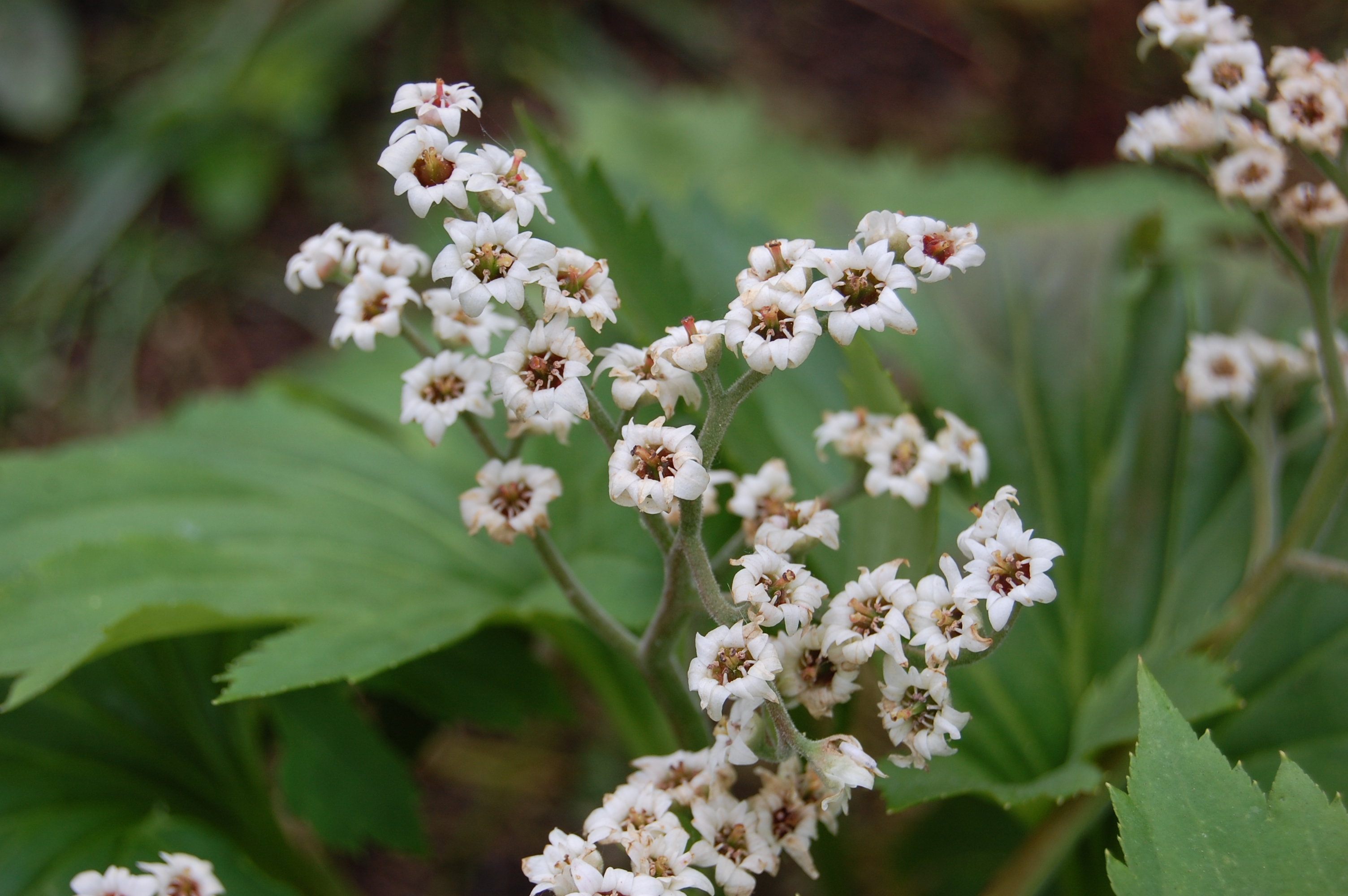

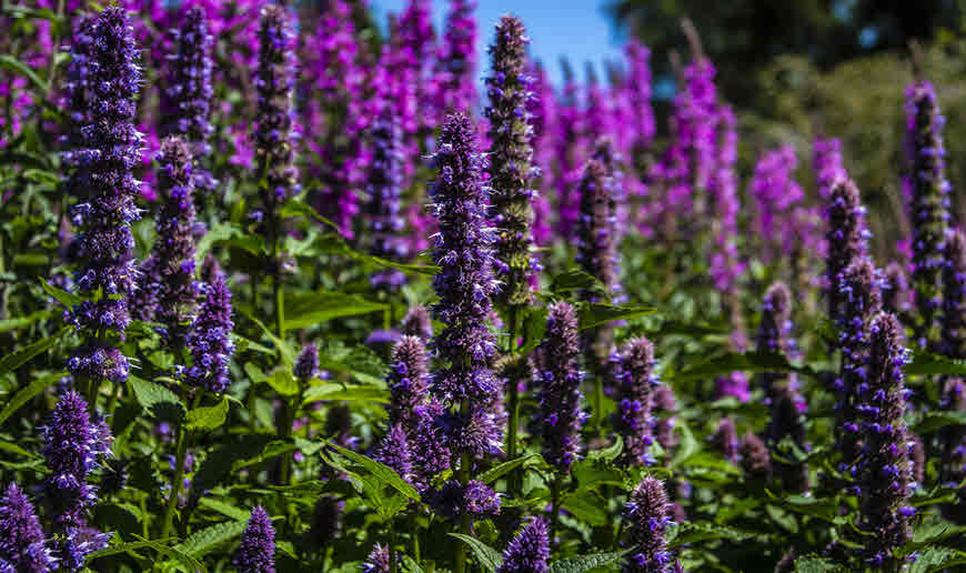

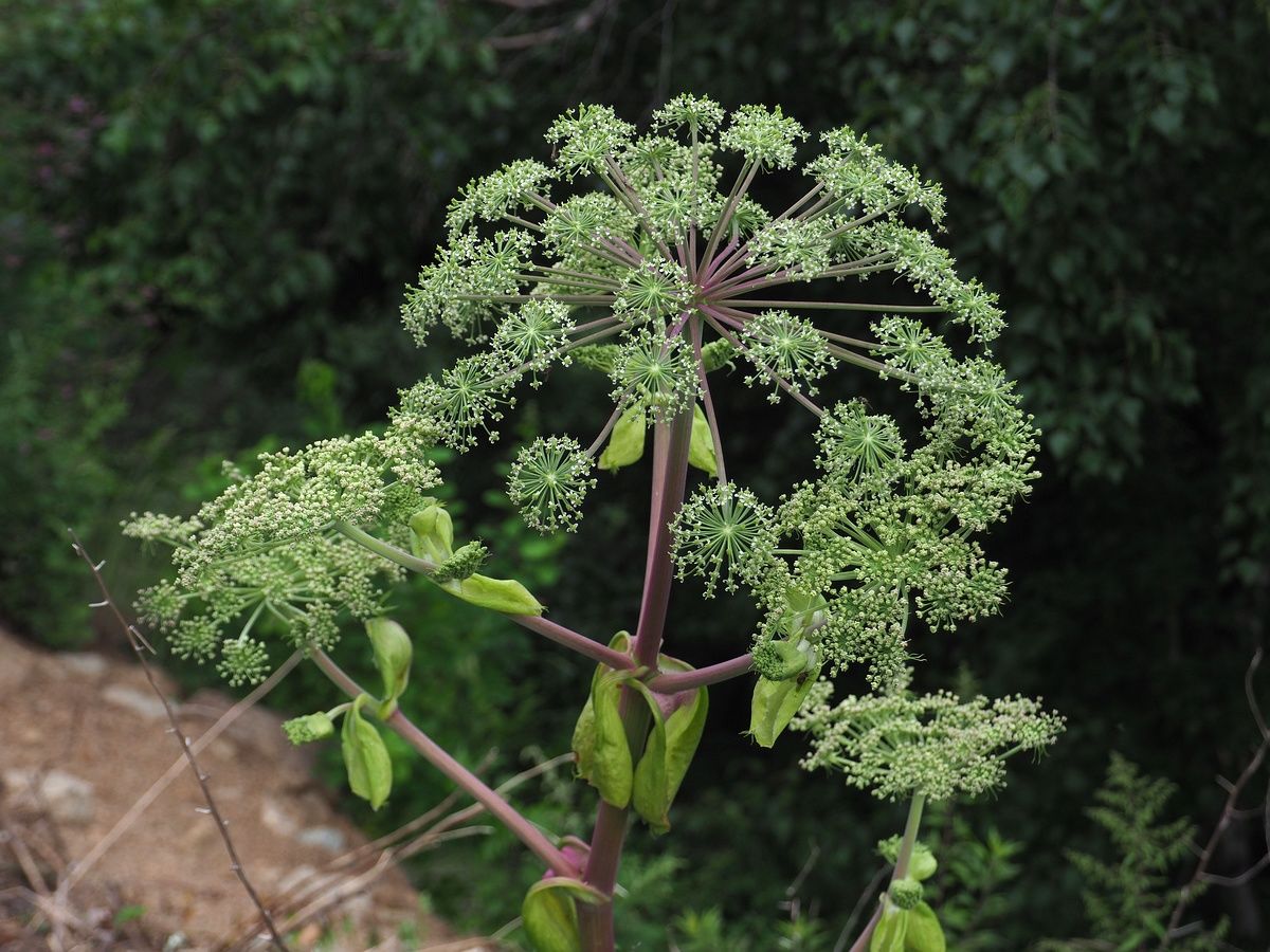
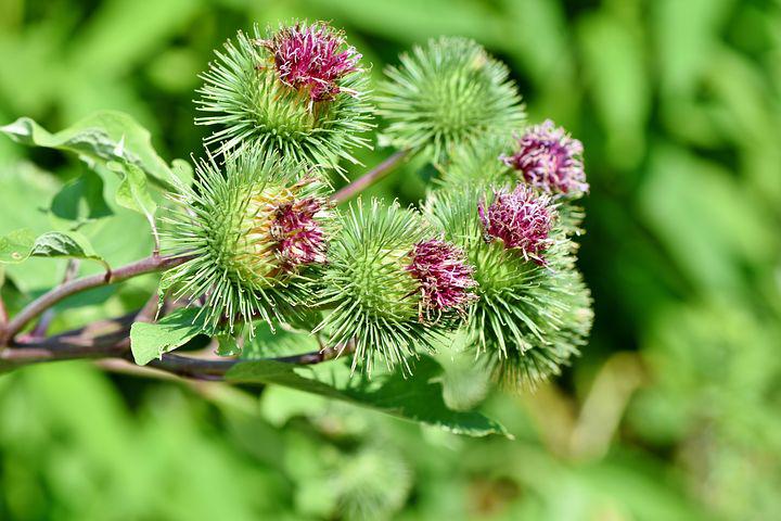
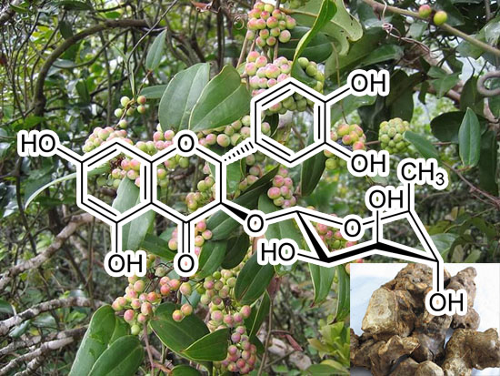
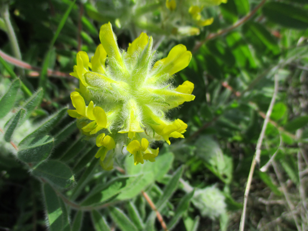


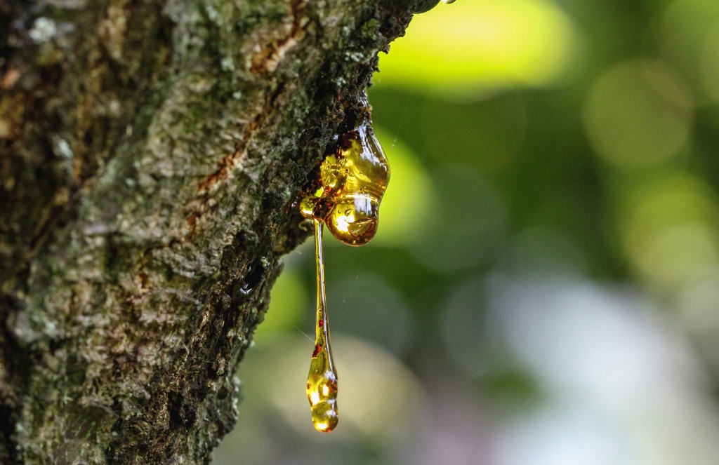
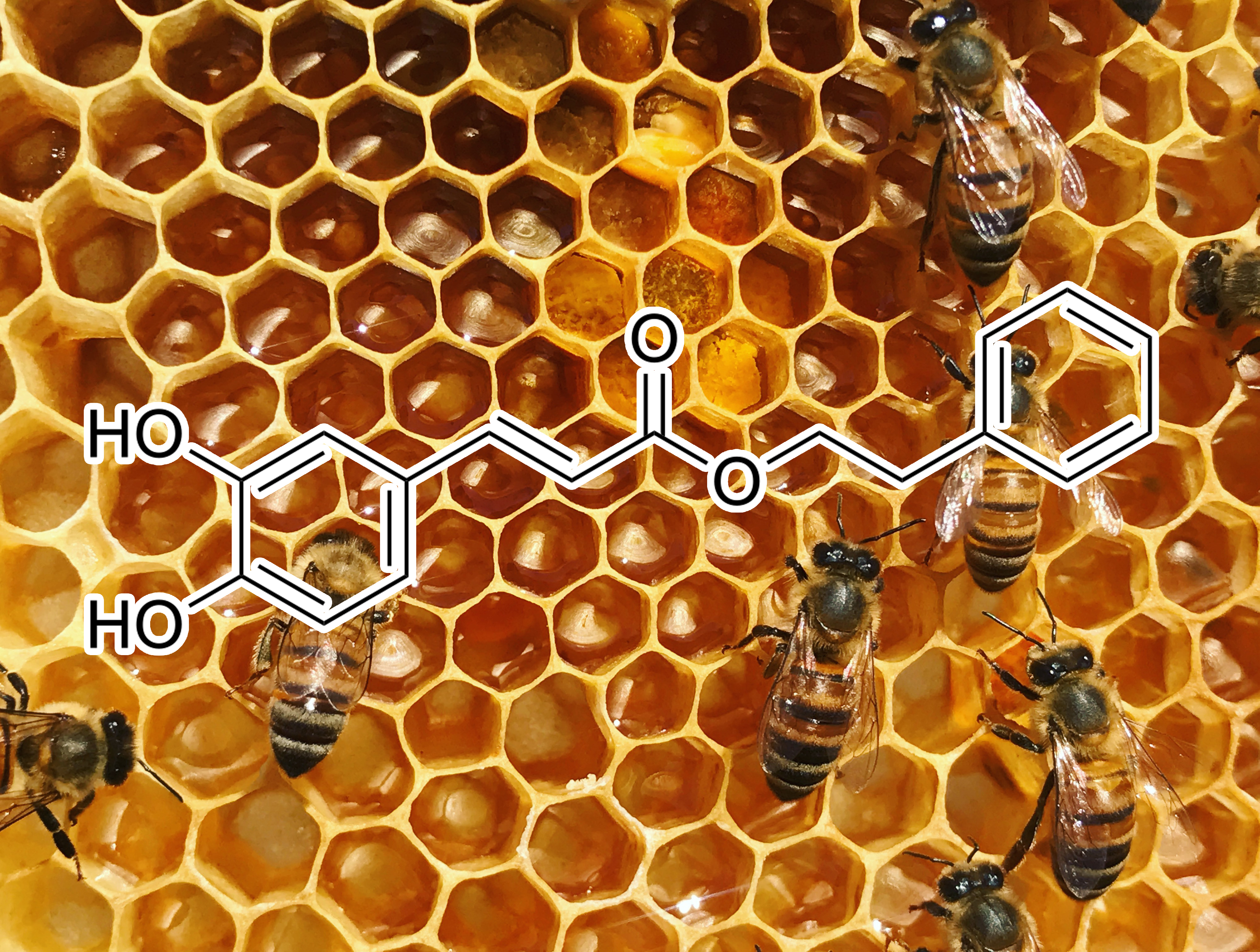

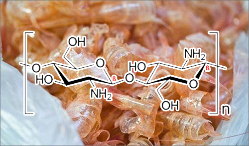

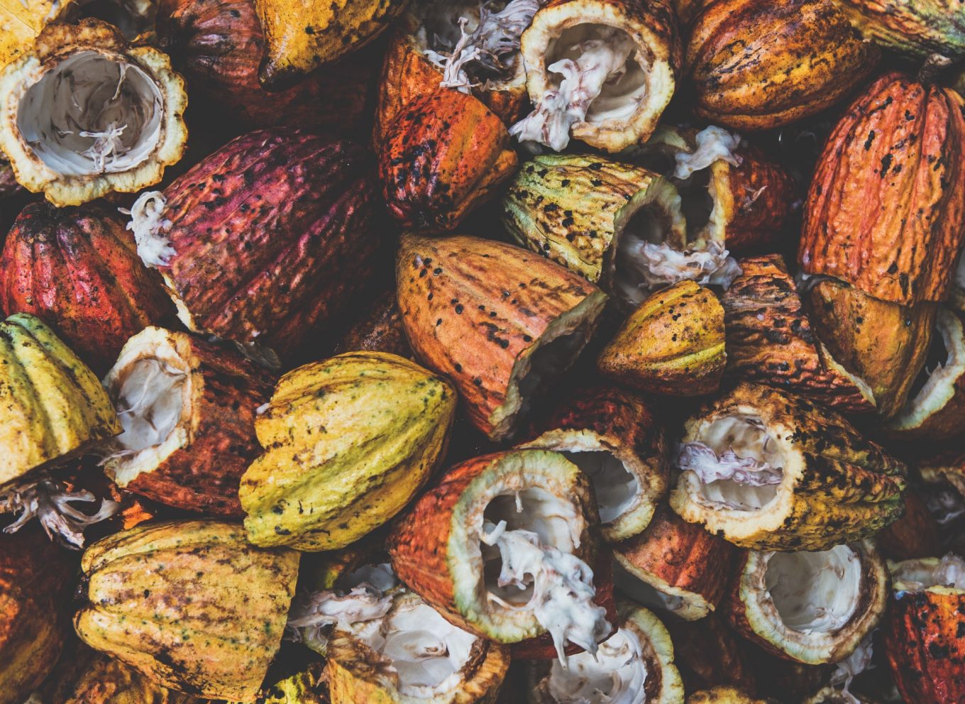
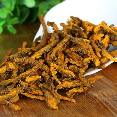


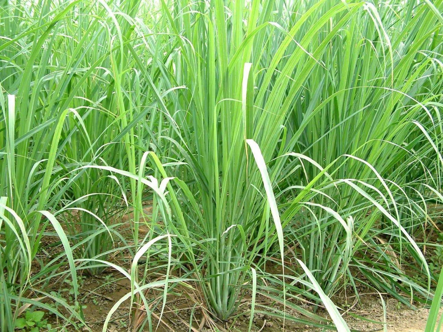
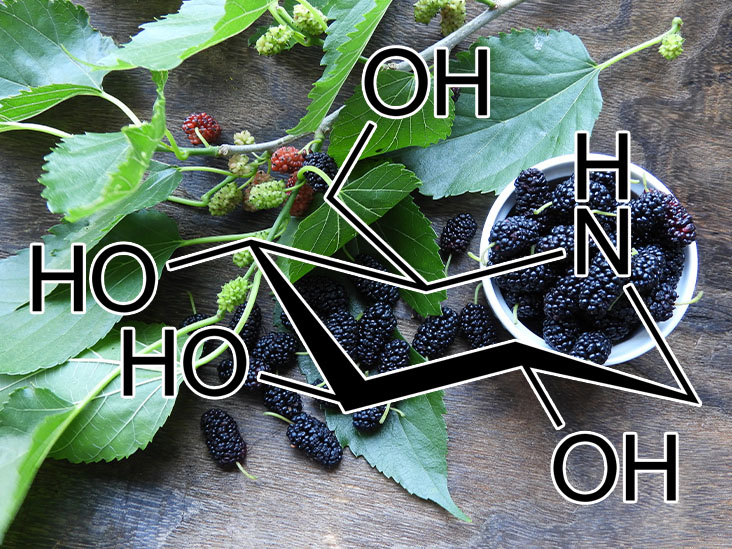
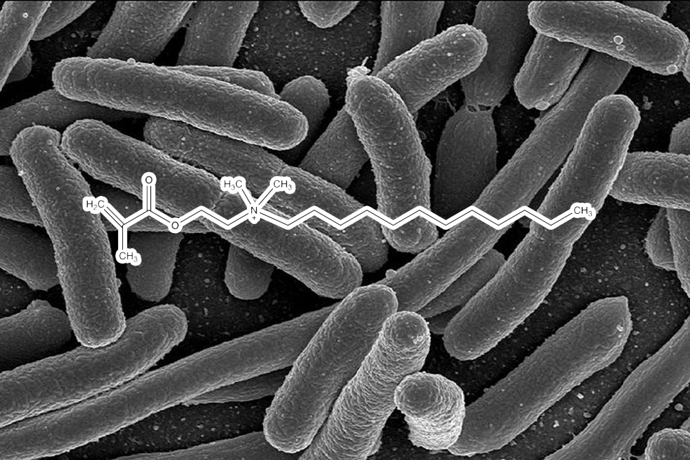
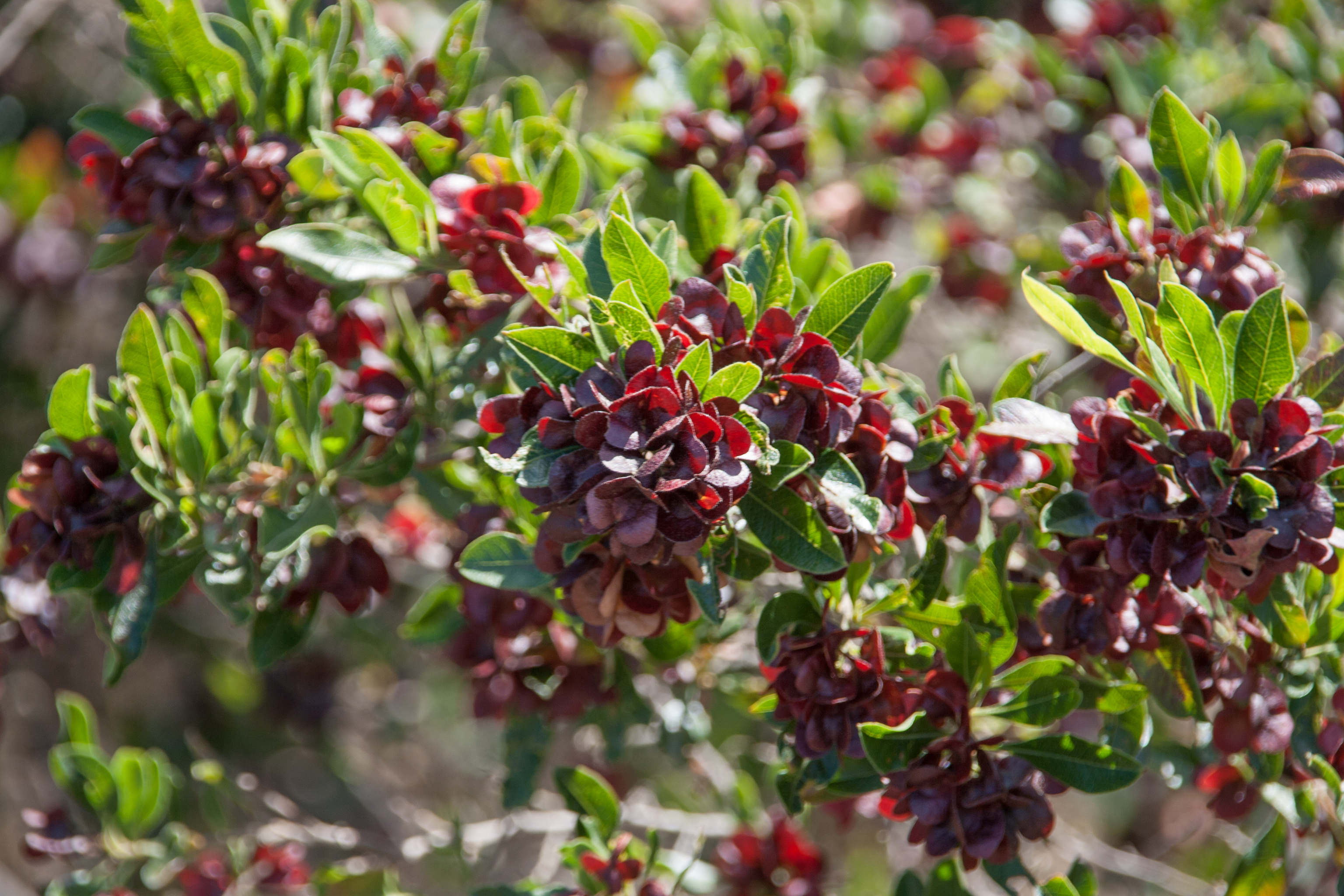

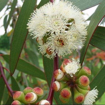

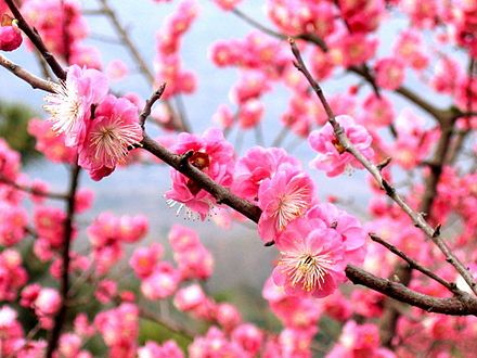

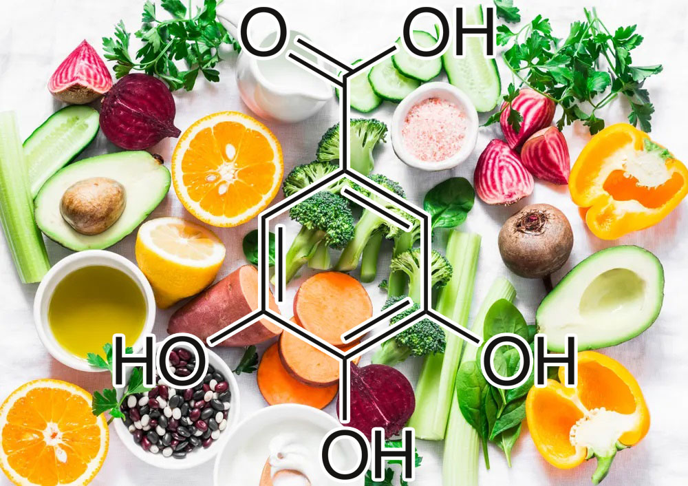

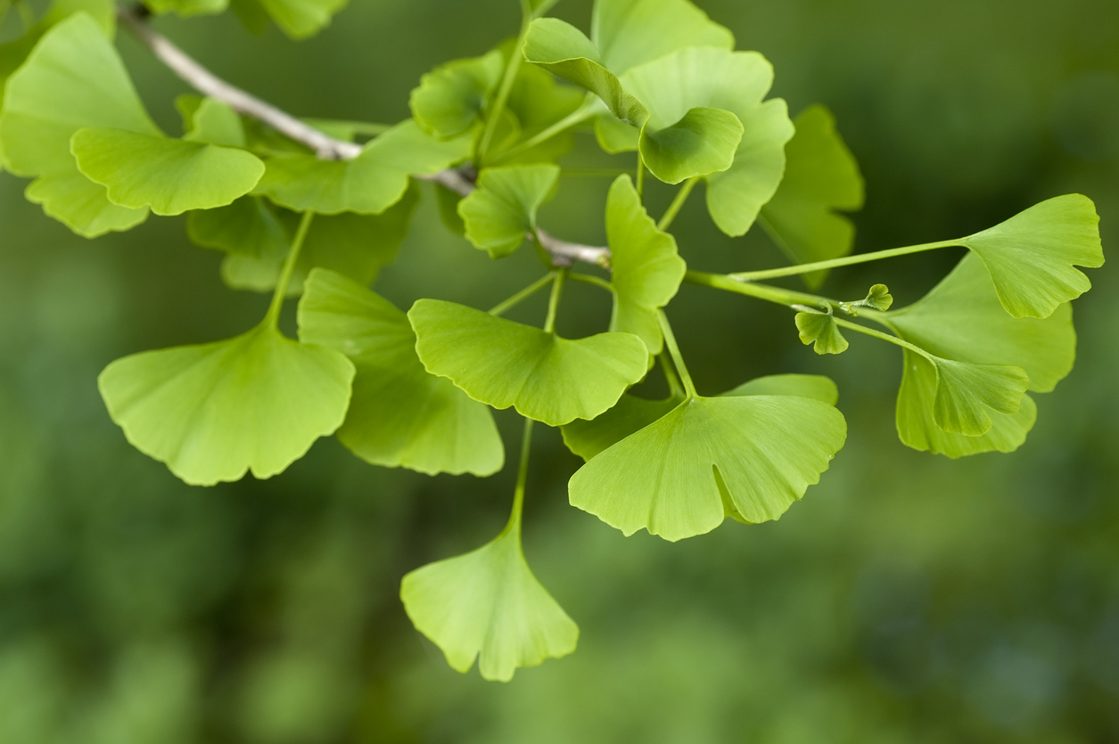
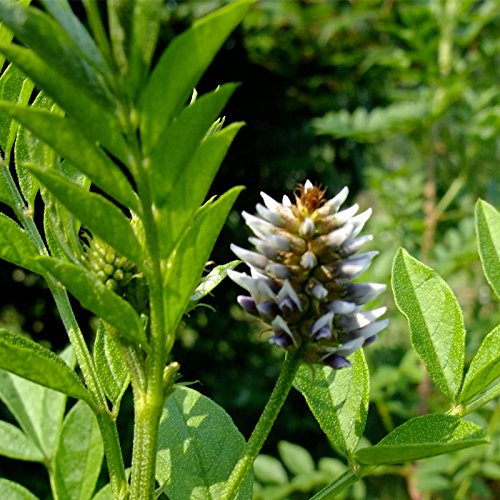
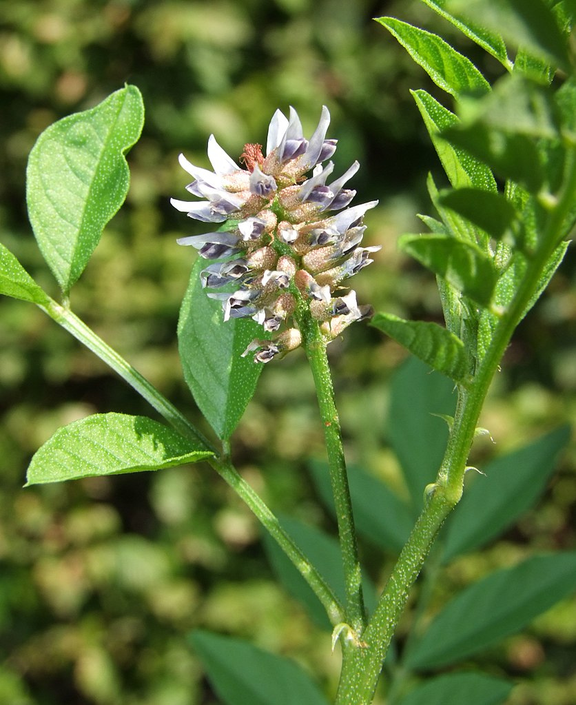
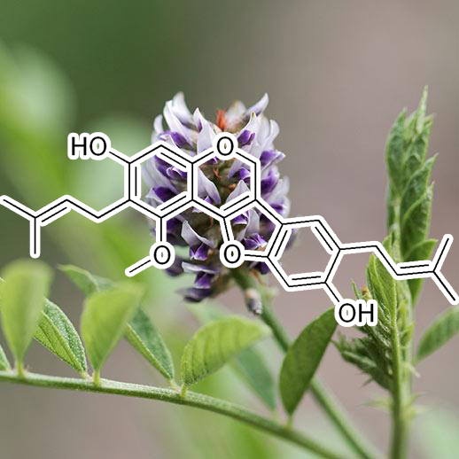


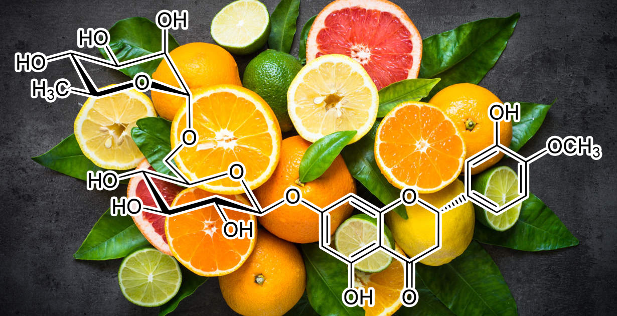


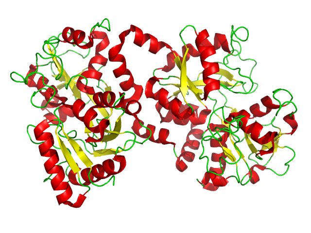

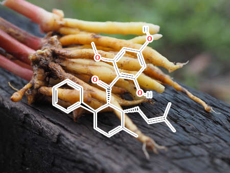


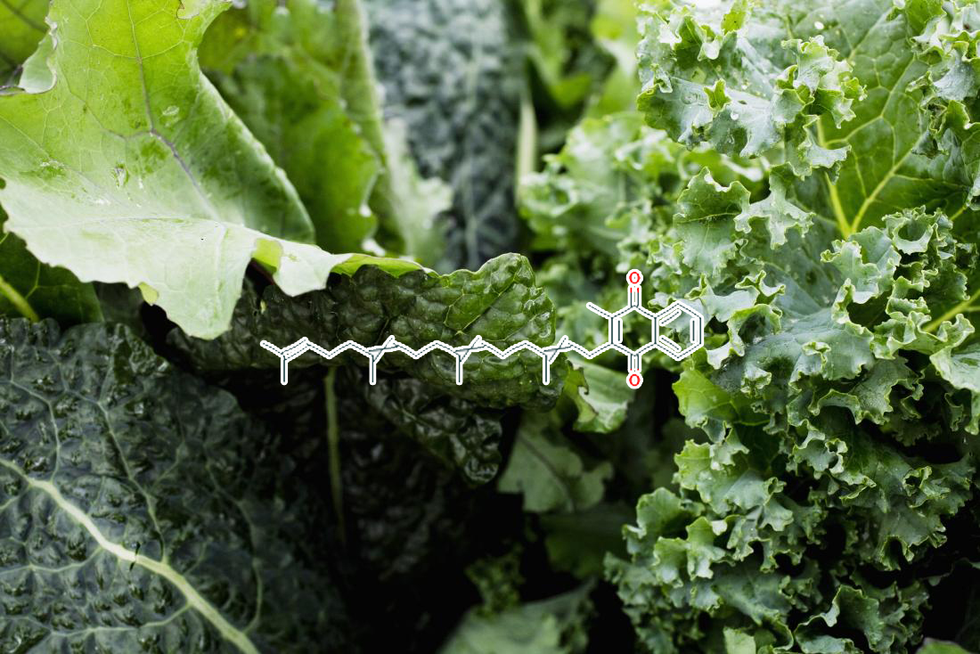
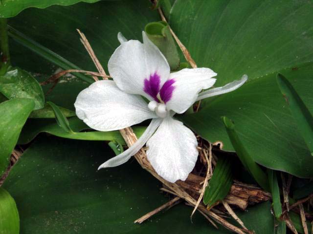

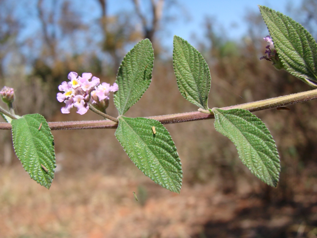

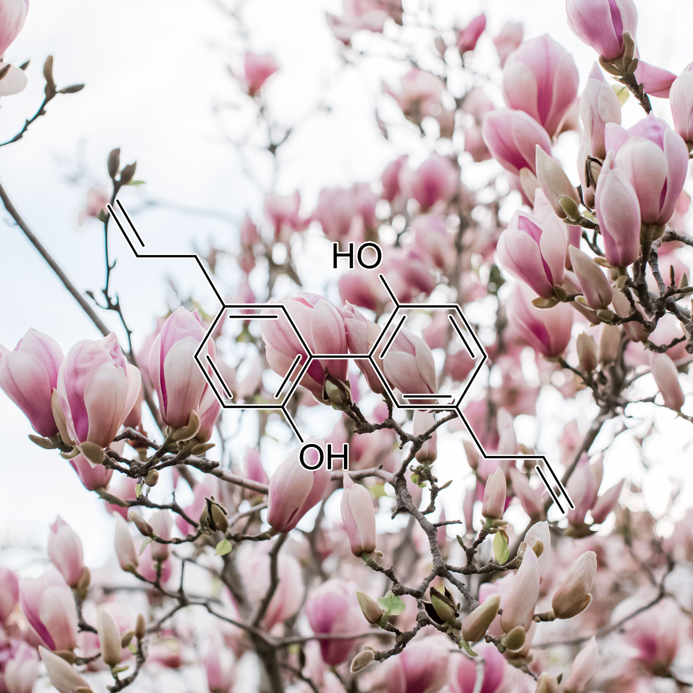
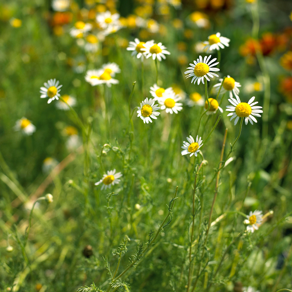
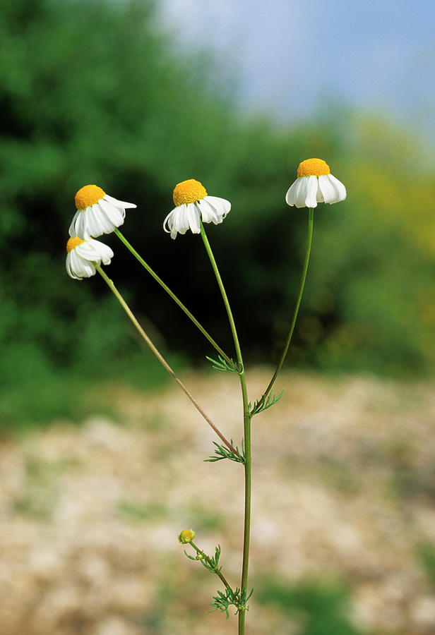


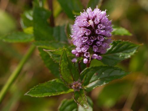

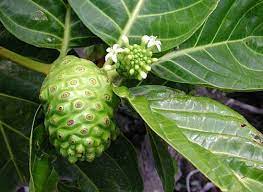
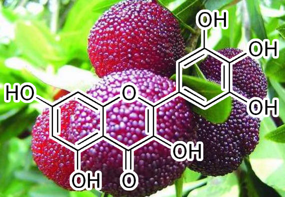
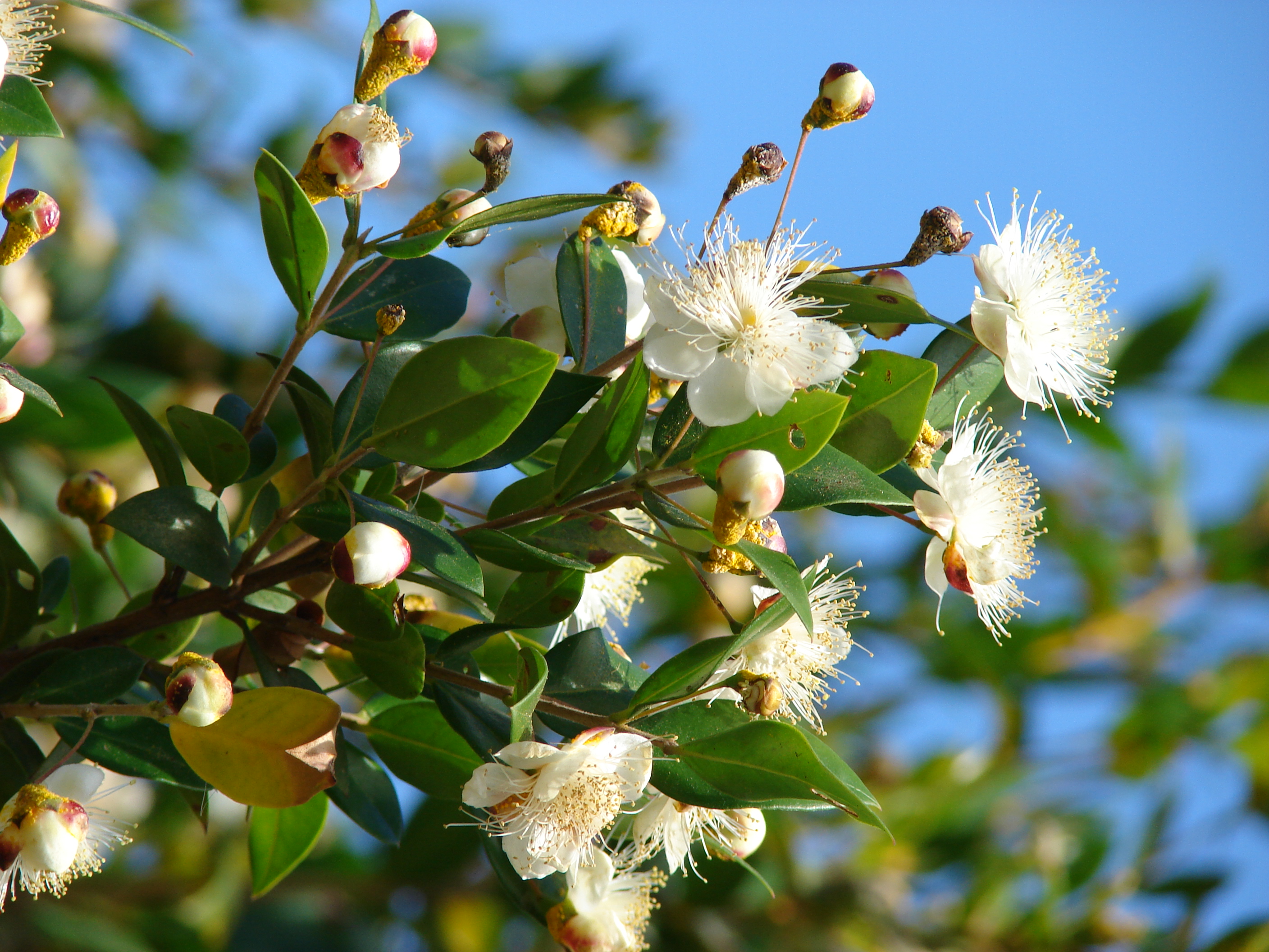

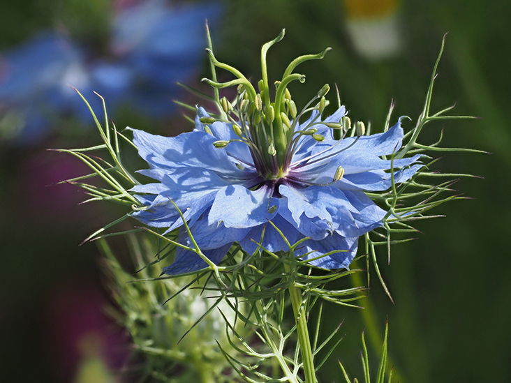
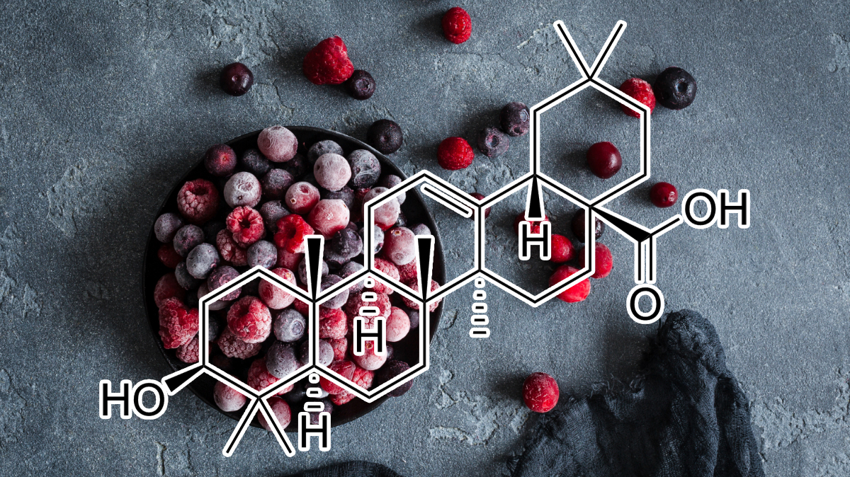
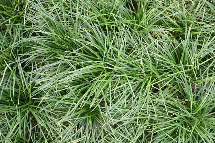


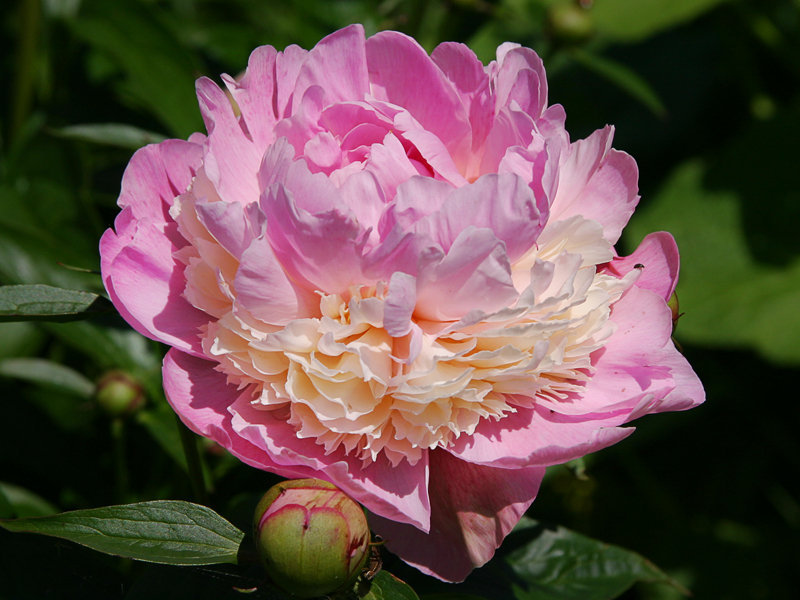

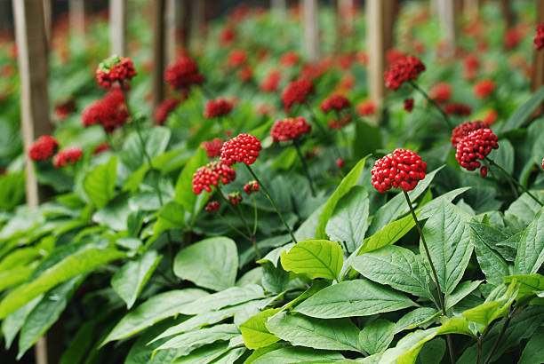

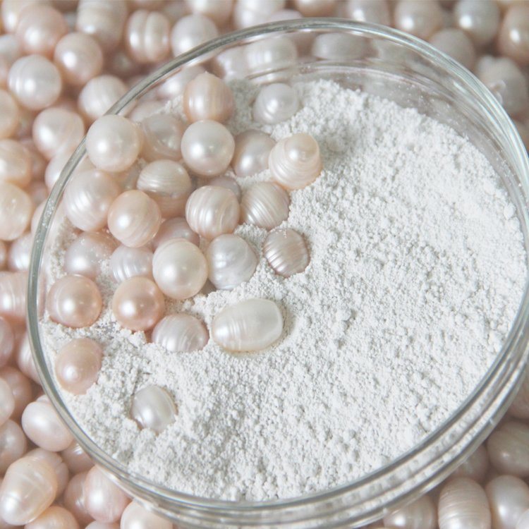
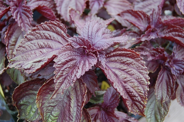

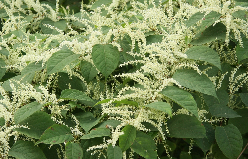


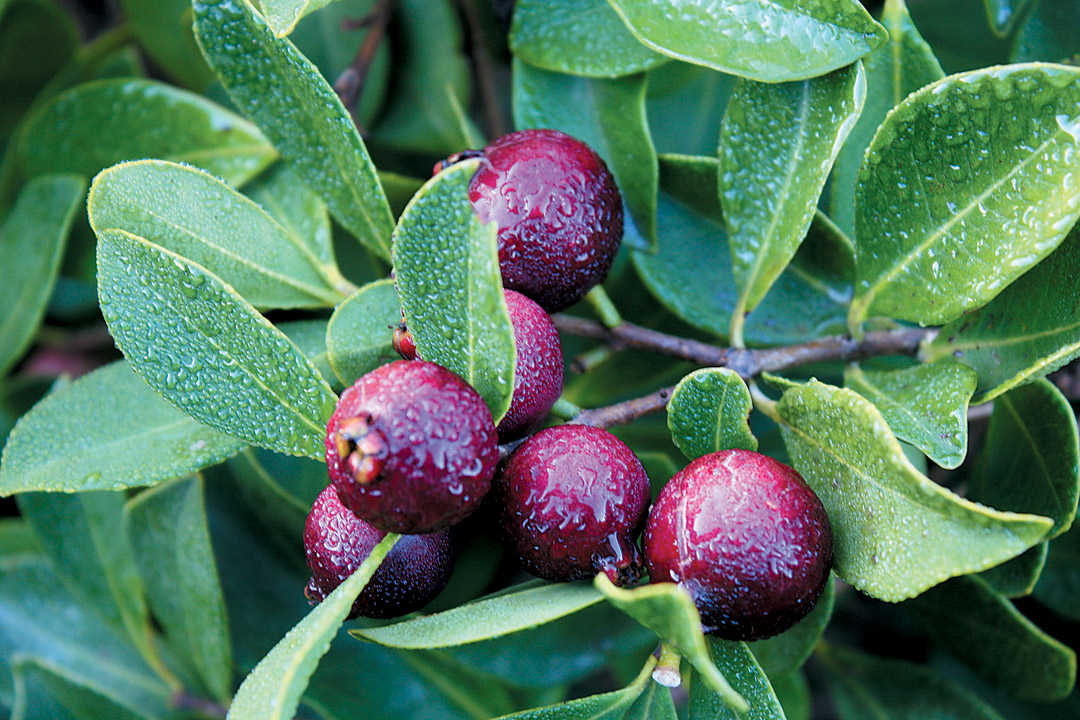
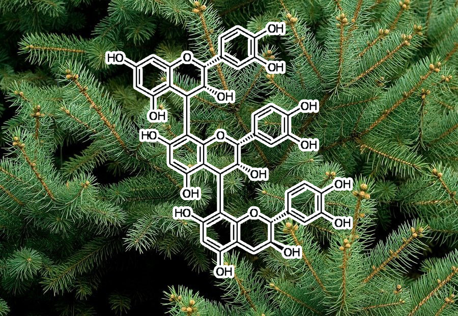
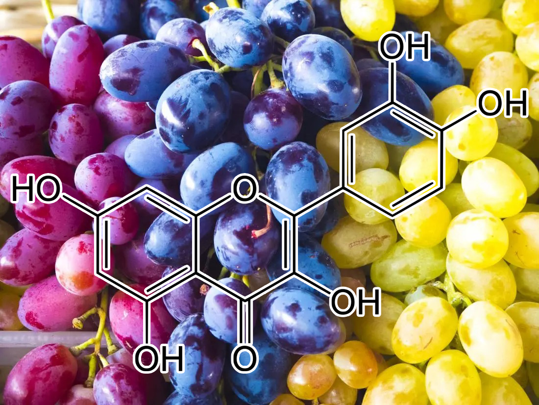
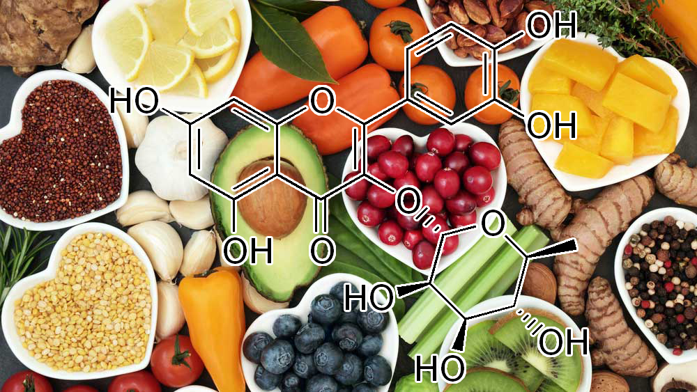


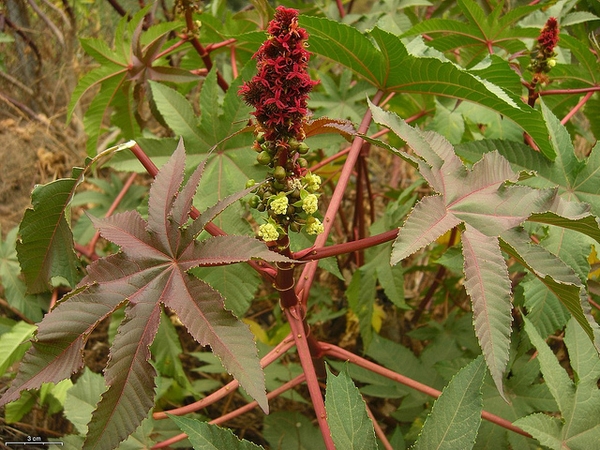
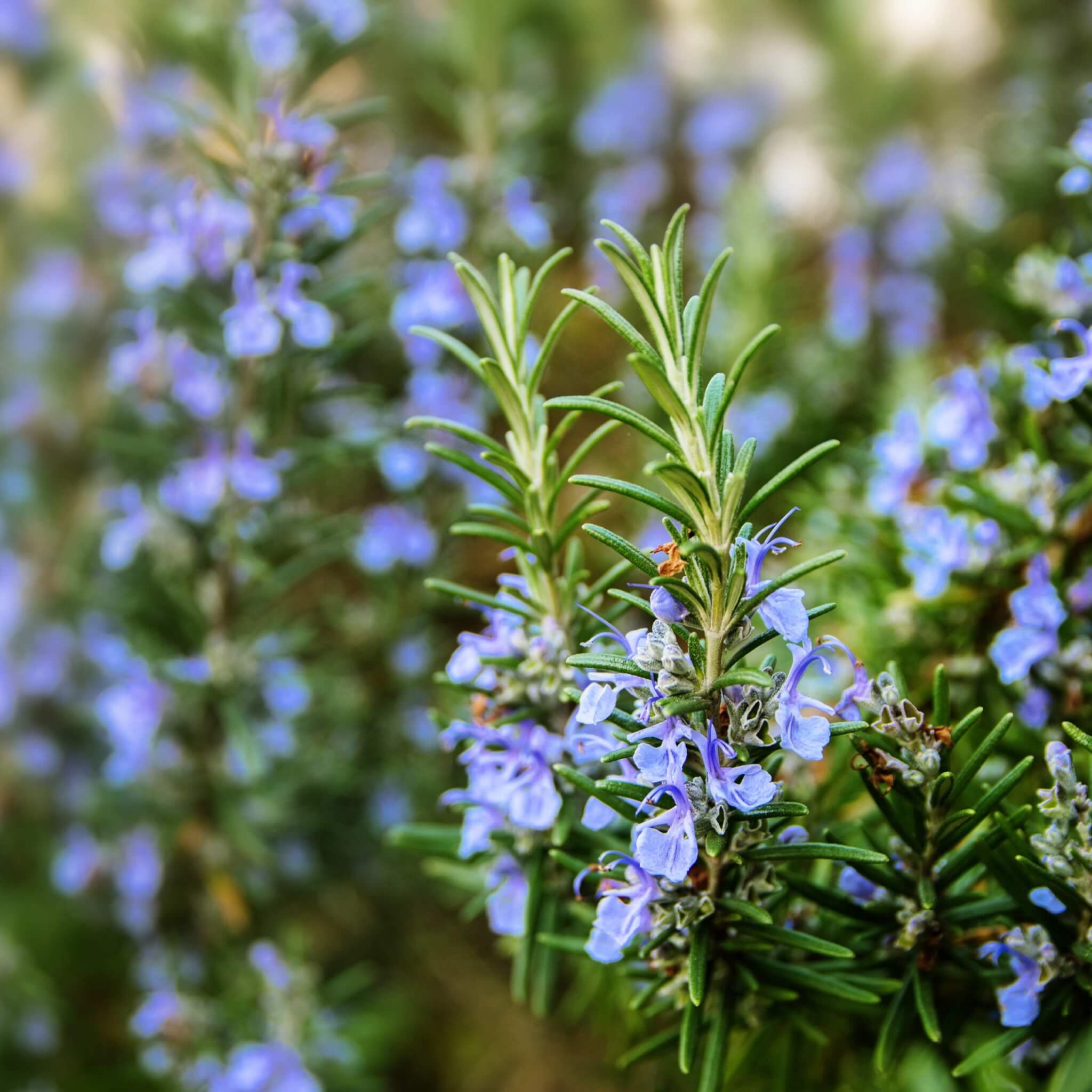


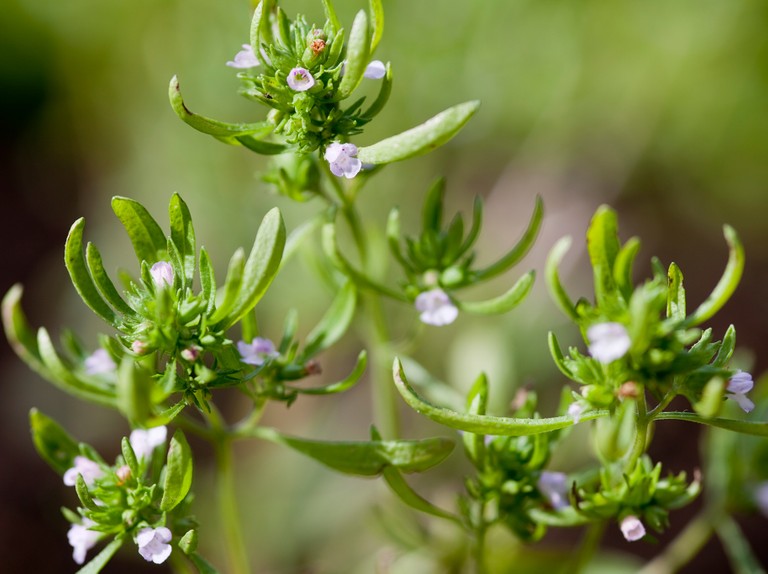
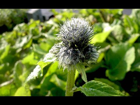
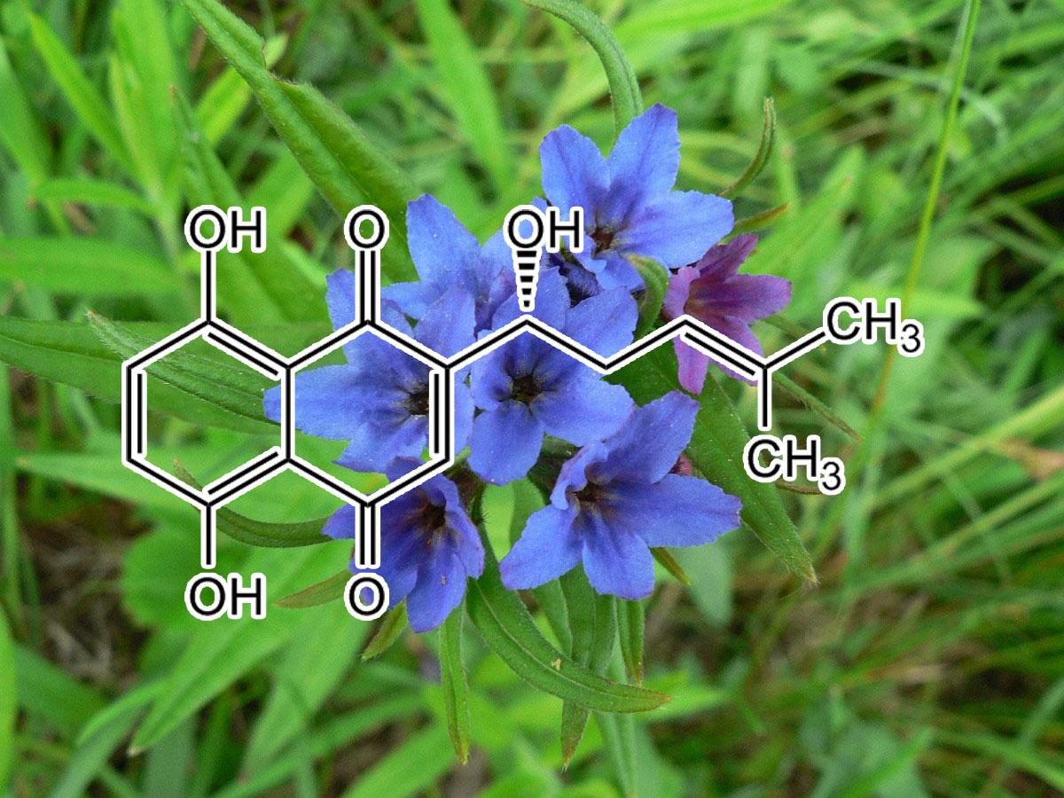

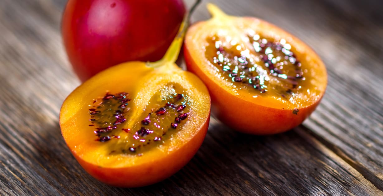
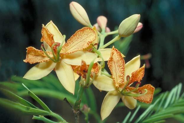
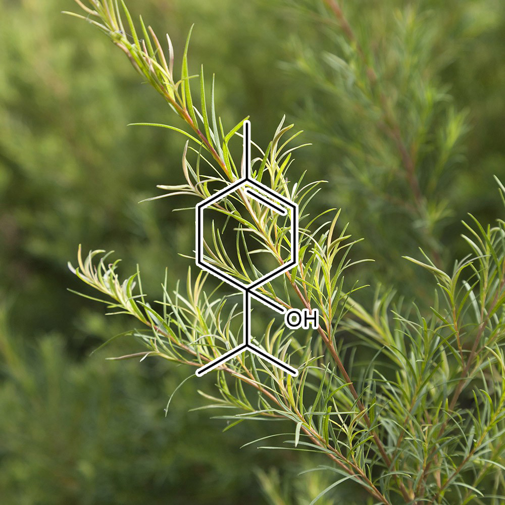

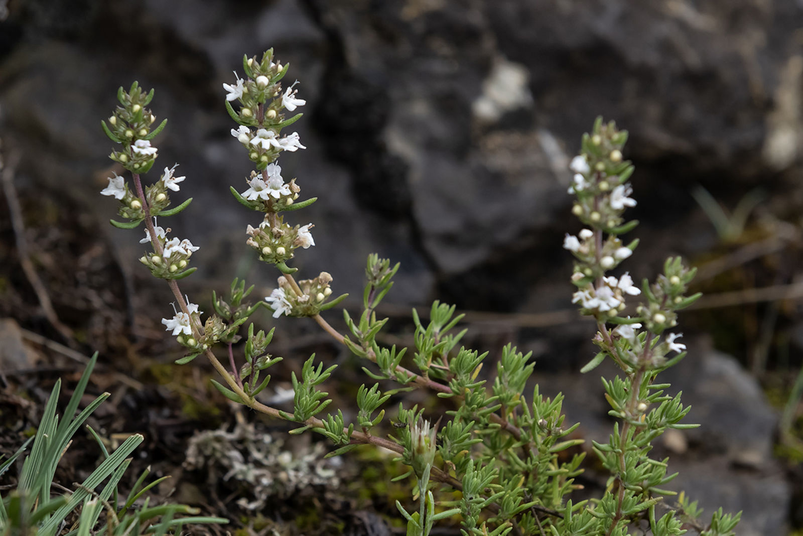





Stephen Vincent Shuman (verified owner) –
This product works fast on gums! Inside of two weeks of following Gavin’s protocol and taking Sabre Tooth both with grapefruit juice and mixing it into my water for my water pic, my gums stopped bleeding when I brushed them! I am excited to see how well it works to re-mineralize teeth!
Jennifer Nix (verified owner) –
SABRETOOTH REVIEW!
“Hey there…. Just sharing some amazing results of yet another blend. I went to my dental cleaning today. I started using Sabertooth prior to dental cleaning even though you recommended to get that first. Anyway, the last time I went they claimed that I had 3 cavities and needed a root canal on top of receding gums. I had only ever had one cavity up until that point at age 49. Soooooooo…… at this appointment my gum line got better by 3 points and miraculously I have zero cavities and do not need a root canal. How bout that??!!!! I would greatly recommend this product to any aging person or persons with gum disease. You’re stuck with me as a client for life!! Lol. I just love every product you’ve put out. Not one of them has disappointed me yet. Thank you so much again. That little potion saved me about $5000 in dental work. BOOM!!
BTW the dental office was shocked as this was only 3 months later. They thought that they had put the wrong films in the wrong chart. LOL”
⭐️⭐️⭐️⭐️⭐️
—Jennifer N
Debra –
I received a sample of Sabretooth to try after getting word from the dentist that my lifelong struggle with gingivitis had turned into full-blown periodontitis. I had 16 gum pockets with a depth of at least five. After a cleaning, and being told I would need to come in at least every 3 months for at least a year for maintenance cleanings, I began brushing with sabretooth and baking soda daily. I returned to the dentist today with only four gum pockets with a depth of four or five (and none deeper!), and the rest of my pockets were measuring back within normal range. I was only told my gums had improved so much that I needed to come back in 5 months, not three. If you have gum disease, this stuff works!!
Tanya Ranney –
Gavin,
Thank you so much for the sample of Sabretooth. All of my life I have fought with my teeth and gums, didn’t seem to matter how much I brushed or flossed they always found gum issues or cavities. I started the Sabretooth supplement and also changed to just using baking soda to brush. I waited until my dental cleaning to write you. I asked her if she thought my teeth were better, same, or worse….and she said better! I can also tell the lack of buildup on my teeth and no gum irritation. I would highly recommend this supplement! Again, thank you Gavin…you never disappoint.
krishna sharma –
I’ve had a recurring infection in my gums from wisdom tooth complications. If anyone has experienced the same, they know how painful it is. I haven’t taken a painkiller or any Pharma pill in years, but I was forced to the dentist by the pain just to ensure it isn’t anything more serious. They gave me a prescription and a cleaning protocol.
I returned from uni my home in London and opened the Sabre Tooth sample that Gavin had actually sent as a free sample from my previous order. Following his advice and incorporation of baking soda, my infection was healed not in a week, or two, but in days.
I am not even surprised at the power of natural remedies, specifically the blends anymore.